Review
. 2021 Mar 16;2(3):100216.
doi: 10.1016/j.xcrm.2021.100216.

Toward a broader view of mechanisms of drug cardiotoxicity

Affiliations
Review

Toward a broader view of mechanisms of drug cardiotoxicity

Polina Mamoshina et al. Cell Rep Med. .

Abstract

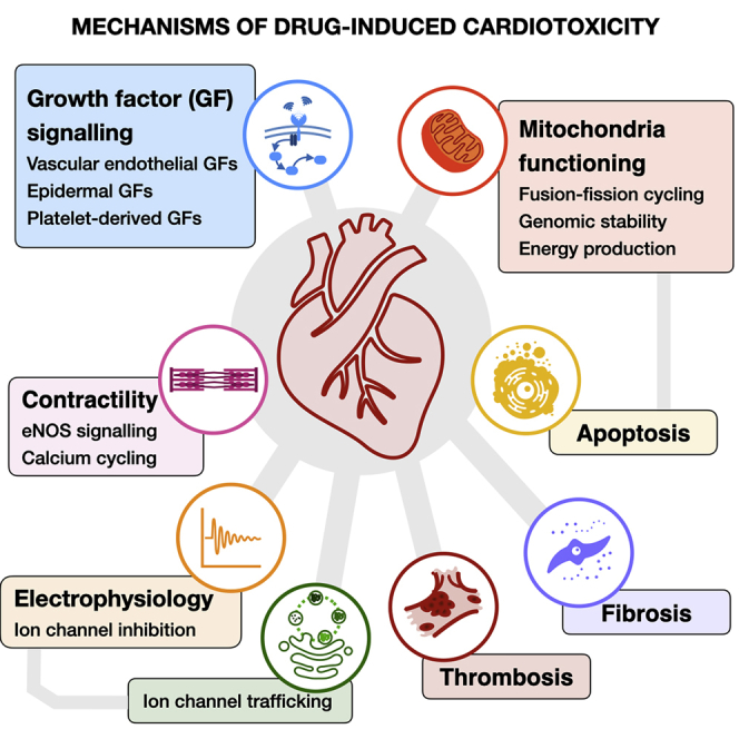
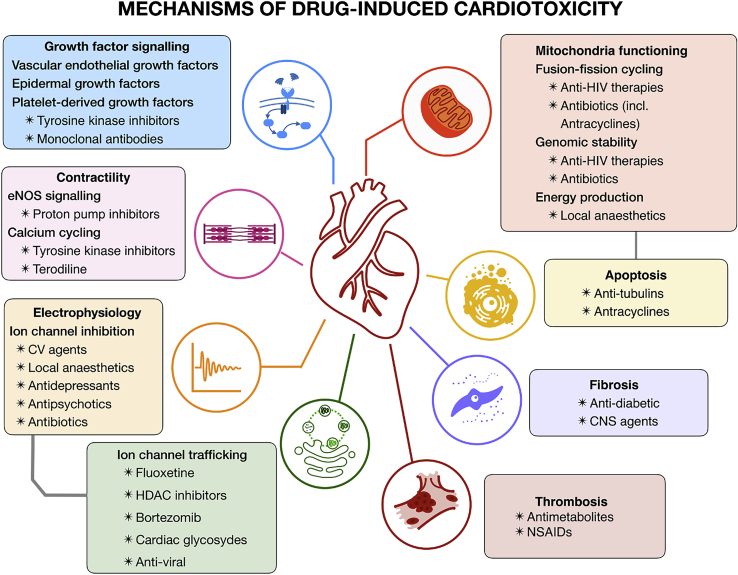
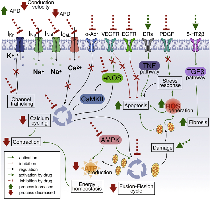
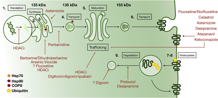
Cardiotoxicity, defined as toxicity that affects the heart, is one of the most common adverse drug effects. Numerous drugs have been shown to have the potential to induce lethal arrhythmias by affecting cardiac electrophysiology, which is the focus of current preclinical testing. However, a substantial number of drugs can also affect cardiac function beyond electrophysiology. Within this broader sense of cardiotoxicity, this review discusses the key drug-protein interactions known to be involved in cardiotoxic drug response. We cover adverse effects of anticancer, central nervous system, genitourinary system, gastrointestinal, antihistaminic, anti-inflammatory, and anti-infective agents, illustrating that many share mechanisms of cardiotoxicity, including contractility, mitochondrial function, and cellular signaling.

Keywords: adverse reactions; cardiotoxicity; cell signaling; mechanisms of toxicity; side effects.


Conflict of interest statement

The authors declare no competing interests.

Figures

None
Graphical abstract
Figure 1
Figure 1
Drugs withdrawn from market due to cardiotoxicity up to May 2020 (A) Number of drugs withdrawn according to therapeutic area. (B) Lifespan of withdrawn agents. CNS, central nervous system.
Figure 2
Figure 2
Mechanisms of drug-induced cardiotoxicity Antibiotics and antiviral therapies can induce mitochondrial dysfunction, leading to an impaired fusion-fission cycle. Local anesthetics increase mitochondrial permeability, affecting function. Cardiotoxicity of tyrosine kinase inhibitors and monoclonal antibodies is primarily linked to inhibition of major signaling pathways for cardiomyocyte survival and maintenance. Different CNS and antidiabetic agents have been associated with the emergence of cardiac fibrosis. Proton pump inhibitors can affect cardiac contractility via inhibition of NO synthesis. Similarly, tyrosine kinases inhibitors have been linked to calcium cycling dysregulation. Multiple neural, cardiovascular, and anti-infective agents also have been shown to interact with cardiac electrophysiology. Equivalently, cardiac adverse effects of fluoxetine, HDAC inhibitors, and bortezomib are primarily linked to affecting ion channel trafficking rather than acute channel block. CV, cardiovascular; NSAIDs, nonsteroidal anti-inflammatory drugs; HDAC, histone deacetylase; eNOS, endothelial nitric oxide synthase.
Figure 3
Figure 3
Overview of mechanisms of cardiotoxicity Abnormalities in action potential duration or conduction velocity are associated with a direct block of ionic currents or inhibition of their trafficking from nucleus to cell membrane. Multiple pathways can trigger apoptosis, including VEGFR or EGFR inhibition (cardiomyocyte survival), PDGF inhibition (compensatory stress response), DR-induced TNF signaling activation, mitochondrial damage, or elevated ROS levels. α-Adr, VEGFR signaling, or eNOS inhibition affects calcium cycling. AMPK signaling inhibition affects both the mitochondrial fusion-fission cycle and the production of ATP. Serotonin-induced activation of the TGF-β pathway is primarily linked to cardiac fibrosis induction. IKr, rapid delayed rectifier current; INa, inward sodium current; INaL, late inward sodium current; ICaL, L-type calcium current; α-Adr, alpha-adrenergic receptor; VEGFR, vascular endothelial growth factor receptor; EGFR, epidermal growth factor receptor; DRs, death receptors; PDGF, platelet-derived growth factor; 5-HT2β, 5-hydroxytryptamine receptor (serotonin receptor 2B); TNF, tumor necrosis factor; TGF-β, transforming growth factor beta; ROS, reactive oxygen species; CaMKII, calcium/calmodulin-dependent protein kinase II; AMPK, AMP-activated protein kinase; ATP, adenosine triphosphate.
Figure 4
Figure 4
Schematic of hERG biogenesis, trafficking, and degradation and pathways of drug-induced IKr deficiency Subcellular processes modulating hERG expression on the cellular membrane include (1) mRNA synthesis, (2) synthesis of polypeptide chain, (3) transport of polypeptide from endoplasmic reticulum to Golgi apparatus with Hsp70 and Hsp90 chaperones attached, (4) transport of the polypeptide in the COPII-mediated vesicle, (5) glycosylation, (6) exocytosis, (7) endocytosis, (8) ubiquitination, and (9) degradation of hERG.

References

    1. Ferri N., Siegl P., Corsini A., Herrmann J., Lerman A., Benghozi R. Drug attrition during pre-clinical and clinical development: understanding and managing drug-induced cardiotoxicity. Pharmacol. Ther. 2013;138:470–484. - PubMed
    1. Ford G.A., Wood S.M., Daly A.K. CYP2D6 and CYP2C19 genotypes of patients with terodiline cardiotoxicity identified through the yellow card system. Br. J. Clin. Pharmacol. 2000;50:77–80. - PMC - PubMed
    1. Garcia-Pavia P., Kim Y., Restrepo-Cordoba M.A., Lunde I.G., Wakimoto H., Smith A.M., Toepfer C.N., Getz K., Gorham J., Patel P. Genetic Variants Associated With Cancer Therapy-Induced Cardiomyopathy. Circulation. 2019;140:31–41. - PMC - PubMed
    1. Sloan J.A., Goldberg R.M., Sargent D.J., Vargas-Chanes D., Nair S., Cha S.S., Novotny P.J., Poon M.A., O’Connell M.J., Loprinzi C.L. Women experience greater toxicity with fluorouracil-based chemotherapy for colorectal cancer. J. Clin. Oncol. 2002;20:1491–1498. - PubMed
    1. Zhao L., Zhang B. Doxorubicin induces cardiotoxicity through upregulation of death receptors mediated apoptosis in cardiomyocytes. Sci. Rep. 2017;7:44735. - PMC - PubMed

Publication types

MeSH terms