

Emerging methods for the characterization of ischemic heart disease: ultrafast Doppler angiography, micro-CT, photon-counting CT, novel MRI and PET techniques, and artificial intelligence
- PMID: 33763754
- PMCID: PMC7991013
- DOI: 10.1186/s41747-021-00207-3
Emerging methods for the characterization of ischemic heart disease: ultrafast Doppler angiography, micro-CT, photon-counting CT, novel MRI and PET techniques, and artificial intelligence
Abstract
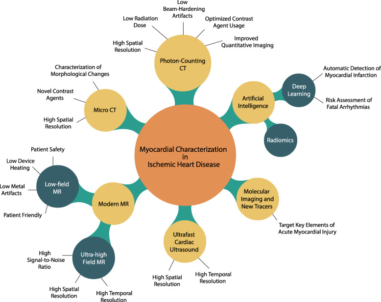
After an ischemic event, disruptive changes in the healthy myocardium may gradually develop and may ultimately turn into fibrotic scar. While these structural changes have been described by conventional imaging modalities mostly on a macroscopic scale-i.e., late gadolinium enhancement at magnetic resonance imaging (MRI)-in recent years, novel imaging methods have shown the potential to unveil an even more detailed picture of the postischemic myocardial phenomena. These new methods may bring advances in the understanding of ischemic heart disease with potential major changes in the current clinical practice. In this review article, we provide an overview of the emerging methods for the non-invasive characterization of ischemic heart disease, including coronary ultrafast Doppler angiography, photon-counting computed tomography (CT), micro-CT (for preclinical studies), low-field and ultrahigh-field MRI, and 11C-methionine positron emission tomography. In addition, we discuss new opportunities brought by artificial intelligence, while addressing promising future scenarios and the challenges for the application of artificial intelligence in the field of cardiac imaging.
Keywords: Artificial intelligence; Coronary artery disease; Myocardial infarction; Myocardial ischemia; Radiology.
Conflict of interest statement
MJW: Activities related to the present article: none. Activities not related to the present article: research grants from American Heart Association (18POST34030192), Philips Healthcare, and Stanford University, consulting for Arterys, Inc, and co-founder/shareholder of Segmed, Inc. Other relationships: disclosed no relevant relationships.
AVS receives institutional research support and/or personal fees from Elucid Bioimaging and Siemens. AVS is one of the Guest Editors of this thematic series. The paper was therefore reviewed and handled by the
UJS receives institutional research support and/or personal fees from Astellas, Bayer, Bracco, Elucid Bioimaging, Guerbet, HeartFlow, and Siemens.
KN received institutional research support from Siemens Healthineers, Bayer healthcare, GE Healthcare, and Heartflow Inc.
DF: Activities related to the present article: none. Activities not related to the present article: received research support from Siemens Healthineers and GE Healthcare; is on the Speakers’ Bureau at Siemens Healthineers; has ownership interest in iSchemaView. Other relationships: disclosed no relevant relationships. The other authors have no conflict of interest to disclose.
Figures
References
-
- GBD 2017 Causes of Death Collaborators (2018) Global, regional, and national age-sex-specific mortality for 282 causes of death in 195 countries and territories, 1980-2017: a systematic analysis for the Global Burden of Disease Study 2017. Lancet 392:1736-1788 10.1016/S0140-6736(18)32203-7 - PMC - PubMed
-
- Pellikka PA, Arruda-Olson A, Chaudhry FA et al (2020) Guidelines for performance, interpretation, and application of stress echocardiography in ischemic heart disease: from the American Society of Echocardiography. J Am Soc Echocardiogr 33:e48 10.1016/j.echo.2019.07.001 - PubMed
Publication types
MeSH terms
Substances
Grants and funding
LinkOut - more resources
Full Text Sources
Other Literature Sources