

Effects of the Competitive Season and Off-Season on Knee Articular Cartilage in Collegiate Basketball Players Using Quantitative MRI: A Multicenter Study
- PMID: 33763929
- PMCID: PMC8817387
- DOI: 10.1002/jmri.27610
Effects of the Competitive Season and Off-Season on Knee Articular Cartilage in Collegiate Basketball Players Using Quantitative MRI: A Multicenter Study
Abstract
Background: Injuries to the articular cartilage in the knee are common in jumping athletes, particularly high-level basketball players. Unfortunately, these are often diagnosed at a late stage of the disease process, after tissue loss has already occurred.
Purpose/hypothesis: To evaluate longitudinal changes in knee articular cartilage and knee function in National Collegiate Athletic Association (NCAA) basketball players and their evolution over the competitive season and off-season.
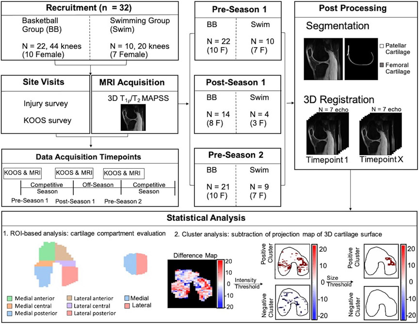
Study type: Longitudinal, multisite cohort study.
Population: Thirty-two NCAA Division 1 athletes: 22 basketball players and 10 swimmers.
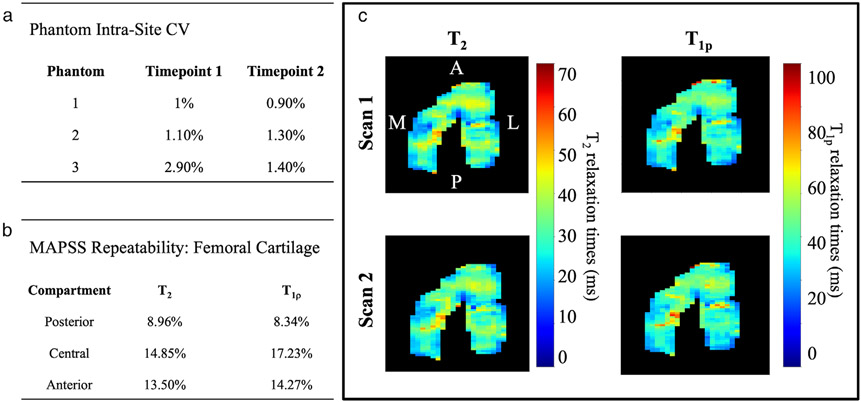
Field strength/sequence: Bilateral magnetic resonance imaging (MRI) using a combined T1ρ and T2 magnetization-prepared angle-modulated portioned k-space spoiled gradient-echo snapshots (MAPSS) sequence at 3T.
Assessment: We calculated T2 and T1ρ relaxation times to compare compositional cartilage changes between three timepoints: preseason 1, postseason 1, and preseason 2. Knee Osteoarthritis Outcome Scores (KOOS) were used to assess knee health.
Statistical tests: One-way variance model hypothesis test, general linear model, and chi-squared test.
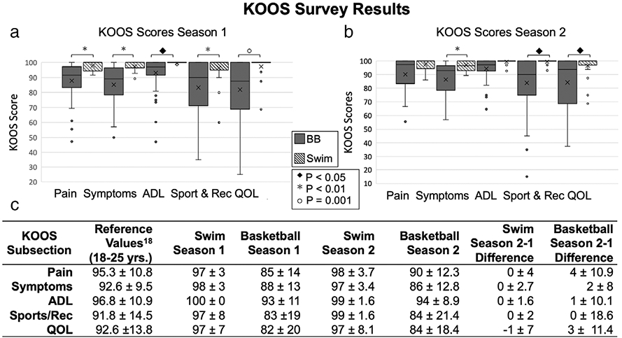
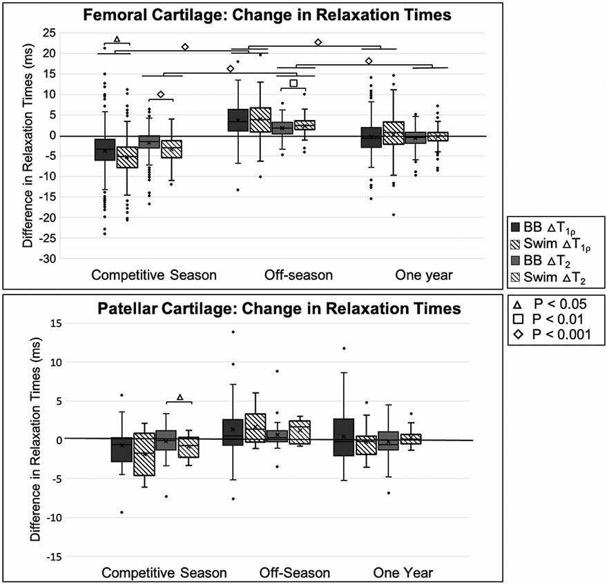
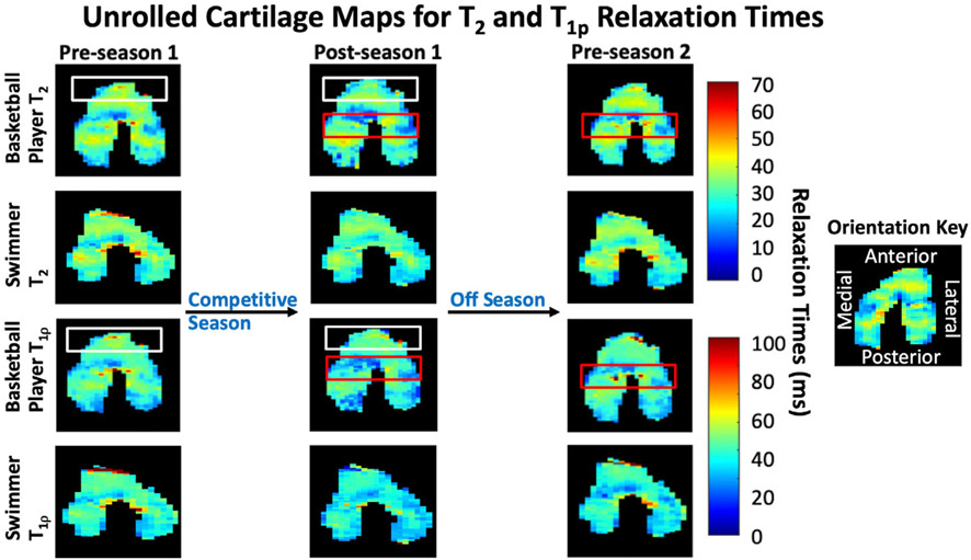
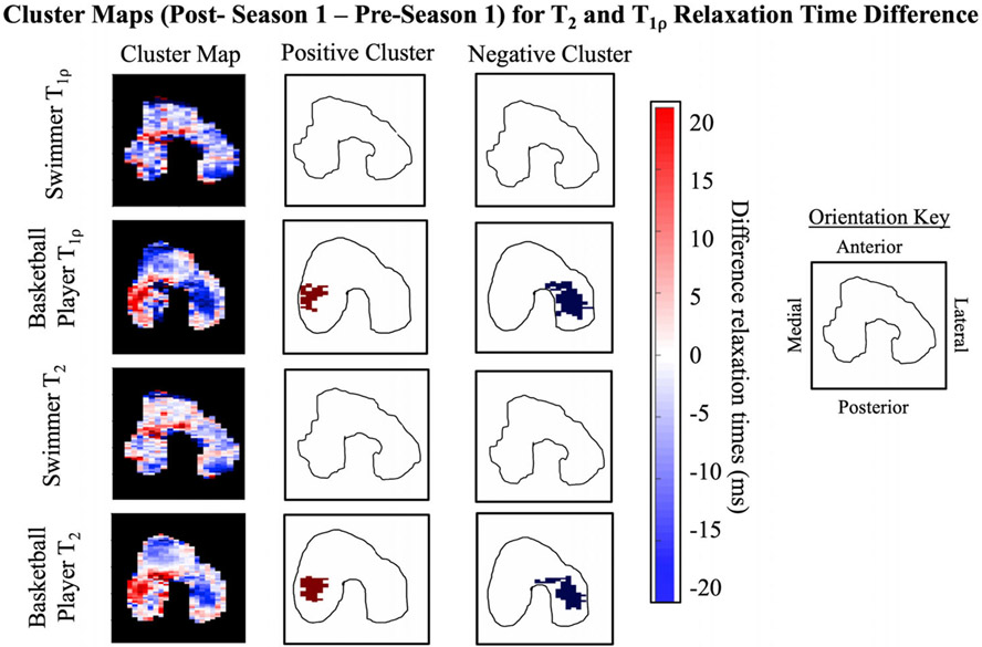
Results: In the femoral articular cartilage of all athletes, we saw a global decrease in T2 and T1ρ relaxation times during the competitive season (all P < 0.05) and an increase in T2 and T1ρ relaxation times during the off-season (all P < 0.05). In the basketball players' femoral cartilage, the anterior and central compartments respectively had the highest T2 and T1ρ relaxation times following the competitive season and off-season. The basketball players had significantly lower KOOS measures in every domain compared with the swimmers: Pain (P < 0.05), Symptoms (P < 0.05), Function in Daily Living (P < 0.05), Function in Sport/Recreation (P < 0.05), and Quality of Life (P < 0.05).
Conclusion: Our results indicate that T2 and T1ρ MRI can detect significant seasonal changes in the articular cartilage of basketball players and that there are regional differences in the articular cartilage that are indicative of basketball-specific stress on the femoral cartilage. This study demonstrates the potential of quantitative MRI to monitor global and regional cartilage health in athletes at risk of developing cartilage problems.
Level of evidence: 2 Technical Efficacy Stage: 2.
Keywords: articular cartilage; basketball; general; imaging; knee; magnetic resonance; relaxometry; swimming.
© 2021 International Society for Magnetic Resonance in Medicine.
Figures
References
-
- Walczak BE, McCulloch PC, Kang RW, Zelazny A, Tedeschi F, Cole BJ. Abnormal findings on knee magnetic resonance imaging in asymptomatic NBA players. J Knee Surg 2008;21(1):27–33. - PubMed
Publication types
MeSH terms
Grants and funding
LinkOut - more resources
Full Text Sources
Other Literature Sources
Research Materials
