. 2021 Jun 1;147(6):518-525.
doi: 10.1001/jamaoto.2021.0176.

Evaluation of the Video Ocular Counter-Roll (vOCR) as a New Clinical Test of Otolith Function in Peripheral Vestibulopathy

Affiliations

Evaluation of the Video Ocular Counter-Roll (vOCR) as a New Clinical Test of Otolith Function in Peripheral Vestibulopathy

Shirin Sadeghpour et al. JAMA Otolaryngol Head Neck Surg. .

Abstract

Importance: Video-oculography (VOG) goggles have been integrated into the assessment of semicircular canal function in patients with vestibular disorders. However, a similar bedside VOG method for testing otolith function is lacking.

Objective: To evaluate the use of VOG-based measurement of ocular counter-roll (vOCR) as a clinical test of otolith function.

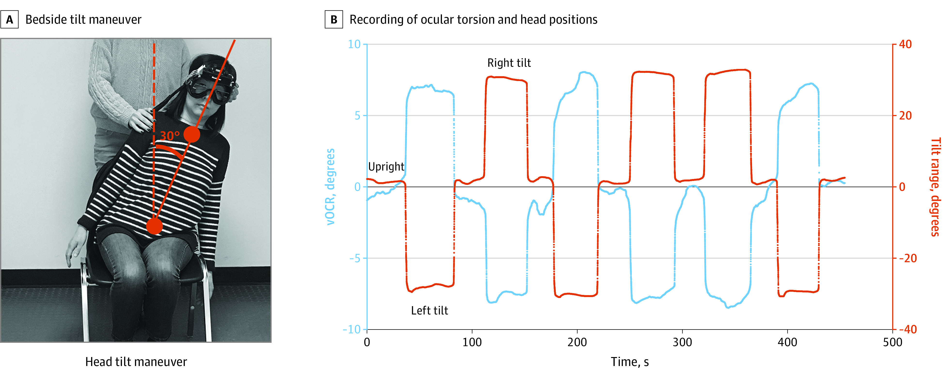
Design, setting, and participants: A case-control study was conducted to compare vOCR measurement among patients at various stages of unilateral loss of vestibular function with healthy controls. The receiver operating characteristic curve method was used to determine the diagnostic accuracy of the vOCR test in detecting loss of otolith function. Participants were recruited at a tertiary center including the Johns Hopkins outpatient clinic and Johns Hopkins Hospital, Baltimore, Maryland. Participants included 56 individuals with acute (≤4 weeks after surgery), subacute (4 weeks-6 months after surgery), and chronic (>6 months after surgery) unilateral vestibular loss as well as healthy controls. A simple bedside maneuver with en bloc, 30° lateral tilt of the head and trunk was used for vOCR measurement. The study was conducted from February 2, 2017, to March 10, 2019.

Intervention: In each participant vOCR was measured during static tilts of the head and trunk en bloc.

Main outcomes and measures: The vOCR measurements and diagnostic accuracy of vOCR in detecting patients with loss of vestibular function from healthy controls.

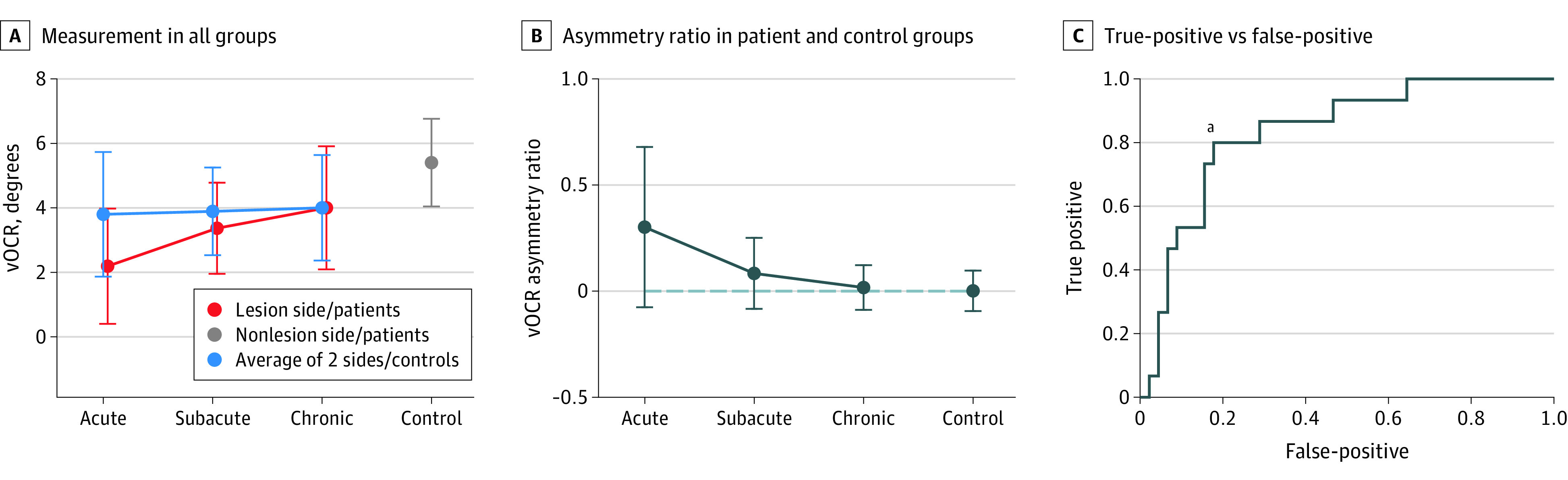
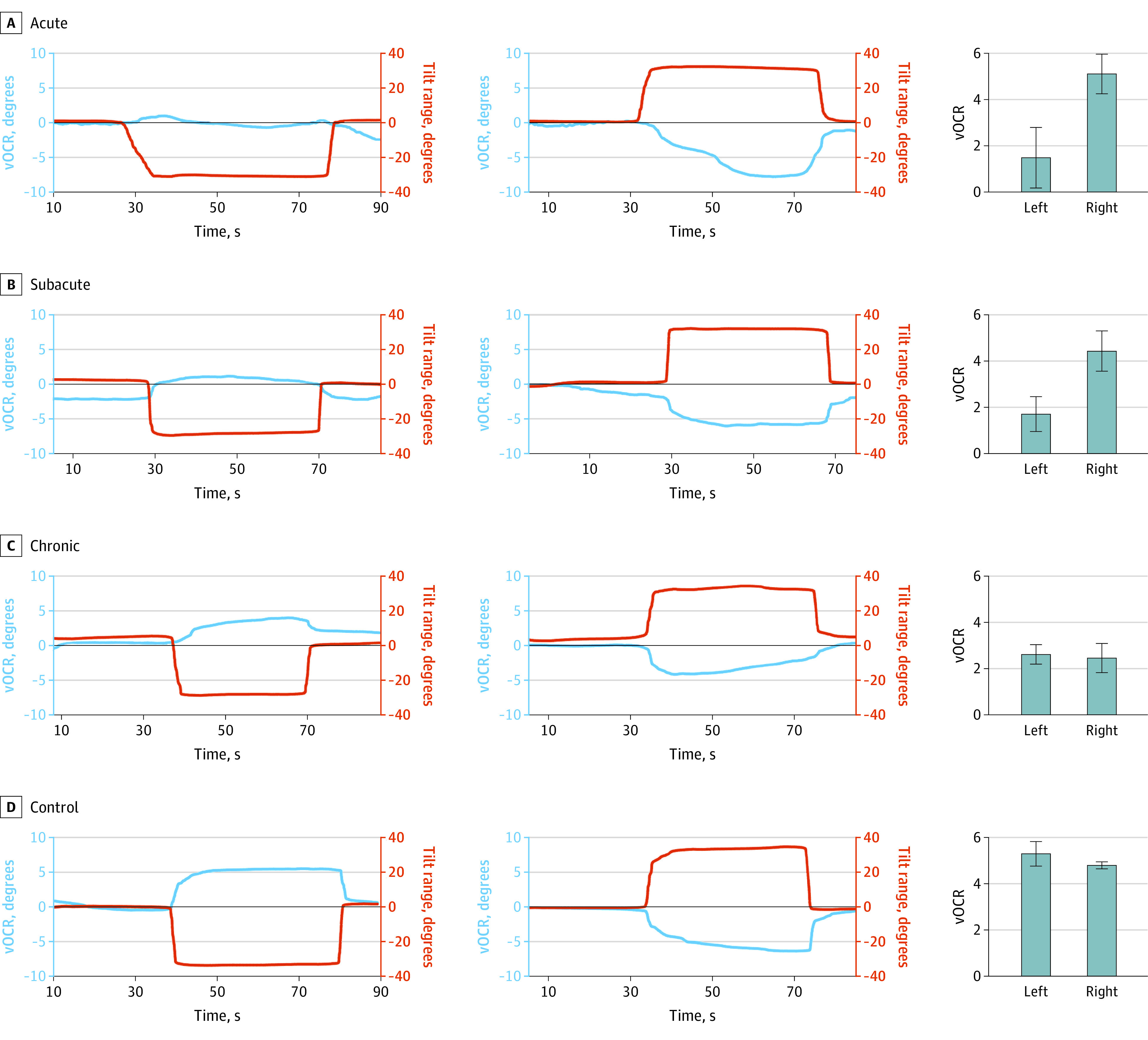
Results: Of the 56 participants, 28 (50.0%) were men; mean (SD) age was 53.5 (11.4) years. The mean (SD) time of acute unilateral vestibular loss was 9 (7) days (range, 2-17 days) in the acute group, 61 (39) days (range, 28-172 days) in the subacute group, and 985 (1066) days (range 185-4200 days) in the chronic group. The vOCR test showed reduction on the side of vestibular loss, and the deficit was greater in patients with acute and subacute vestibular loss than in patients with chronic loss and healthy controls (acute vs chronic: -1.81°; 95% CI, -3.45° to -0.17°; acute vs control: -3.18°; 95% CI, -4.83° to -1.54°; subacute vs chronic: -0.63°; 95% CI, -2.28° to 1.01°; subacute vs control: -2.01°; 95% CI, -3.65° to -0.36°; acute vs subacute: -1.17°; 95% CI, -2.88° to 0.52°; and chronic vs control: -1.37°; 95% CI, -2.96° to 0.21°). The asymmetry in vOCR between the side of vestibular loss and healthy side was significantly higher in patients with acute vs chronic loss (0.28; 95% CI, 0.06-0.51). Overall, the performance of the vOCR test in discriminating between patients with vestibular loss and healthy controls was 0.83 (area under the receiver operating characteristic curve). The best vOCR threshold to detect vestibular loss at the 30° tilt was 4.5°, with a sensitivity of 80% (95% CI, 0.62%-0.88%) and specificity of 82% (95% CI, 0.57%-1.00%).

Conclusions and relevance: The findings of this case-control study suggest that the vOCR test can be performed with a simple bedside maneuver and may be used to detect or track loss of otolith function.


Conflict of interest statement

Conflict of Interest Disclosures: Dr Otero-Millan reported receiving grants from the National Institutes of Health (NIH) during the conduct of the study and software used in this study (not patented) was licensed to Labyrinth Devices, LLC in 2016 and Interacoustics A/S in 2018. Dr Zee reported receiving royalties from Oxford University Press outside the submitted work. No other disclosures were reported.

Figures

Figure 1.
Figure 1.. Tilt Maneuver as a Passive 30° Tilt of the Head and Trunk While the Participant Is Sitting on a Chair
The recording started in the upright position followed by 3 right and 3 left tilts in random order separated by a return to the upright position during which video-oculography measurement of ocular counter-roll (vOCR) and head tilt angle were measured simultaneously. Note the different scales on the ordinate for the tilt angle and vOCR. The right panel shows an example of the vOCR recording (blue trace/left axis) during the en bloc tilt maneuver (tilt angle shown by the orange trace/right axis). The positive value on the graph indicates a rightward direction and the negative value indicates a leftward direction.
Figure 2.
Figure 2.. General Structure of the Video-Oculography (VOG) Method Used to Detect Ocular Torsion and Measure Ocular Counter-Roll (OCR)
This torsion method is based on template matching of the entire iris and tracks the eyelids to account for partial occlusion of the iris and pupil. A polar transformation is applied to the iris pattern and the image is optimized to enhance the iris features and mask the parts covered by the eyelids. For OCR measurement, the iris pattern is set as the baseline reference in the upright position and a template-matching method is implemented to compare the iris pattern at any given point in time during head tilt with the baseline reference in the upright position.
Figure 3.
Figure 3.. Video-Oculography Measurement of Ocular Counter-Roll (vOCR) Results
A, Mean vOCR measurements in all participant groups. B, Mean vOCR asymmetry ratios in patient and control groups. Error bars represent SD. C, Receiver operating characteristic curve from the average vOCR measurements from both sides in all patients and healthy controls. The area under the curve was 0.83. aThe best vOCR threshold to detect vestibular loss was 4.5° with a sensitivity of 80% and specificity of 82%.
Figure 4.
Figure 4.. Examples of Video-Oculography Measurement of Ocular Counter-Roll (vOCR) in Individual Patients With Acute, Subacute, and Chronic Vestibular Loss as Well as a Healthy Control Individual
The vOCR recording is shown as the blue trace/axis and the tilt angle is shown as the orange trace/axis. The negative values on the graphs indicate leftward direction and the positive values indicate rightward direction. The mean vOCR values form 3 measurements on each side are shown in the bar graphs. The lower vOCR values correspond to the side of the vestibular loss with an overall lower asymmetry in the patient with chronic vestibular loss. A, In the patient with acute vestibular loss, there is an asymmetry with a vOCR of 1.5° on the side of vestibular loss and 5° on the healthy side. B, In the patient with subacute vestibular loss, there is also an asymmetry with a vOCR of 1.7° on the side of vestibular loss and 4.4° on the healthy side. C, In the patient with chronic right-side vestibular loss, there is no significant asymmetry and vOCR values are reduced by approximately 2.4° and 2.6°.D, In the healthy control individuals, vOCR values are 5.3° and 4.7°.

References

    1. MacDougall HG, Weber KP, McGarvie LA, Halmagyi GM, Curthoys IS. The video head impulse test: diagnostic accuracy in peripheral vestibulopathy. Neurology. 2009;73(14):1134-1141. doi:10.1212/WNL.0b013e3181bacf85 - DOI - PMC - PubMed
    1. Halmagyi GM, Chen L, MacDougall HG, Weber KP, McGarvie LA, Curthoys IS. The video head impulse test. Front Neurol. 2017;8:258. doi:10.3389/fneur.2017.00258 - DOI - PMC - PubMed
    1. Diamond SG, Markham CH. Ocular counterrolling as an indicator of vestibular otolith function. Neurology. 1983;33(11):1460-1469. doi:10.1212/WNL.33.11.1460 - DOI - PubMed
    1. Ramat S, Zee DS. Ocular motor responses to abrupt interaural head translation in normal humans. J Neurophysiol. 2003;90(2):887-902. doi:10.1152/jn.01121.2002 - DOI - PubMed
    1. Bronstein AM, Gresty MA. Short latency compensatory eye movement responses to transient linear head acceleration: a specific function of the otolith-ocular reflex. Exp Brain Res. 1988;71(2):406-410. doi:10.1007/BF00247500 - DOI - PubMed

Publication types

LinkOut - more resources