

Association of Near-Infrared and Short-Wavelength Autofluorescence With the Retinal Sensitivity in Eyes With Resolved Central Serous Chorioretinopathy
- PMID: 33764400
- PMCID: PMC7995351
- DOI: 10.1167/iovs.62.3.36
Association of Near-Infrared and Short-Wavelength Autofluorescence With the Retinal Sensitivity in Eyes With Resolved Central Serous Chorioretinopathy
Abstract
Purpose: The purpose of this study was to compare the results of near-infrared autofluorescence (NIRAF) and short-wavelength autofluorescence (SWAF) imaging of eyes with resolved central serous chorioretinopathy (CSC) and to assess the retinal sensitivity (RS) in areas with abnormal autofluorescence (AF) using white-on-white (WW) and blue-on-yellow (BY) perimetries.
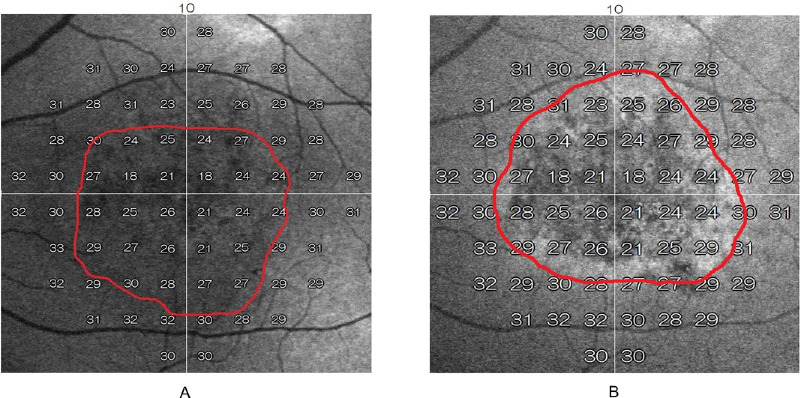
Methods: We examined 20 consecutive eyes with resolved CSC. We calculated the areas of abnormal AF detected by SWAF and NIRAF imaging as SWAF_area and NIRAF_area, respectively, and the number of measurement points within and outside abnormal SWAF and NIRAF regions were counted. The results of WW and BY perimetries were superimposed on the AF images, and the mean overall RS within and outside abnormal SWAF and NIRAF regions were calculated using both WW and BY perimetries (W-RSin_SWAF, W-RSout_SWAF, W-RSin_NIRAF, W-RSout_NIRAF, B-RSin_SWAF, B-RSout_SWAF, B-RSin_NIRAF, and B-RSout_NIRAF, respectively).
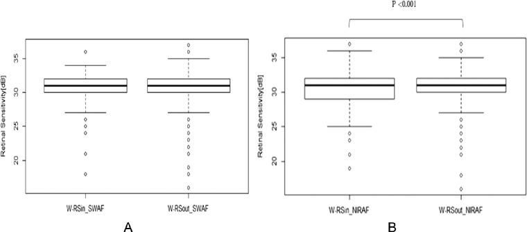
Results: The mean age of the participants was 54.1 years. The SWAF_area was significantly smaller than the NIRAF_area (P < 0.0001, Wilcoxon signed rank test). A χ2 test suggested a significant relationship between the number of measurement points within/outside abnormal SWAF and NIRAF regions (P < 0.0001). In the results of measurement by WW perimetry, there was a significant difference between W-RSin_NIRAF and W-RSout_NIRAF (P < 0.0001), but not between W-RSin_SWAF and W-RSout_SWAF (P = 0.060, Wilcoxon rank sum test). In contrast, on BY perimetry, there were significant differences between both B-RSin_SWAF and B-RSout_SWAF and between B-RSin_NIRAF and B-RSout_NIRAF (P < 0.0001).
Conclusions: NIRAF was useful for predicting impaired RS in eyes with resolved CSC.
Conflict of interest statement
Disclosure:
Figures

References
-
- Daruich A, Matet A, Dirani A, et al.. Central serous chorioretinopathy: RECENT findings and new physiopathology hypothesis. Prog Retin Eye Res. 2015; 48: 82–118. - PubMed
-
- Liew G, Quin G, Gillies M, Fraser-Bell S. Central serous chorioretinopathy: a review of epidemiology and pathophysiology. Clin Exp Ophthalmol. 2013; 41(2): 201–214. - PubMed
-
- Imamura Y, Fujiwara T, Spaide RF. Fundus autofluorescence and visual acuity in central serous chorioretinopathy. Ophthalmology. 2011; 118: 700–705. - PubMed
Publication types
MeSH terms
LinkOut - more resources
Full Text Sources
Other Literature Sources
