

Fetal and placental infection with SARS-CoV-2 in early pregnancy
- PMID: 33764543
- PMCID: PMC8250563
- DOI: 10.1002/jmv.26965
Fetal and placental infection with SARS-CoV-2 in early pregnancy
Abstract
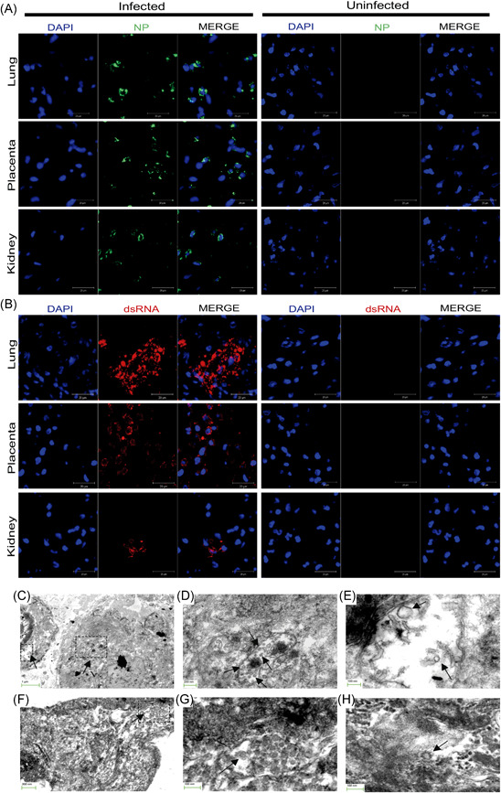
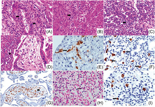
To date, mother-to-fetus transmission of severe acute respiratory syndrome coronavirus 2 (SARS-CoV-2), responsible for the coronavirus disease 2019 (COVID-19) pandemic, remains controversial. Although placental COVID-19 infection has been documented in some cases during the second- and third-trimesters, no reports are available for the first trimester of pregnancy, and no SARS-CoV-2 protein has been found in fetal tissues. We studied the placenta and fetal organs from an early pregnancy miscarriage in a COVID-19 maternal infection by immunohistochemical, reverse transcription quantitative real-time polymerase chain reaction, immunofluorescence, and electron microscopy methods. SARS-CoV-2 nucleocapsid protein, viral RNA, and particles consistent with coronavirus were found in the placenta and fetal tissues, accompanied by RNA replication revealed by double-stranded RNA (dsRNA) positive immunostain. Prominent damage of the placenta and fetal organs were associated with a hyperinflammatory process identified by histological examination and immunohistochemistry. The findings provided in this study document that congenital SARS-CoV-2 infection is possible during the first trimester of pregnancy and that fetal organs, such as lung and kidney, are targets for coronavirus. The infection and multi-organic fetal inflammation produced by SARS-CoV-2 during early pregnancy should alert clinicians in the assessment and management of pregnant women for possible fetal consequences and adverse perinatal outcomes.
Keywords: COVID-19; fetus; first trimester; miscarriage; placenta; pregnancy.
© 2021 Wiley Periodicals LLC.
Conflict of interest statement
The authors declare that there are no conflict of interests.
Figures
Similar articles
-
Investigating the risk of maternal-fetal transmission of SARS-CoV-2 in early pregnancy.Placenta. 2021 Mar;106:25-29. doi: 10.1016/j.placenta.2021.02.006. Epub 2021 Feb 14. Placenta. 2021. PMID: 33610934 Free PMC article.
-
Persistence of SARS-CoV-2 in the first trimester placenta leading to transplacental transmission and fetal demise from an asymptomatic mother.Hum Reprod. 2021 Mar 18;36(4):899-906. doi: 10.1093/humrep/deaa367. Hum Reprod. 2021. PMID: 33346816 Free PMC article.
-
Histologic and Immunohistochemical Evaluation of 65 Placentas From Women With Polymerase Chain Reaction-Proven Severe Acute Respiratory Syndrome Coronavirus 2 (SARS-CoV-2) Infection.Arch Pathol Lab Med. 2021 Jun 1;145(6):648-656. doi: 10.5858/arpa.2020-0793-SA. Arch Pathol Lab Med. 2021. PMID: 33596304
-
Impact of SARS-CoV-2 infection during pregnancy on the placenta and fetus.Semin Perinatol. 2024 Jun;48(4):151919. doi: 10.1016/j.semperi.2024.151919. Epub 2024 Jun 6. Semin Perinatol. 2024. PMID: 38897829 Free PMC article. Review.
-
Placental Pathology of COVID-19 with and without Fetal and Neonatal Infection: Trophoblast Necrosis and Chronic Histiocytic Intervillositis as Risk Factors for Transplacental Transmission of SARS-CoV-2.Viruses. 2020 Nov 15;12(11):1308. doi: 10.3390/v12111308. Viruses. 2020. PMID: 33203131 Free PMC article. Review.
Cited by
-
Integrated Analysis Reveals the Characteristics and Effects of SARS-CoV-2 Maternal-Fetal Transmission.Front Microbiol. 2022 Jan 27;13:813187. doi: 10.3389/fmicb.2022.813187. eCollection 2022. Front Microbiol. 2022. PMID: 35154056 Free PMC article.
-
Prediction of Non-canonical Routes for SARS-CoV-2 Infection in Human Placenta Cells.Front Mol Biosci. 2021 Nov 8;8:614728. doi: 10.3389/fmolb.2021.614728. eCollection 2021. Front Mol Biosci. 2021. PMID: 34820418 Free PMC article.
-
COVID-19: the possibility, ways, mechanisms, and interruptions of mother-to-child transmission.Arch Gynecol Obstet. 2023 Jun;307(6):1687-1696. doi: 10.1007/s00404-022-06639-5. Epub 2022 Jun 4. Arch Gynecol Obstet. 2023. PMID: 35665849 Free PMC article. Review.
-
Preeclampsia in the Context of COVID-19: Mechanisms, Pathophysiology, and Clinical Outcomes.Am J Reprod Immunol. 2024 Aug;92(2):e13915. doi: 10.1111/aji.13915. Am J Reprod Immunol. 2024. PMID: 39132825 Free PMC article. Review.
-
Evidence of vertical transmission of SARS-CoV-2 and interstitial pneumonia in second-trimester twin stillbirth in asymptomatic woman. Case report and review of the literature.Am J Obstet Gynecol MFM. 2022 May;4(3):100589. doi: 10.1016/j.ajogmf.2022.100589. Epub 2022 Feb 4. Am J Obstet Gynecol MFM. 2022. PMID: 35131495 Free PMC article. Review.
References
Publication types
MeSH terms
Substances
Grants and funding
LinkOut - more resources
Full Text Sources
Other Literature Sources
Medical
Miscellaneous
