. 2021 Aug;86(2):866-880.
doi: 10.1002/mrm.28761. Epub 2021 Mar 25.

A multi-inversion multi-echo spin and gradient echo echo planar imaging sequence with low image distortion for rapid quantitative parameter mapping and synthetic image contrasts

Affiliations

A multi-inversion multi-echo spin and gradient echo echo planar imaging sequence with low image distortion for rapid quantitative parameter mapping and synthetic image contrasts

Mary Kate Manhard et al. Magn Reson Med. 2021 Aug.

Abstract

Purpose: Brain imaging exams typically take 10-20 min and involve multiple sequential acquisitions. A low-distortion whole-brain echo planar imaging (EPI)-based approach was developed to efficiently encode multiple contrasts in one acquisition, allowing for calculation of quantitative parameter maps and synthetic contrast-weighted images.

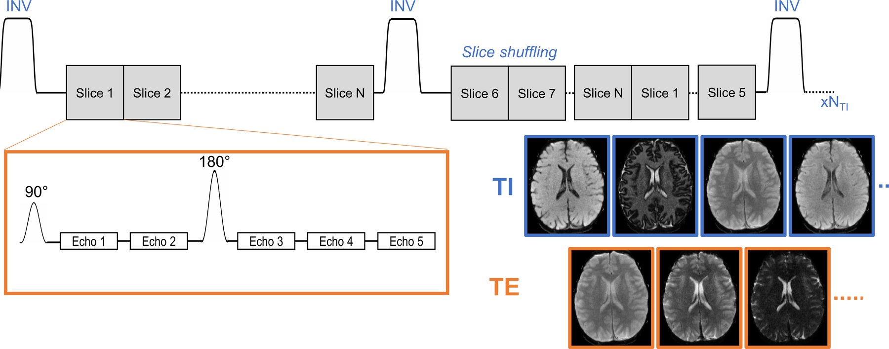
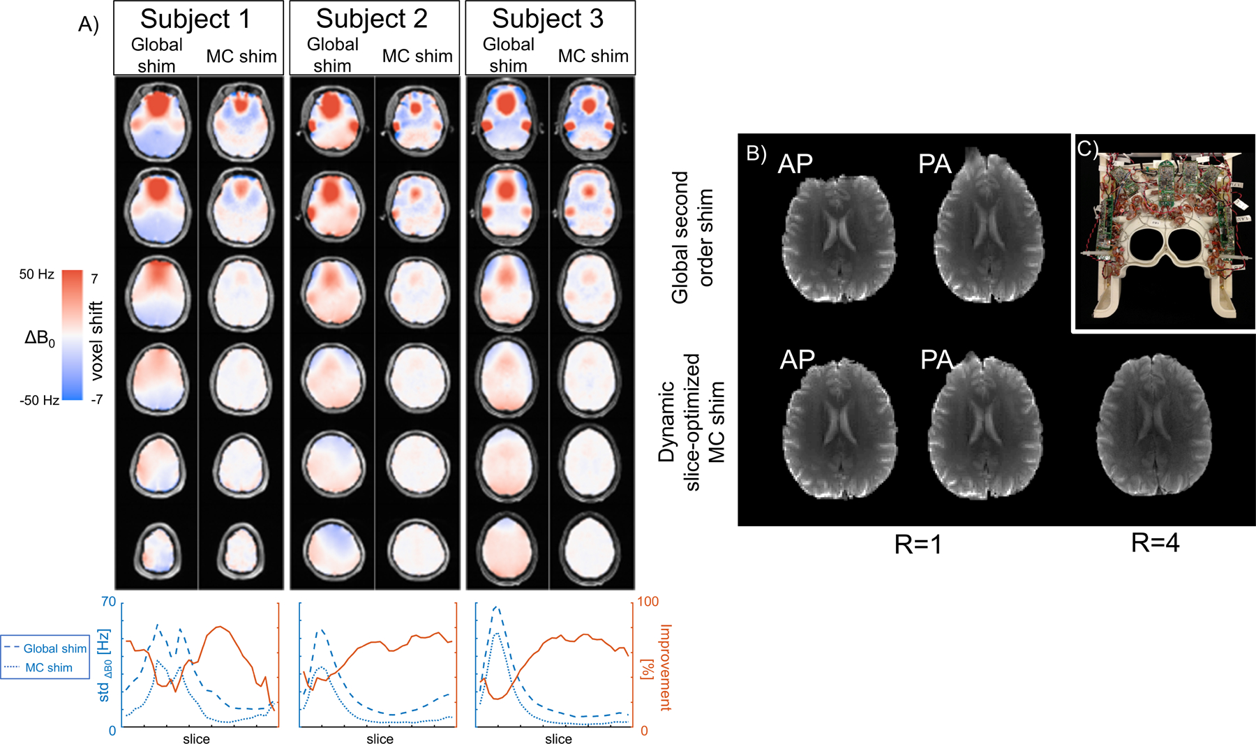
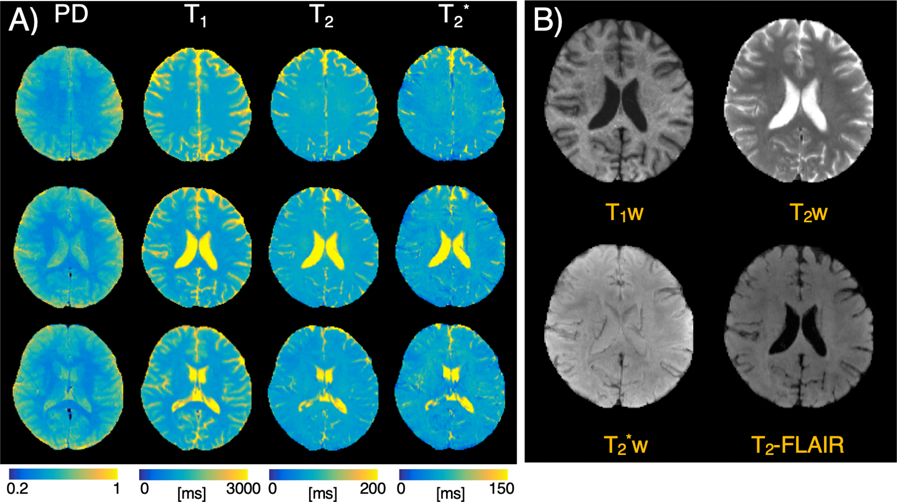
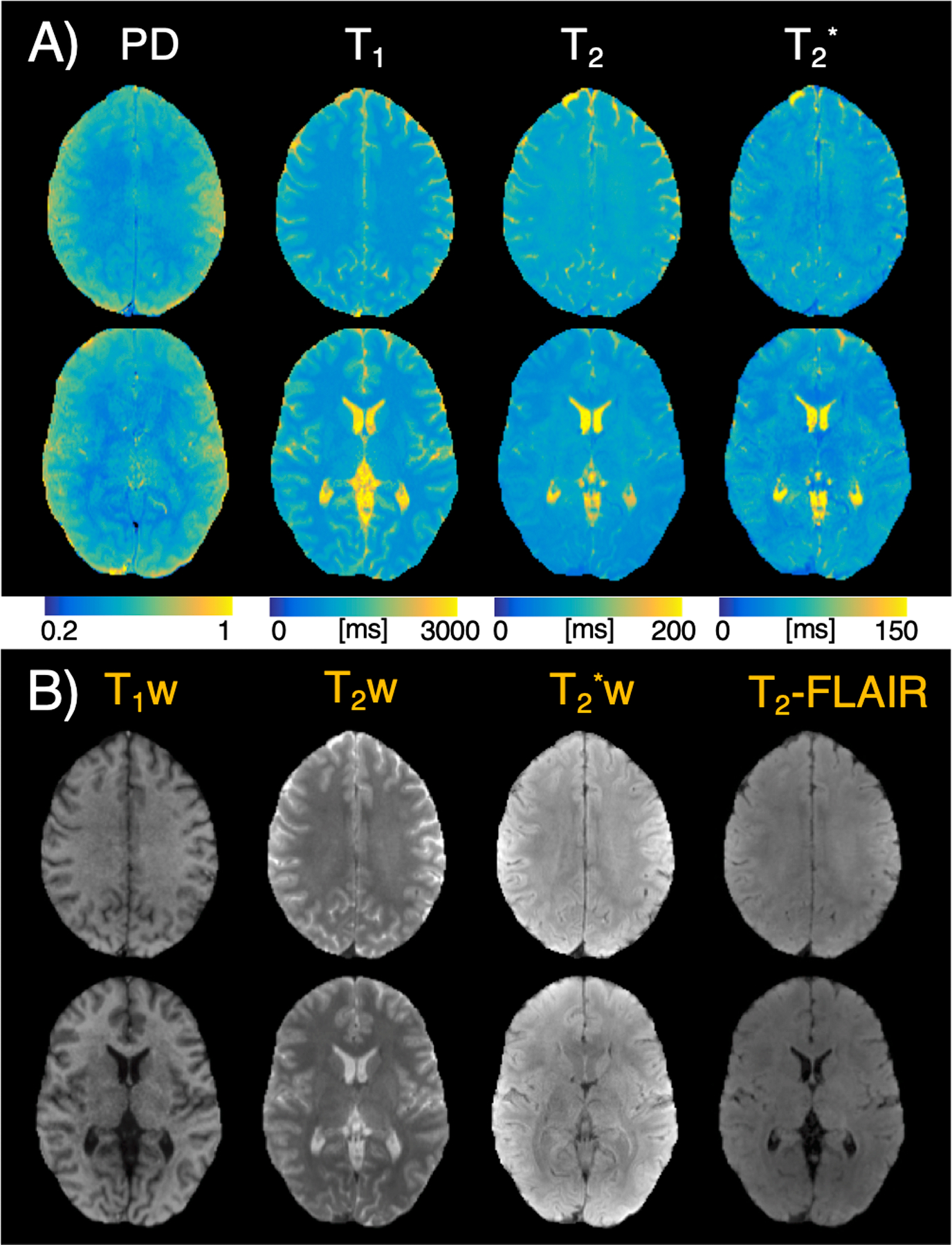
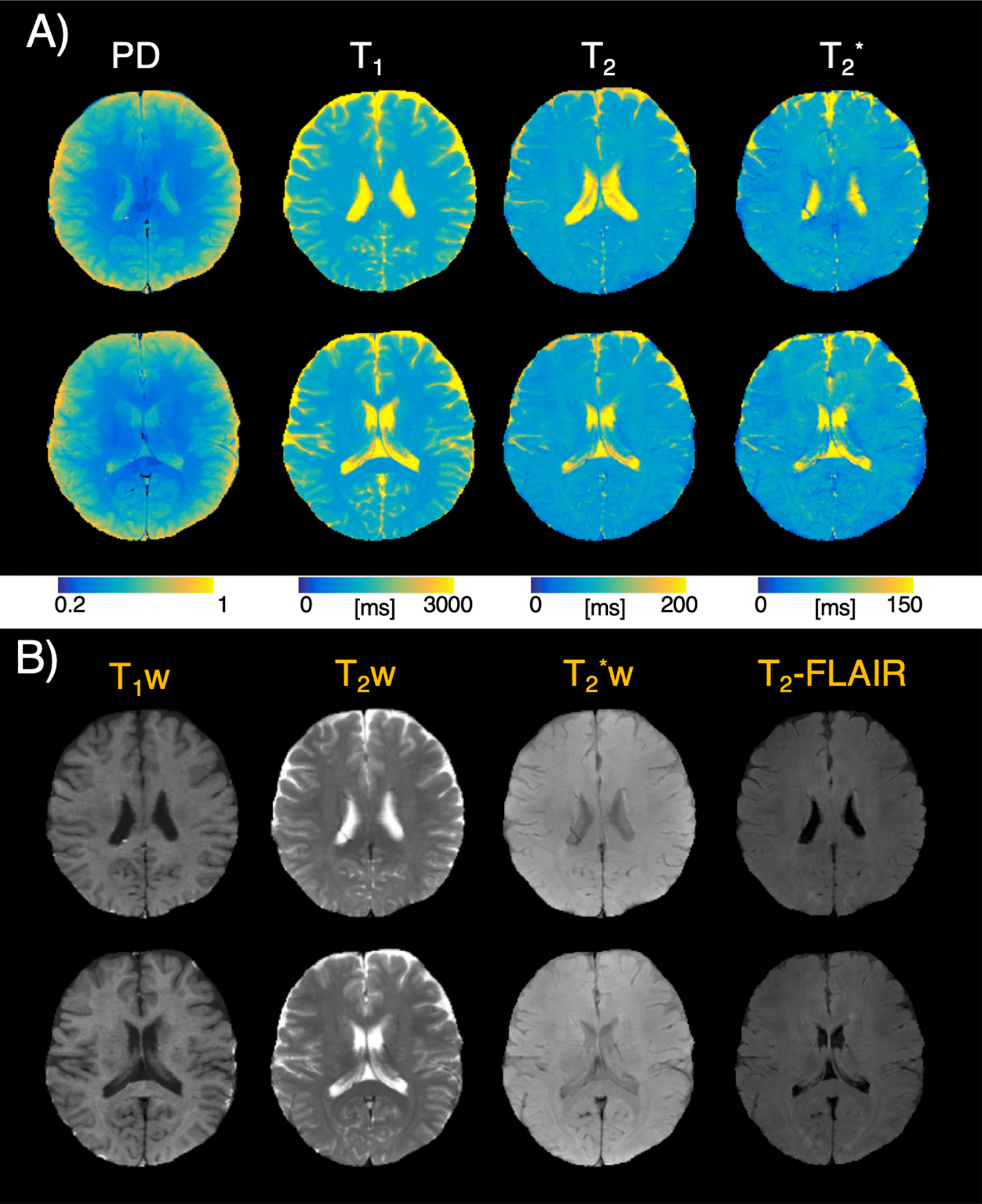
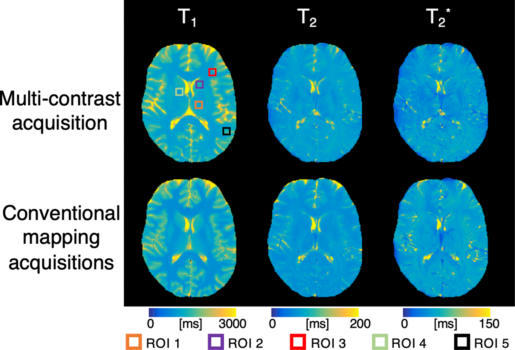
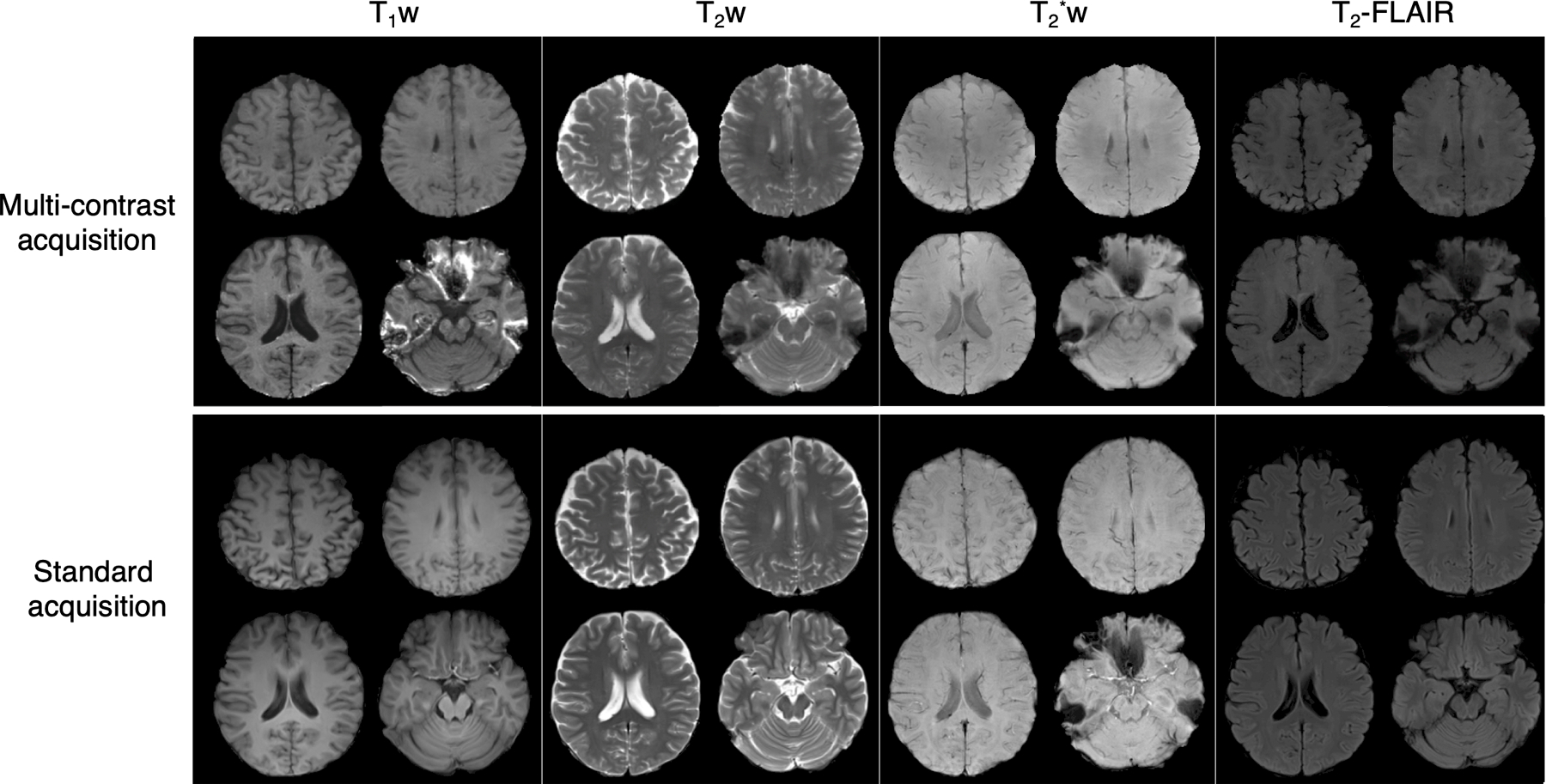
Methods: Inversion prepared spin- and gradient-echo EPI was developed with slice-order shuffling across measurements for efficient acquisition with T1 , T2 , and T2 weighting. A dictionary-matching approach was used to fit the images to quantitative parameter maps, which in turn were used to create synthetic weighted images with typical clinical contrasts. Dynamic slice-optimized multi-coil shimming with a B0 shim array was used to reduce B0 inhomogeneity and, therefore, image distortion by >50%. Multi-shot EPI was also implemented to minimize distortion and blurring while enabling high in-plane resolution. A low-rank reconstruction approach was used to mitigate errors from shot-to-shot phase variation.

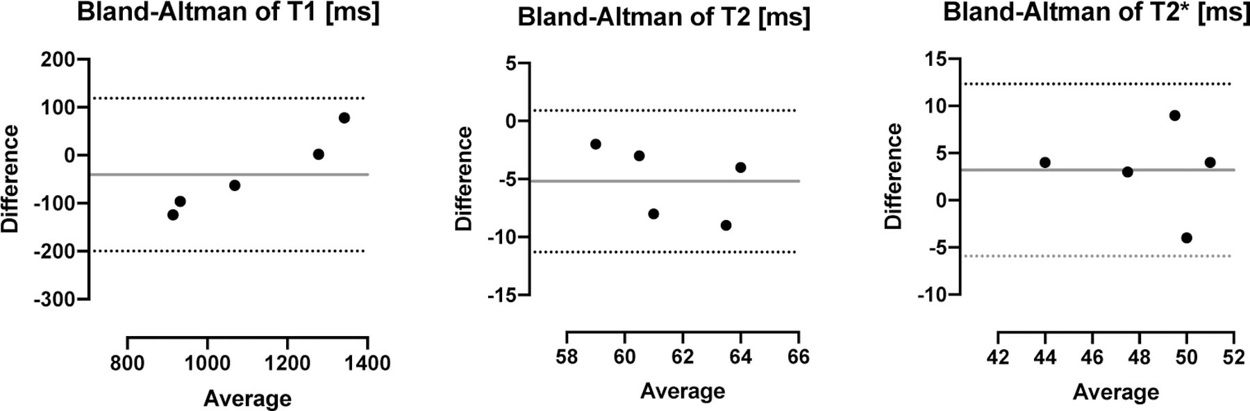
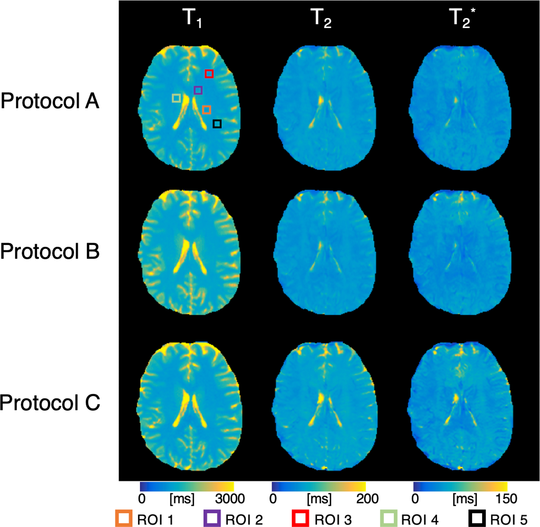
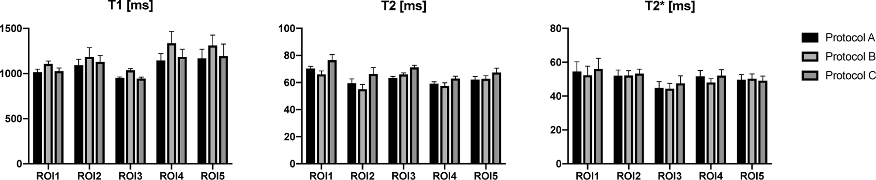
Results: The slice-optimized shimming approach was combined with in-plane parallel-imaging acceleration of 4× to enable single-shot EPI with more than eight-fold distortion reduction. The proposed sequence efficiently obtained 40 contrasts across the whole-brain in just over 1 min at 1.2 × 1.2 × 3 mm resolution. The multi-shot variant of the sequence achieved higher in-plane resolution of 1 × 1 × 4 mm with good image quality in 4 min. Derived quantitative maps showed comparable values to conventional mapping methods.

Conclusion: The approach allows fast whole-brain imaging with quantitative parameter maps and synthetic weighted contrasts. The slice-optimized multi-coil shimming and multi-shot reconstruction approaches result in minimal EPI distortion, giving the sequence the potential to be used in rapid screening applications.

Keywords: low distortion EPI; multi-shot acquisitions; quantitative mapping; synthetic imaging.


Figures

Figure 1 –
Figure 1 –
Pulse sequence for multi-inversion, multi-echo EPI acquisition. A nonselective inversion pulse is used, followed by the acquisition of all slices. This is repeated up to NTI repetitions, with a shuffled slice order for each repetition. Therefore, each slice sees a range of different inversion times, as shown in example images in the blue boxes. In addition, each slice acquisition consists of multiple spin and gradient echoes, as shown in the orange box, resulting in images with different echo times, shown in the images with orange boxes on the right.
Figure 2 –
Figure 2 –
Demonstration of EPI distortion improvements using a dynamic shim coil. A) B0 maps overlaid on anatomical images from three subjects in several slices, comparing a global second order shim and a predicted dynamic slice-optimized multi-coil (MC) shim. The B0 maps also show the corresponding voxel shift that would occur with a single shot acquisition with no acceleration (1.2 mm acquisition with echo spacing = 0.76, number of phase encode lines = 188). The B0 inhomogeneity and voxel shift is visibly improved using the dynamic shim approach. Below, the standard deviation (std) of B0 is shown across slices from foot to head for both the global shim and MC shim approaches in the three subjects in blue, with the percent improvement shown in red. The improvement in image distortion is demonstrated in one slice in B), where a single shot EPI acquisition with no acceleration acquired in both the AP and PA directions shows severe distortion when using a global shim (top), compared to a much-improved distortion using a dynamic slice-optimized MC shim (bottom). The distortion with R=4 and the dynamic MC shimming is very low. The 32-channel AC/DC coil is pictured in C).
Figure 3 –
Figure 3 –
A) Quantitative parameter maps from protocol A, single-shot R=4, acquired with dynamic slice-optimized MC shimming, in three representative slices. B) Synthetic weighted contrasts derived from the middle slice in A), with T1w, T2w, T2*w, and T2-FLAIR images.
Figure 4 –
Figure 4 –
A) Quantitative parameter maps from two slices from protocol B, a multiband acquisition (MB=2, in-plane R=4). B) Synthetic weighted contrasts from both slices show T1w, T2w, T2*w, and T2-FLAIR images.
Figure 5 –
Figure 5 –
A) Quantitative parameter maps from two slices from protocol C, a multi-shot acquisition (R=8, 3 shots). B) Synthetic weighted contrasts derived from parameter maps of both slices, showing T1w, T2w, T2*w, and T2-FLAIR images.
Figure 6 –
Figure 6 –
Quantitative maps from a multi-shot acquisition of the multi-contrast protocol (top) are compared with quantitative maps derived from conventional mapping approaches (bottom). ROIs from the slice are shown by colored boxes, with values displayed in Figure 7.
Figure 7 –
Figure 7 –
Bland-Altman plots showing the mean and difference for the ROIs in Figure 6 for T1, T2, and T2* values. Though some biases remain, all values are within the limits of agreement.
Figure 8 –
Figure 8 –
Quantitative maps in the same slice from protocols A, B, and C acquired with a global shim in the standard 32-ch coil. ROIs from the slice are shown by colored boxes, with values displayed in Figure 9.
Figure 9 –
Figure 9 –
Plots showing the mean and standard deviation from the five ROIs shown in Figure 8 for T1, T2, and T2* values.
Figure 10 –
Figure 10 –
Synthetic images created from the proposed protocol (top) are compared to standard brain acquisition methods (bottom) in four slices across the brain. Good agreement is seen between the two methods, though the proposed protocol was acquired 3× faster. The T2-TSE and T2-FLAIR images from the proposed method have limited MT weighting compared to the standard acquisitions. Intravoxel dephasing can be seen in the bottom slice in all contrasts in the EPI-based acquisition, as observed in the standard T2* image.

Similar articles

Cited by

References

    1. Truong T-K, Darnell D, Song AW. Integrated RF/shim coil array for parallel reception and localized B 0 shimming in the human brain. Neuroimage. 2014;103:235–240. doi:10.1016/j.neuroimage.2014.09.052 - DOI - PMC - PubMed
    1. Hoffmann J, Shajan G, Scheffler K, Pohmann R. Numerical and experimental evaluation of RF shimming in the human brain at 9.4 T using a dual-row transmit array. Magn Reson Mater Physics, Biol Med 2014;27(5):373–386. doi:10.1007/s10334-013-0419-y - DOI - PubMed
    1. Stockmann JP, Witzel T, Keil B, et al. A 32-channel combined RF and B 0 shim array for 3T brain imaging. Magn Reson Med 2016;75(1):441–451. doi:10.1002/mrm.25587 - DOI - PMC - PubMed
    1. Chen N, Guidon A, Chang H-C, Song AW. A robust multi-shot scan strategy for high-resolution diffusion weighted MRI enabled by multiplexed sensitivity-encoding (MUSE). Neuroimage 2013;72:41–47. doi:10.1016/j.neuroimage.2013.01.038 - DOI - PMC - PubMed
    1. Haldar JP. Low-Rank Modeling of Local k-Space Neighborhoods (LORAKS) for Constrained MRI. IEEE Trans Med Imaging 2014;33(3):668–681. doi:10.1109/TMI.2013.2293974 - DOI - PMC - PubMed

Publication types

LinkOut - more resources