. 2021 Dec;17(12):4083-4101.
doi: 10.1080/15548627.2021.1901204. Epub 2021 Mar 25.

Long noncoding RNA (lncRNA) EIF3J-DT induces chemoresistance of gastric cancer via autophagy activation

Affiliations

Long noncoding RNA (lncRNA) EIF3J-DT induces chemoresistance of gastric cancer via autophagy activation

Yuhao Luo et al. Autophagy. 2021 Dec.

Abstract

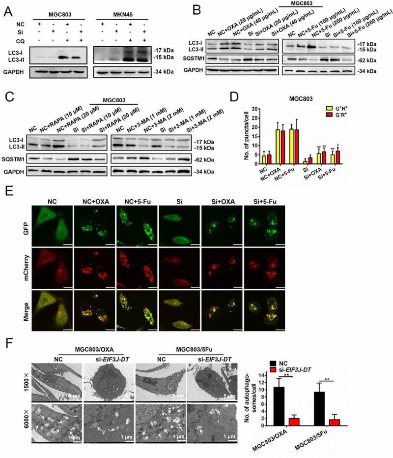
Chemotherapy is currently the main treatment for unresectable or advanced postoperative gastric cancers. However, its efficacy is negatively affected by the occurrence of chemoresistance, which severely affects patient prognosis. Recently, dysregulation in autophagy has been suggested as a potential mechanism for chemoresistence, and long noncoding RNA (lncRNA) also shows its regulatory role in cancer drug resistance. Using RNA sequencing, we found that lncRNA EIF3J-DT was highly expressed in drug-resistant gastric cancer cells. In-vitro and in-vivo experiments showed that EIF3J-DT activated autophagy and induced drug resistance in gastric cancer cells by targeting ATG14. Bioinformatics and experimental results showed that EIF3J-DT regulated the expression of ATG14 through direct binding to enhance stabilization of ATG14 mRNA and via blocking the degradation of ATG14 mRNA through competitively binding with microRNA (miRNA) MIR188-3p. Therefore, EIF3J-DT increased the expression of ATG14, contributing to activation of autophagy and chemoresistance. Furthermore, it was confirmed that EIF3J-DT and ATG14 were highly expressed in gastric cancer patients resistant to chemotherapy, and this was closely associated with patient prognosis. In conclusion, EIF3J-DT is involved in the regulation of autophagy and chemoresistance in gastric cancer cells by targeting ATG14. It may be a suitable new target for enhancing chemosensitivity and improving prognosis.Abbreviations: 3-MA: 3-methyladenine; 5-Fu: 5-fluorouracil; ATG: autophagy related; C-CASP3: cleaved caspase 3; C-CASP7: cleaved caspase 7; C-PARP: cleaved PARP; CQ: chloroquine; CR: complete response; DIG: digoxigenin; ESR1: estrogen receptor 1; FBS: fetal bovine serum; FISH: fluorescence in situ hybridization; IHC: immunohistochemistry; ISH: in situ hybridization; lncRNA: long noncoding RNA; miRNA: microRNA; MUT: mutant; NC: negative control; OXA: oxaliplatin; PBS: phosphate-buffered saline; PD: progressive disease; PFA: paraformaldehyde; PR: partial response; qPCR: quantitative polymerase chain reaction; RAPA: rapamycin; SD: stable disease; TEM: transmission electron microscopy; WT: wild type.

Keywords: ATG14; EIF3J-DT; gastric cancer; autophagy; chemotherapy resistance.


Conflict of interest statement

The authors declare no competing interests.

Figures

Figure 1.
Figure 1.
OXA and 5-Fu treatment activated autophagy in gastric cancer cells. (A and B) Western blotting performed on the MGC803 cells to evaluate the expression levels of LC3-I, LC3-II and SQSTM1 after being treated with OXA (10 μg/mL) (A) and 5-Fu (20 μg/mL) (B) over time. (C and D) Western blotting performed on the MGC803 cells to evaluate the expression levels of LC3-I and LC3-II after being treated with OXA (10 μg/mL) (C) and 5-Fu (20 μg/mL) (D) combined with or without CQ (10 μM) treatment over time. (E) Cell viability of the MGC803, MGC803/OXA R1 and MGC803/OXA R2 cells 24 h after being treated with OXA (left), and cell viability of the MGC803, MGC803/5Fu R1 and MGC803/5Fu R2 cells 24 h after being treated with 5-Fu (right) detected by MTT assay. (F) Western blotting performed on the MGC803, MGC803/OXA R1 and MGC803/OXA R2 cells (left), and the MGC803, MGC803/5Fu R1 and MGC803/5Fu R2 cells (right) against LC3-I, LC3-II and SQSTM1. (G) Representative TEM images of the MGC803, MGC803/OXA and MGC803/5Fu cells (left). The white arrows indicated the autophagosomes in the cytoplasm. Magnification: 4 × . The graph on the right summarized the numbers of autophagosomes in the cytoplasm of different cells. (H) Cell viability of the MGC803/OXA (left) and MGC803/5Fu (right) cells 24 h after treatment with OXA and 5-Fu combined with or without CQ (10 μM) detected by MTT assay. Data were quantified as mean ± SEM (n = 3). **P < 0.01, ***P < 0.001, vs the relative control
Figure 2.
Figure 2.
EIF3J-DT was highly expressed in the drug-resistant gastric tumor cells and EIF3J-DT silencing inhibited tumor growth. (A) Heatmap of differential expressed lncRNAs (fold change > 2) in the MGC803, MGC803/OXA, and MGC803/5Fu cells. (B) mRNA expression levels of PAX8-AS1, EIF3J-DT, AKT3-IT1, MALAT1, and TXNP6 with OXA (10 μg/mL) (upper) or 5-Fu (20 μg/mL) (lower) treatment over time detected by qPCR. (C) mRNA expression levels of EIF3J-DT (upper) and MALAT1 (lower) between the MGC803 and MGC803/OXA cells, or between the MGC803 and MGC803/5Fu cells detected by qPCR. (D) MTT assay comparing the cell viability between the NC and si-EIF3J-DT MGC803 cells 24 h after being treated with OXA (left) and 5-Fu (right). (E) MTT assay comparing the cell viability between the NC and si-EIF3J-DT MGC803/OXA cells or MGC803/5Fu cells 24 h after being treated with OXA (left) or 5-Fu (right). (F) Numbers of colony formation of the LV-NC and LV-Sh transfected MGC803 (left) and MKN45 (right) cells 14 d after being treated with PBS, OXA (10 μg/mL), and 5-Fu (20 μg/mL). LV-NC indicated the MGC803 and MKN45 cells with stable mock-vehicle transfection. LV-Sh indicated the MGC803 and MKN45 cells with sh-EIF3J-DT transfection. (G) Apoptosis rates of the LV-NC and LV-Sh MGC803 (left) and MKN45 (right) cells 24 h after being treated with PBS, OXA (10 μg/mL), and 5-Fu (20 μg/mL). (H) Western blotting performed on the NC, NC+OXA (20 and 40 μg/mL), Si, and Si+OXA (20 and 40 μg/mL) MGC803 cells (left), or the NC, NC+5-Fu (100 and 200 μg/mL), Si, and Si+5-Fu (100 and 200 μg/mL) MGC803 cells (right) with 24-h treatment against C-PARP, PARP, C-CASP3, and CASP3. Si indicated si-EIF3J-DT transfection. (I) Changes in tumor volumes (left) and tumor weight (right) of mice from the six groups (including NC, Sh, NC+OXA, Sh+OXA, NC+5-Fu, and Sh+5-Fu). Sh indicated sh-EIF3J-DT transfection. For OXA treatment, 0.1 mg/kg, intraperitoneal injection, three times per week; for 5-Fu treatment, 0.5 mg/kg, intraperitoneal injection, three times per week. (J) Representative images from the in vivo living imaging showing the subcutaneous tumors in the six groups (left) on day 28. The graph on the right calculated the total flux of tumors from different groups. Data were quantified as mean ± SEM (n = 3). *P < 0.05, **P < 0.01, ***P < 0.001, vs the relative control
Figure 3.
Figure 3.
EIF3J-DT activated autophagy in the gastric cancer cells. (A) Expression levels of LC3-I and LC3-II in the NC and Si MGC803 (left) or MKN45 (right) cells treated with or without CQ (10 μM) detected by western blotting. (B) Expression levels of LC3-I, LC3-II and SQSTM1 in the NC and Si MGC803 cells treated with OXA (20 and 40 μg/mL, left) or 5-Fu (100 and 200 μg/mL, right) detected by western blotting. (C) Expression levels of LC3-I, LC3-II and SQSTM1 in the NC and Si MGC803 cells treated with RAPA (10 and 20 μM, left) or 3-MA (1 and 2 mM, right) detected by western blotting. (D and E) Representative immunofluorescence images of the GFP-mCherry-LC3 transfected NC and Si MGC803 cells 24 h after treatment with OXA (10 μg/mL) and 5-Fu (20 μg/mL) (E). Scale bar: 10 μm. Numbers of the G+R+ and GR+ fluorescent puncta in different treated groups were calculated in D. (F) TEM performed on the MGC803/OXA and MGC803/5Fu cells transfected with NC or si-EIF3J-DT (left). The white arrows indicated the autophagosomes in the cytoplasm. Magnification: 4 × . The graph summarized the numbers of autophagosomes in different groups (right). Data were quantified as mean ± SEM (n = 3). *P < 0.05, **P < 0.01, vs the relative control
Figure 4.
Figure 4.
The expression of ATG14 affected cell apoptosis and autophagy. (A) TCGA Nature 2014 and TCGA Provisional database analyses showing the correlation between EIF3J-DT and ATG14 mRNA expression levels. (B) mRNA expression level of ATG14 in the MGC803 cells with overexpressed (left), silenced (middle), or overexpressed+silenced (right) EIF3J-DT detected by qPCR. (C) Cell viability among the NC, siATG14 #1, and siATG14 #2 MGC803 cells 24 h after being treated with OXA (left) and 5-Fu (right) detected by MTT assay. (D) Cell viability among the NC, siATG14 #1, and siATG14 #2 drug-resistant MGC803 cells 24 h after being treated with OXA (left) and 5-Fu (right) detected by MTT assay. (E) Numbers of colony formation of the NC, siATG14 #1, and siATG14 #2 MGC803 (left) and MKN45 (right) cells 14 d after being treated with PBS, OXA (10 μg/mL) and 5-Fu (20 μg/mL). (F) Apoptosis rates of the NC, siATG14 #1, and siATG14 #2 MGC803 (left) and MKN45 (right) cells 24 h after being treated with PBS, OXA (10 μg/mL) and 5-Fu (20 μg/mL) detected by flow cytometry. (G) Western blotting performed on the NC, siATG14 #1, and siATG14 #2 MGC803 cells 24 h after being treated with OXA (20 μg/mL) (left) and 5-Fu (20 μg/mL) (right) against C-PARP, PARP, C-CASP3, CASP3, C- CASP7, and CASP7. (H) Expression levels of ATG14, LC3-I, LC3-II and SQSTM1 in the NC, siATG14 #1, and siATG14 #2 MGC803 (left) and MKN45 (right) cells detected by western blotting. (I) Expression levels of LC3-I and LC3-II in the NC, siATG14 #1, and siATG14 #2 MGC803 cells with or without CQ treatment (10 μM) detected by western blotting. (J) Representative fluorescence images of the GFP-mCherry-LC3 transfected NC and siATG14 (siRNA2) MGC803 cells 24 h after being treated with PBS, OXA (10 μg/mL) and 5-Fu (20 μg/mL) (left). Scale bar: 10 μm. The statistics was shown on the right. Data were quantified as mean ± SEM (n = 3). **P < 0.01, ***P < 0.001, vs the relative control
Figure 5.
Figure 5.
ATG14 silencing reversed the chemotherapy resistance induced by EIF3J-DT. (A) Cell viability of the four groups (including pcDNA, EIF3J-DT, EIF3J-DT+siATG14 #1 and EIF3J-DT+siATG14 #2) with OXA (left) and 5-Fu (right) treatment for 24 h detected by MTT assay. (B) Clone formation assay performed on the cells from the four groups 14 d after being treated with PBS, OXA (10 μg/mL), and 5-Fu (20 μg/mL) (left). The statistics was shown on the right. (C) Expression levels of ATG14, LC3-I, LC3-II and SQSTM1 in the cells from the four groups detected by western blotting. (D) Expression levels of LC3-I and LC3-II in the MGC803 (upper) and MKN45 (lower) cells from the four groups treated with or without CQ (10 μM) detected by western blotting. Data were quantified as mean ± SEM (n = 3). *P < 0.05, **P < 0.01, ***P < 0.001, vs the relative control
Figure 6.
Figure 6.
EIF3J-DT promoted the mRNA stability of ATG14 via direct binding to induce cell autophagy and chemotherapy resistance. (A) Representative images of FISH assay showing the localization of EIF3J-DT (red). The cell nuclei were stained with DAPI (blue). Scale bar: 30 μm. (B) Table showing the potential binding sites between EIF3J-DT and ATG14 mRNA. (C and D) Changes in mRNA expression levels of ATG14 (C) and GAPDH (D) in the pcDNA or EIF3J-DT transfected MGC803 cells with α-amanitin (50 mM, left) and actinomycin D (10 μg/mL, right) treatment over time detected by qPCR assay. (E) Changes in mRNA expression level of ATG14 in the pcDNA, EIF3J-DT WT, and EIF3J-DT MUT1 transfected MGC803 cells with α-amanitin (50 mM, left) and actinomycin D (10 μg/mL, right) treatment over time detected by qPCR assay. (F) MTT assay performed on the pcDNA, EIF3J-DT WT, and EIF3J-DT MUT1 transfected MGC803 cells showing the cell viability 24 h after OXA (left) and 5-Fu (right) treatment. (G) Numbers of colony formation of the pcDNA, EIF3J-DT WT, and EIF3J-DT MUT1 transfected MGC803 (left) and MKN45 (right) cells 14 d after PBS, OXA (10 μg/mL) and 5-Fu (20 μg/mL) treatment. (H) Western blotting performed on the pcDNA, EIF3J-DT WT, and EIF3J-DT MUT1 transfected MGC803 (left) and MKN45 (right) cells against ATG14, LC3-I, LC3-II and SQSTM1. (I) Relative luciferase activity of the indicated luciferase reporter vectors in MGC803 (left) and 293 T (right) cells co-transfected with EIF3J-DT or pcDNA plasmid. (J) Interaction between ATG14 mRNA and EIF3J-DT WT or EIF3J-DT MUT1 detected by RNA affinity-isolation assay (left). The statistics was shown on the right. Data were quantified as mean ± SEM (n = 3). *P < 0.05, **P < 0.01, ***P < 0.001, vs the relative control
Figure 7.
Figure 7.
MIR188-3p inhibited the expression of ATG14 and reversed the autophagy and chemotherapy resistance induced by EIF3J-DT. (A) RegRNA2.0 and Targetscan database analyzing the binding sites between MIR188-3p and EIF3J-DT, or ATG14 mRNA. (B) Relative luciferase activity of psi-CHECK2, ATG14 WT and ATG14 MUT2 in the MGC803 (left) and 293 T (right) cells co-transfected with MIR188-3p AgomiR. (C) Relative luciferase activity of EIF3J-DT WT and EIF3J-DT MUT2 in the MGC803 (left) and 293 T (left) cells co-transfected with MIR188-3p AgomiR or NC. (D) Expression of MIR188-3p and GAPDH isolated by EIF3J-DT WT or EIF3J-DT MUT2 from MGC803 (left) and MKN45 (right) cells detected by qPCR. (E) Representative fluorescence images of MGC803/OXA and MGC803/5Fu cells when NC or MIR188-3p AgomiR co-transfected with the GFP-mCherry-LC3. Scale bar: 10 μm. The statistics was shown on the right. (F) Western blotting performed on the NC and anti-MIR188-3p transfected MGC803 cells against LC3-I and LC3-II with treatment of OXA (10 μg/mL, left) and 5-Fu (20 μg/mL, right) with or without CQ (10 μM). (G) Cell viability of the MGC803 cells transfected with pcDNA, EIF3J-DT, EIF3J-DT+MIR188-3p AgomiR 24 h after OXA (left) and 5-Fu (right) treatment by MTT assay. (H) Western blotting performed on the pcDNA, EIF3J-DT WT, EIF3J-DT MUT2, and EIF3J-DT WT+MIR188-3p AgomiR transfected MGC803 (upper) and MKN45 (lower) cells against ATG14, LC3-I, LC3-II and SQSTM1. Data were quantified as mean ± SEM (n = 3). *P < 0.05, **P < 0.01, ***P < 0.001, vs the relative control
Figure 8.
Figure 8.
The expression of EIF3J-DT and ATG14 related to the prognosis of gastric cancer patients with chemotherapy treatment. (A and B) Expression of EIF3J-DT (A) and ATG14 (B) in fresh tumor tissues collected from gastric cancer patients sensitive (n = 24) or resistant (n = 19) to chemotherapy detected by qPCR. *P < 0.05, **P < 0.01 (C) Correlation between EIF3J-DT and ATG14 mRNA expression levels. Two-tailed Spearman’s correlation analysis (n = 43). P < 0.001. (D) Representative images showing the expression of EIF3J-DT (detected by ISH) and ATG14 (detected by IHC) in paraffin-embedded gastric tumor tissues (n = 107). Magnification: 2 × . Scale bar: 50 μm. (E) Table showing the classification of 107 paraffin-embedded gastric tumor tissues by different expression levels of EIF3J-DT (high or low) and ATG14 (high or low) according to ISH and IHC. (F-H) Retrospective analysis of Kaplan-Meier plots for EIF3J-DT (F), ATG14 (G), and the two (H) expression in association with disease-free time

References

    1. Feng RM, Zong YN, Cao SM, et al. Current cancer situation in China: good or bad news from the 2018 Global Cancer Statistics? Cancer Commun (Lond). 2019;39(1):22. - PMC - PubMed
    1. Chandra R, Balachandar N, Wang S, et al. The changing face of gastric cancer: epidemiologic trends and advances in novel therapies. Cancer Gene Ther. 2020. DOI:10.1038/s41417-020-00234-z. - DOI - PubMed
    1. Shen L, Shan Y-S, Hu H-M, et al. Management of gastric cancer in Asia: resource-stratified guidelines. Lancet Oncol. 2013;14(12):e535–47. - PubMed
    1. Ruan T, Liu W, Tao K, et al. A review of research progress in multidrug-resistance mechanisms in gastric cancer. Onco Targets Ther. 2020;13:1797–1807. - PMC - PubMed
    1. Singletary K, Milner J.. Diet, autophagy, and cancer: a review. Cancer Epidemiol Biomarkers Prev. 2008;17(7):1596–1610. - PubMed

Publication types

MeSH terms