

Macular pigment changes after cataract surgery with yellow-tinted intraocular lens implantation
- PMID: 33764992
- PMCID: PMC7993776
- DOI: 10.1371/journal.pone.0248506
Macular pigment changes after cataract surgery with yellow-tinted intraocular lens implantation
Abstract
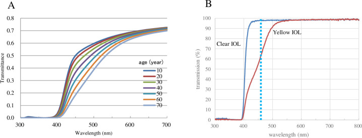
Purpose: We previously reported that macular pigment optical density (MPOD) levels decreased during a long follow-up period after clear intraocular lens (IOL) implant surgery presumably due to excessive light exposure. We examined changes in MPOD levels in the eyes that received yellow-tinted IOL implant surgery.
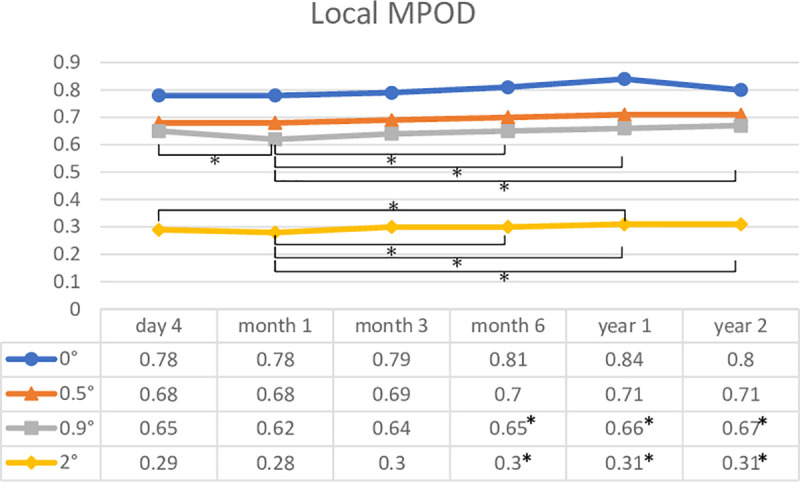
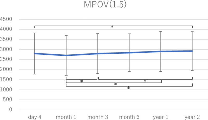
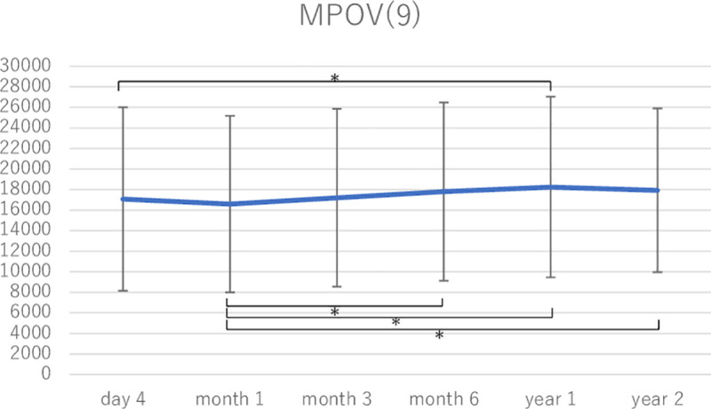
Subjects and methods: This was a prospective, observational study. Fifty-five eyes of 35 patients were studied. MPOD levels were measured with a dual-wavelength autofluorescence technique on day 4; months 1, 3, and 6; and years 1 and 2 postoperatively. The average optical densities at 0°- 2° eccentricities (local MPODs) and total volumes of MPOD (MPOVs) in the area within 1.5° and 9° eccentricities were analyzed.
Results: The mean local MPOD at baseline (on day 4) was 0.79 at 0°, 0.71 at 0.5°, 0.68 at 0.9°, and 0.32 at 2°. The mean MPOV within 1.5° and 9° at baseline was 2950 and 18,897, respectively. Local MPOD at 0.9° and 2° and MPOVs were slightly decreased at month 1 and increased after that. The increase reached statistical significance in local MPOD at 0.5° and 2° and MPOVs (Tukey-Kramer test). The changes in MPOV within 9° at year 2 [(MPOV on year 2 - MPOV on day 4) / MPOV on day 4] were from -0.21 to 1.18 (mean and standard deviation: 1.14 ± 0.28). The MPOV of 15 eyes increased more than 10% from the initial value, was maintained within 10% in 21 eyes, and deteriorated more than 10% in only 3 eyes.
Conclusions: Local MPOD and MPOV tended to slightly decrease month 1 postoperatively and gradually increased after that, but the rates of increases in MPOD levels were small. Yellow-tinted IOLs that have a lower transmittance of blue light might be preferable for preserving MPOD levels after surgery.
Conflict of interest statement
The authors have declared that no competing interests exist.
Figures
References
-
- Sparrow JR, Nakanishi K, Parish CA. The lipofuscin fluorophore A2E mediates blue light-induced damage to retinal pigmented epithelial cells. Invest Ophthalmol Vis Sci. 2000;41(7):1981–9. Epub 2000/06/14. . - PubMed
Publication types
MeSH terms
Substances
LinkOut - more resources
Full Text Sources
Other Literature Sources
