. 2021 Sep;74(3):1560-1577.
doi: 10.1002/hep.31831. Epub 2021 Jun 21.

CD47-Mediated Hedgehog/SMO/GLI1 Signaling Promotes Mesenchymal Stem Cell Immunomodulation in Mouse Liver Inflammation

Affiliations

CD47-Mediated Hedgehog/SMO/GLI1 Signaling Promotes Mesenchymal Stem Cell Immunomodulation in Mouse Liver Inflammation

Mingwei Sheng et al. Hepatology. 2021 Sep.

Abstract

Background and aims: The cluster of differentiation 47 (CD47)-signal regulatory protein alpha (SIRPα) signaling pathway plays important roles in immune homeostasis and tissue inflammatory response. Activation of the Hedgehog/smoothened (SMO)/GLI family zinc finger 1 (Gli1) pathway regulates cell growth, differentiation, and immune function. However, it remains unknown whether and how the CD47-SIRPα interaction may regulate Hedgehog/SMO/Gli1 signaling in mesenchymal stem cell (MSC)-mediated immune regulation during sterile inflammatory liver injury.

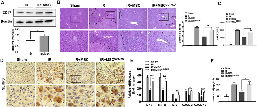
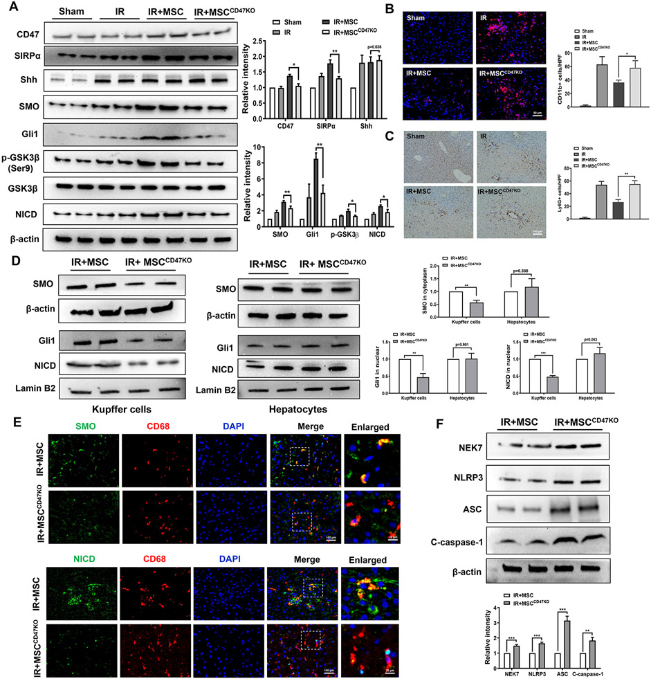
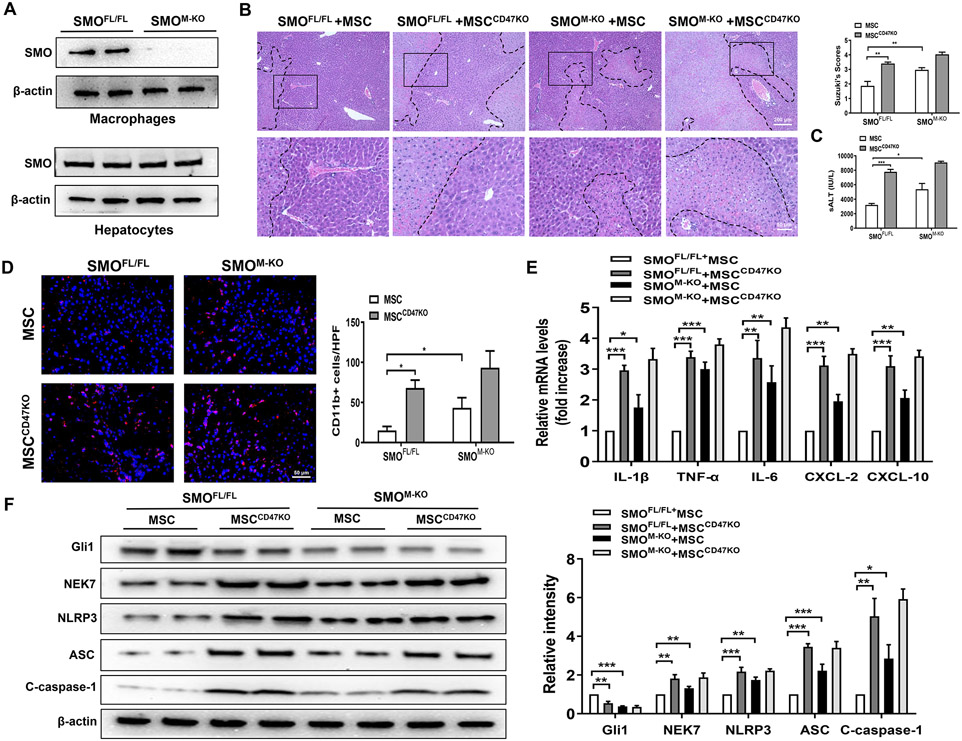
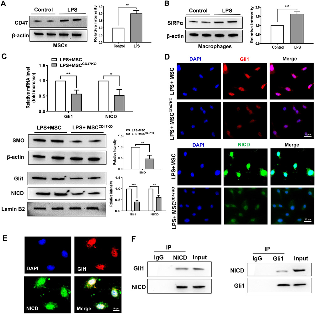
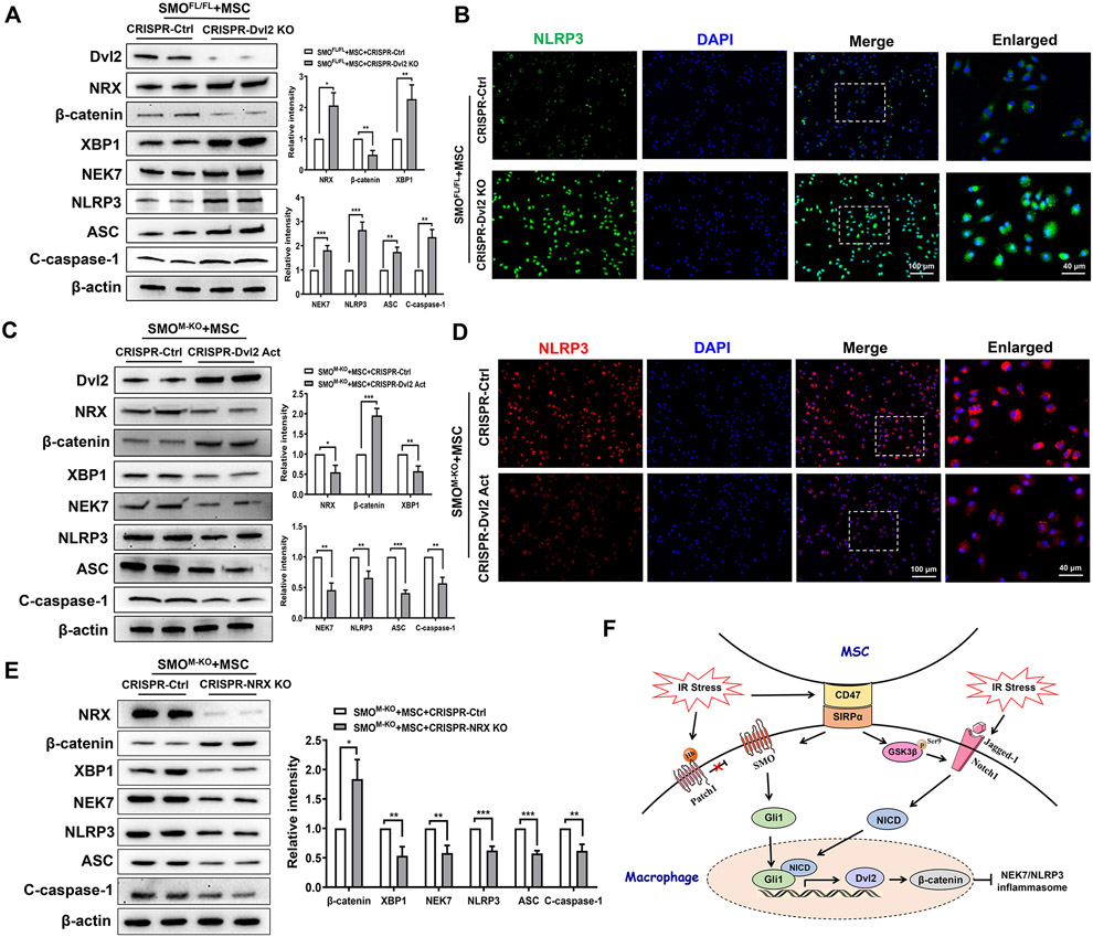
Approach and results: In a mouse model of ischemia/reperfusion (IR)-induced sterile inflammatory liver injury, we found that adoptive transfer of MSCs increased CD47 expression and ameliorated liver IR injury. However, deletion of CD47 in MSCs exacerbated IR-induced liver damage, with increased serum ALT levels, macrophage/neutrophil infiltration, and pro-inflammatory mediators. MSC treatment augmented SIRPα, Hedgehog/SMO/Gli1, and Notch1 intracellular domain (NICD), whereas CD47-deficient MSC treatment reduced these gene expressions in IR-stressed livers. Moreover, disruption of myeloid SMO or Notch1 increased IR-triggered liver inflammation with diminished Gli1 and NICD, but enhanced NIMA related kinase 7 (NEK7) and NLR family pyrin domain containing 3 (NLRP3) activation in MSC-transferred mice. Using a MSC/macrophage co-culture system, we found that MSC CD47 and macrophage SIRPα expression were increased after LPS stimulation. The CD47-SIRPα interaction increased macrophage Gli1 and NICD nuclear translocation, whereby NICD interacted with Gli1 and regulated its target gene Dvl2 (dishevelled segment polarity protein 2), which in turn inhibited NEK7/NLRP3 activity.

Conclusions: The CD47-SIRPα signaling activates the Hedgehog/SMO/Gli1 pathway, which controls NEK7/NLRP3 activity through a direct interaction between Gli1 and NICD. NICD is a coactivator of Gli1, and the target gene Dvl2 regulated by the NICD-Gli1 complex is crucial for the modulation of NLRP3-driven inflammatory response in MSC-mediated immune regulation. Our findings provide potential therapeutic targets in MSC-mediated immunotherapy of sterile inflammatory liver injury.


Figures

Fig. 1.
Fig. 1.. Disruption of CD47 in MSCs exacerbates IR-induced liver injury and augments proinflammatory mediators.
WT mice were adoptive transferred MSCs or CD47-deficient MSCs (1x10⁶) 24h prior to liver ischemic insult. (A) The CD47 expression was detected by Western blot assay in ischemic livers. The representative of three experiments. (B) Representative histological staining (H&E) of ischemic liver tissue (n=4-6 mice/group) and Suzuki's histological score. Scale bars, 200μm and 50μm. (C) Liver function was evaluated by serum ALT levels (IU/L) (n=4-6 samples/group). (D) Immunohistochemistry staining of NLRP3 in ischemic livers (n=4-6 mice/group). Scale bars, 50μm and 20μm. (E) qRT-PCR analysis of IL-1β, TNF-α, IL-6, CXCL-2, and CXCL-10 in ischemic livers (n=3-4 samples/group). (F) ELISA analysis of serum IL-1β levels (n=3-4 samples/group). All data represent the mean±SD. *p<0.05, **p<0.01, ***p<0.001.
Fig. 2.
Fig. 2.. The CD47-SIRPα interaction activates Hedgehog/SMO/Gli1 pathway, Notch1 signaling, and inhibits NEK7/NLRP3 activation in IR-stressed livers.
WT mice were adoptive transferred MSCs or CD47-deficient MSCs (1x10⁶) 24h prior to liver ischemic insult. (A) Western blot analysis and relative density ratio of CD47, SIRPα, Shh, SMO, Gli1, p-GSK3β, GSK3β, and NICD in ischemic livers. (B) Immunofluorescence staining of CD11b+ macrophages in ischemic livers (n=4-6 mice/group). Quantification of CD11b+ macrophages, Scale bars, 50μm. (C) Immunohistochemistry staining of Ly6G+ neutrophils in ischemic livers (n=4-6 mice/group). Quantification of Ly6G+ neutrophils, Scale bars, 100μm. (D) Western blot analysis and relative density ratio of SMO, Gli1, and NICD in Kupffer cells and hepatocytes. (E) Immunofluorescence staining for SMO or NICD expression in macrophages from the WT liver tissues (n=3-4 samples/group). DAPI was used to visualize nuclei. Scale bars, 100μm and 20μm. (F) Western blot analysis and relative density ratio of NEK7, NLRP3, ASC, and cleaved caspase-1 in ischemic livers. All Western blots represent three experiments, and the data represent the mean±SD. *p<0.05, **p<0.01, ***p<0.001.
Fig. 3.
Fig. 3.. Myeloid SMO deficiency in MSC-treated livers aggravates IR-induced hepatocellular damage and promotes NLRP3 inflammasome activation in IR-stressed livers.
The SMOFL/FL and SMOM-KO mice were adoptive transferred MSCs or CD47-deficient MSCs (1x10⁶) 24h prior to liver ischemic insult. (A) Western blot analysis and relative density ratio of SMO in ischemic livers. (B) Representative histological staining (H&E) of ischemic liver tissue (n=4-6 mice/group) and Suzuki's histological score. Scale bars, 200μm and 50μm. (C) Serum ALT levels (IU/L) (n=4-6 samples/group). (D) Immunofluorescence staining of CD11b+ macrophages in ischemic livers (n=4-6 mice/group). Quantification of CD11b+ macrophages, Scale bars, 50μm. (E) qRT-PCR analysis of IL-1β, TNF-α, and IL-6, CXCL-2, and CXCL-10 (n=3-4 samples/group). (F) Western blot analysis and relative density ratio of Gli1, NEK7, NLRP3, ASC, and cleaved caspase-1 in ischemic livers. All Western blots represent three experiments, and the data represent the mean±SD. *p<0.05, **p<0.01, ***p<0.001.
Fig. 4.
Fig. 4.. Disruption of myeloid Notch1 in MSC-treated livers enhances NEK7/NLRP3 function in IR-stressed livers.
The Notch1FL/FL and Notch1M-KO mice were adoptive transferred MSCs or CD47-deficient MSCs (1x10⁶) 24h prior to liver ischemic insult. (A) Representative histological staining (H&E) of ischemic liver tissue (n=4-6 mice/group) and Suzuki's histological score. Scale bars, 200μm and 50μm. (B) Serum ALT levels (IU/L) (n=4-6 samples/group). (C) Immunofluorescence staining of CD11b+ macrophages in ischemic livers (n=4-6 mice/group). Quantification of CD11b+ macrophages, Scale bars, 50μm. (D) Immunohistochemistry staining of Ly6G+ neutrophils in ischemic livers (n=4-6 mice/group). Quantification of Ly6G+ neutrophils, Scale bars, 50μm. (E) Western blot analysis and relative density ratio of NICD, NEK7, NLRP3, ASC, and cleaved caspase-1 ischemic livers. (F) qRT-PCR analysis of IL-1β, TNF-α, IL-6, CXCL-2, and CXCL-10 (n=3-4 samples/group). All Western blots represent three experiments, and the data represent the mean±SD. *p<0.05, **p<0.01, ***p<0.001.
Fig. 5.
Fig. 5.. The CD47-SIRPα signaling regulates the interaction between macrophage Gli1 and NICD in MSC-mediated immune regulation.
Bone marrow-derived macrophages (BMMs, 1x10⁶) were co-cultured with MSCs or CD47-deficient MSCs (2x10⁵) for 6h followed by LPS (100 ng/ml) stimulation. (A) (B) Western blot analysis and relative density ratio of CD47 and SIRPα in LPS-stimulated macrophages. (C) Analysis of mRNA levels and protein expression of Gli1 and NICD in macrophages after co-culture with MSCs or CD47-deficient MSCs by qRT-PCR and Western blot assay. (D) Immunofluorescence staining of nuclear Gli1 (red) and NICD (green) in macrophages after co-culture with MSCs or CD47-deficient MSCs. DAPI was used to visualize nuclei (blue). Scale bars, 20μm. (E) Immunofluorescence staining for macrophage Gli1 (red) and NICD (green) co-localization in the nucleus after co-culture with MSCs. DAPI was used to visualize nuclei (blue). Scale bars, 10μm. (F) Immunoprecipitation analysis of NICD and Gli1 in macrophages after co-culture with MSCs. All Western blots represent three experiments, and the data represent the mean±SD. *p<0.05, **p<0.01, ***p<0.001.
Fig. 6.
Fig. 6.. The Gli1-NICD interaction targets Dvl2 and modulates NEK7/NLRP3 function in MSC-mediated immune regulation.
(A) Experimental design of Gli1 ChIP-seq analysis. BMMs were collected and fixed after co-culture with MSCs. Following chromatin shearing and Gli1 antibody selection, the precipitated DNA fragments bound by Gli1-containing protein complexes were used for sequencing. (B) Localization of Gli1-binding sites on the mouse Dvl2 gene. The 15 exons, 14 introns, 3' untranslated region (UTR), 5’ UTR, and transcription start sites (TSS) of the mouse Dvl2 gene on chromosome 11 are shown. (C) ChIP-PCR analysis of Gli1 and NICD binding to the Dvl2 promoter. Protein-bound chromatin was prepared from BMMs and immunoprecipitated with Gli1 or NICD antibodies. For sequential ChIP, the protein-bound chromatin was first immunoprecipitated with the Gli1 antibody followed by elution with a second immunoprecipitation using NICD antibody, and then the immunoprecipitated DNA was analyzed by PCR. The normal IgG was used as a negative control. (D) RNA in situ hybridization for Dvl2 transcripts in LPS-stimulated macrophages after co-culture with MSCs or CD47-deficient MSCs (n=3-4 samples/group). (E) Western blot analysis and relative density ratio of SMO, Gli1, NICD, Dvl2, NRX, NEK7, NLRP3, ASC, and cleaved caspase-1 in macrophages after co-culture with MSCs or CD47-deficient MSCs. (F) qRT-PCR analysis of IL-1β, TNF-α, IL-6, CXCL-2, and CXCL-10 in LPS-stimulated macrophages (n=3-4 samples/group). All Western blots represent three experiments, and the data represent the mean±SD. *p<0.05, **p<0.01, ***p<0.001.
Fig. 7.
Fig. 7.. Notch1 signaling is required for the Hedgehog/SMO/Gli1 pathway-mediated Dvl2 activation and NEK7/NLRP3 inhibition in MSC-mediated immune regulation.
(A) BMMs were isolated from SMOFL/FL mice and co-cultured with MSCs or CD47-deficient MSCs followed by LPS stimulation. Immunofluorescence staining for Gli1 expression in macrophages (n=3-4 samples/group). DAPI was used to visualize nuclei. Scale bars, 40μm and 20μm. (B) Western blot analysis and relative density ratio of Gli1, Dvl2, NRX, β-catenin, NEK7, and NLRP3 in LPS-stimulated macrophages. (C) BMMs were isolated from SMOFL/FL mice and transfected with CRISPR/Cas9-Notch1 KO or control vector, and then co-cultured with MSCs followed by LPS stimulation. Immunofluorescence staining for Dvl2 expression in macrophages (n=3-4 samples/group). DAPI was used to visualize nuclei. Scale bars, 100μm and 40μm. (D) Western blot analysis and relative density ratio of NICD, Dvl2, NRX, β-catenin, NEK7, and NLRP3 in LPS-stimulated macrophages. (E) Caspase-1 activity (U) in macrophages after co-culture (n=3-4 samples/group). (F) qRT-PCR analysis of TNF-α, IL-1β, IL-6, CXCL-2, and CXCL-10 (n=3-4 samples/group). All Western blots represent three experiments, and the data represent the mean±SD. *p<0.05, **p<0.01, ***p<0.001.
Fig. 8.
Fig. 8.. Dvl2 is crucial to regulate NLRP3-driven inflammatory response in MSC-mediated immune regulation.
BMMs were isolated from SMOFL/FL and SMOM-KO mice and transfected with the CRISPR/Cas9-Dvl2 KO, CRISPR-Dvl2 activation, CRISPR/Cas9-NRX KO or control vector, and then co-cultured with MSCs followed by LPS stimulation. (A) Western blots analysis and relative density ratio of Dvl2, NRX, β-catenin, XBP1, NEK7, NLRP3, ASC, and cleaved caspase-1 in LPS-stimulated macrophages after transfection of CRISPR/Cas9-Dvl2 KO or control vector. (B) Immunofluorescence staining for NLRP3 expression in macrophages (n=3-4 samples/group). DAPI was used to visualize nuclei. Scale bars, 100μm and 40μm. (C) Western blots analysis and relative density ratio of Dvl2, NRX, β-catenin, XBP1, NEK7, NLRP3, ASC, and cleaved caspase-1 in LPS-stimulated macrophages after transfection of CRISPR-Dvl2 activation or control vector. (D) Immunofluorescence staining for NLRP3 expression in macrophages (n=3-4 samples/group). DAPI was used to visualize nuclei. Scale bars, 100μm and 40μm. (E) Western blots analysis and relative density ratio of NRX, β-catenin, XBP1, NEK7, NLRP3, ASC, and cleaved caspase-1 in LPS-stimulated macrophages after transfection of CRISPR/Cas9-NRX KO or control vector. (F) The schematic figure depicts putative molecular mechanisms by which CD47-mediated Hedgehog/SMO/Gli1 signaling regulates NEK7/NLRP3 activation in MSC-mediated immune regulation. All Western blots represent three experiments, and the data represent the mean±SD. *p<0.05, **p<0.01, ***p<0.001.

Similar articles

Cited by

References

    1. Contreras RA, Figueroa FE, Djouad F, Luz-Crawford P. Mesenchymal Stem Cells Regulate the Innate and Adaptive Immune Responses Dampening Arthritis Progression. Stem Cells Int 2016;2016:3162743. - PMC - PubMed
    1. Wang LT, Ting CH, Yen ML, Liu KJ, Sytwu HK, Wu KK, et al. Human mesenchymal stem cells (MSCs) for treatment towards immune- and inflammation-mediated diseases: review of current clinical trials. J Biomed Sci 2016;23:76. - PMC - PubMed
    1. Galipeau J, Sensebe L. Mesenchymal Stromal Cells: Clinical Challenges and Therapeutic Opportunities. Cell Stem Cell 2018;22:824–833. - PMC - PubMed
    1. Lindberg FP, Gresham HD, Schwarz E, Brown EJ. Molecular cloning of integrin-associated protein: an immunoglobulin family member with multiple membrane-spanning domains implicated in alpha v beta 3-dependent ligand binding. J Cell Biol 1993;123:485–496. - PMC - PubMed
    1. Jaiswal S, Jamieson CH, Pang WW, Park CY, Chao MP, Majeti R, et al. CD47 is upregulated on circulating hematopoietic stem cells and leukemia cells to avoid phagocytosis. Cell 2009;138:271–285. - PMC - PubMed

Publication types

MeSH terms