

Review
doi: 10.1177/19714009211004185.
Epub 2021 Mar 26.
Hydrophilic polymer embolization following flow diversion of cerebral aneurysms
Affiliations
- PMID: 33765885
- PMCID: PMC8447820
- DOI: 10.1177/19714009211004185
Item in Clipboard
Review
Hydrophilic polymer embolization following flow diversion of cerebral aneurysms
Neuroradiol J.
2021 Aug.
Abstract
Foreign body embolization is a rare and potentially under-recognized complication of neuroendovascular procedures. This complication should be considered in the differential diagnosis for clinical or radiological deterioration following neurovascular interventions. We report a case of foreign body hydrophilic coating embolization that occurred following an attempted flow diversion of an intracranial aneurysm with dramatic flare-up after repeat exposure. We also provide a literature review of all reported cases of hydrophilic polymer embolization following flow diversion procedures.
Keywords: Aneurysms; embolization; flow diversion; hydrophilic.
Figures
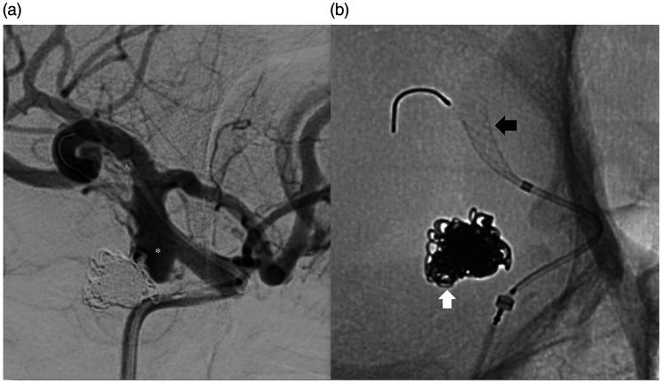
Angiographic images at the time of attempted flow diverter placement. Digital
subtraction angiogram (right anterior oblique projection) of the right
internal carotid artery (a) shows the recurrence of the previously coiled
right posterior communicating aneurysm (asterisk). Unsubtracted image (b)
shows the previous coil mass (white arrow) and the partially deployed flow
diverter in the supraclinoid internal carotid artery (black arrow).
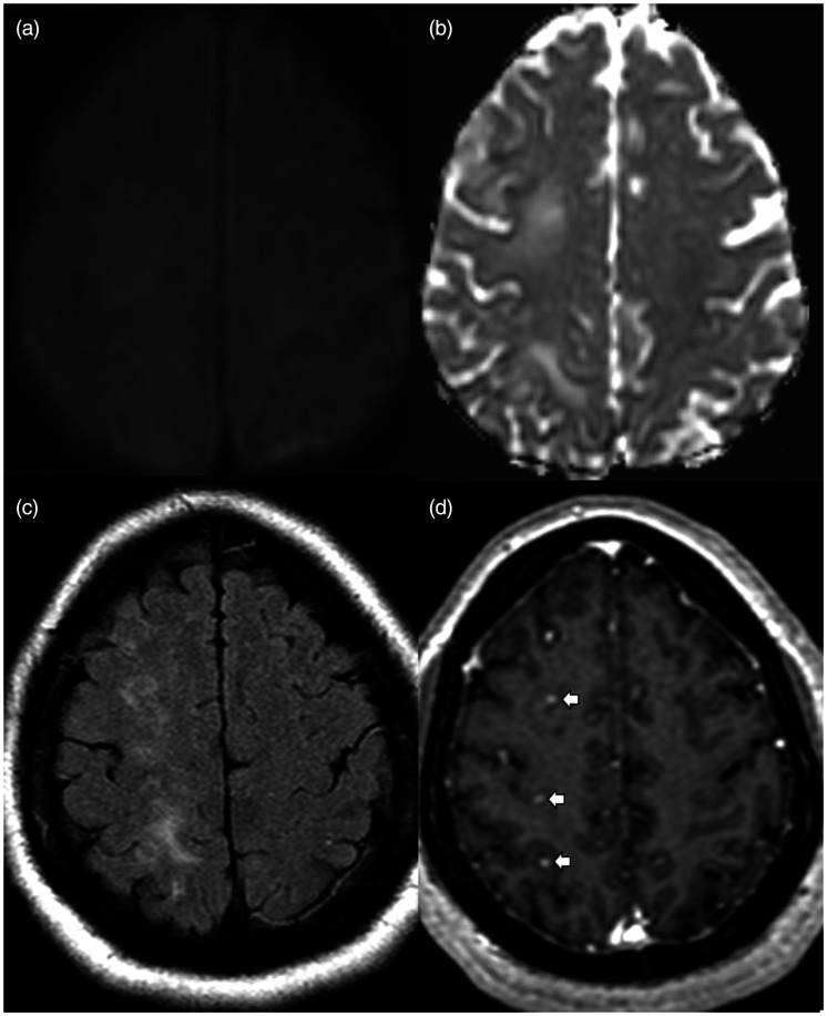
MRI brain exam performed 1 month after aborted flow diverter placement. Axial
diffusion weighted images (a) with apparent diffusion coefficient (b)
showing no restricted diffusion. Axial FLAIR image (c) shows multifocal
hyperintensities in the subcortical white matter of the right frontal and
parietal lobes. Axial gadolinium-enhanced T1 image (d) shows multiple small
enhancing lesions (white arrows).
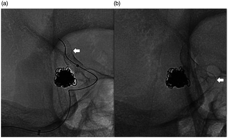
Angiographic images at the time of subsequent flow diverter placement.
Unsubstracted angiograms (right anterior oblique projection) of the right
internal carotid artery (a) and (b) showing successful deployment of two
telescoping flow diverters (white arrows) across the neck of the aneurysm
with the distal end of the distal flow diverter noted in the proximal M1
segment to ensure stability.
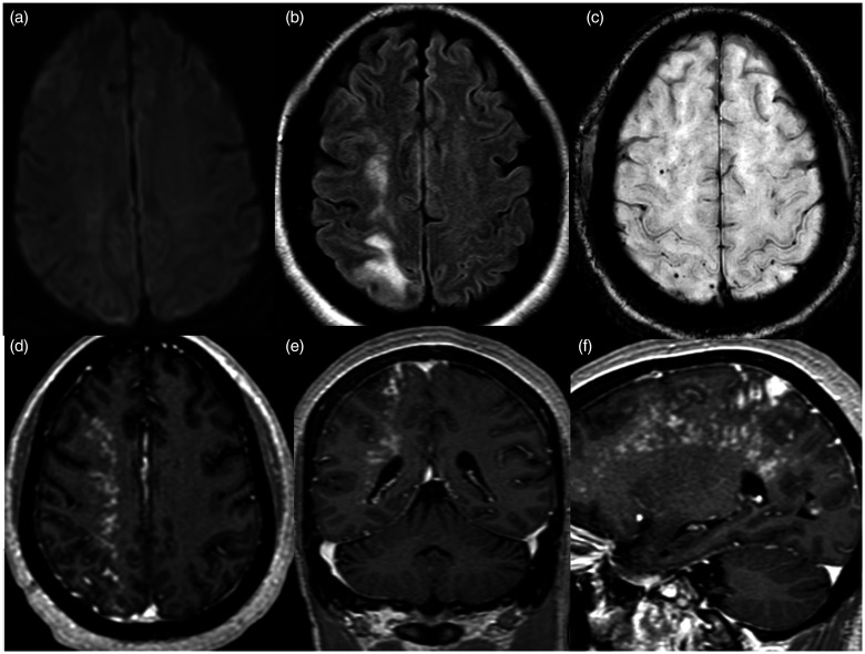
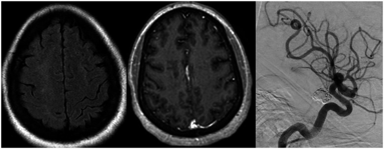
MRI brain exam performed two days after successful placement of the flow
diverters. Axial diffusion weighted images (a) showing no restricted
diffusion. Axial FLAIR image (b) shows worsened hyperintensities in the
right frontal and parietal subcortical white matter. Axial susceptibility
weighted image (c) shows a few associated punctate signal voids and axial
(d), coronal (e) and sagittal (f) gadolinium-enhanced T1 images show a
dramatic increase in extent of multinodular enhancement.
Follow up MRI brain exam and angiogram performed after flow diverter
placement. Axial FLAIR (a) and gadolinium-enhanced T1 (b) images 2.5 months
later show near-complete resolution of the subcortical hyperintensities and
enhancement in the right frontal and parietal lobes. Digital subtraction
angiogram (lateral projection) of the right internal carotid artery (c) 6
months later shows occlusion of the residual aneurysm with reconstruction of
the supraclinoid carotid artery.
References
Publication types
MeSH terms
Substances
Grants and funding
LinkOut - more resources
Full Text Sources
Other Literature Sources
Medical
