. 2021 Jul:156:7-19.
doi: 10.1016/j.yjmcc.2021.03.007. Epub 2021 Mar 22.

Impact of etiology on force and kinetics of left ventricular end-stage failing human myocardium

Affiliations

Impact of etiology on force and kinetics of left ventricular end-stage failing human myocardium

Mohammed A Mashali et al. J Mol Cell Cardiol. 2021 Jul.

Abstract

Background: Heart failure (HF) is associated with highly significant morbidity, mortality, and health care costs. Despite the significant advances in therapies and prevention, HF remains associated with poor clinical outcomes. Understanding the contractile force and kinetic changes at the level of cardiac muscle during end-stage HF in consideration of underlying etiology would be beneficial in developing targeted therapies that can help improve cardiac performance.

Objective: Investigate the impact of the primary etiology of HF (ischemic or non-ischemic) on left ventricular (LV) human myocardium force and kinetics of contraction and relaxation under near-physiological conditions.

Methods and results: Contractile and kinetic parameters were assessed in LV intact trabeculae isolated from control non-failing (NF; n = 58) and end-stage failing ischemic (FI; n = 16) and non-ischemic (FNI; n = 38) human myocardium under baseline conditions, length-dependent activation, frequency-dependent activation, and response to the β-adrenergic stimulation. At baseline, there were no significant differences in contractile force between the three groups; however, kinetics were impaired in failing myocardium with significant slowing down of relaxation kinetics in FNI compared to NF myocardium. Length-dependent activation was preserved and virtually identical in all groups. Frequency-dependent activation was clearly seen in NF myocardium (positive force frequency relationship [FFR]), while significantly impaired in both FI and FNI myocardium (negative FFR). Likewise, β-adrenergic regulation of contraction was significantly impaired in both HF groups.

Conclusions: End-stage failing myocardium exhibited impaired kinetics under baseline conditions as well as with the three contractile regulatory mechanisms. The pattern of these kinetic impairments in relation to NF myocardium was mainly impacted by etiology with a marked slowing down of kinetics in FNI myocardium. These findings suggest that not only force development, but also kinetics should be considered as a therapeutic target for improving cardiac performance and thus treatment of HF.

Keywords: Contraction; Heart failure; Kinetics; Left ventricle; Myocardium; Relaxation.


Conflict of interest statement

Disclosures

The authors disclose no conflict of interest.

Figures

Fig. 1.
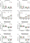
Fig. 1.. Demographic characteristics of donors and end-stage HF patients.
(A-F) Pie charts showing gender and race distribution in donors and HF patients. (G) Donors (n=57) were significantly younger than patients with ischemic (n=16) and non-ischemic HF (n=38) with no significant age difference between both HF groups. (H) BMI was not significantly different between donors (n=55) and patients with ischemic (n=15) or non-ischemic (n=37) HF. (I) No significant difference in HW between donors (n=57) and patients with ischemic (n=15) or non-ischemic (n=38) HF. (J) Left ventricular wall of donor hearts (n=53) had significantly greater thickness than that of ischemic (n=15) and non-ischemic hearts (n=38). Data are presented as means ± SD;-*P<0.05; **P<0.01; ***P<0.001; One-way ANOVA followed by Tukey’s multiple comparisons post hoc test (G-J). A, Asian; AA, African American; BMI, body mass index; C, Caucasian; C/AA, Caucasian/African American; FI, failing ischemic; FNI, failing non-ischemic; g, gram; H, Hispanic; HF, heart failing; LV, left ventricle; NF, non-failing; V, Vietnamese.
Fig. 2.
Fig. 2.. Baseline contractile force and contraction/relaxation kinetics of LV trabeculae isolated from NF (n=58) and failing (n=54) human hearts of ischemic (FI, n=16) and non-ischemic (FNI, n=38) etiology at 1 Hz.
(A) Fdev of NF trabeculae was significantly higher than failing trabeculae (P=0.024), while there was no significant difference in Fdia between both groups. (B) Fdev and Fdia were not significantly different between NF trabeculae and trabeculae from both HF groups. (C) RT90 and TT90 were significantly prolonged in failing trabeculae compared to NF trabeculae (P=0.005 and P=0.044, respectively), while there was no significant difference in TTP or RT50 between both groups. (D) RT90 was significantly prolonged in FNI trabeculae compared to NF trabeculae (P=0.005). (E, G) dF/dt, -dF/dt, and -dF/dt/Fdev were significantly slower in failing myocardium vs. NF myocardium (P=0.011, P=0.004, and P=0.008, respectively), while there was no significant difference in dF/dt/Fdev between both groups. (F, H) dF/dt, -dF/dt, and -dF/dt/Fdev were significantly slower in FNI trabeculae when compared to NF trabeculae (P=0.049, P=0.008, P=0.006, respectively). Data are presented as means ± SEM; *P<0.05; **P<0.01; Student t test (A, C, E, G); One-way ANOVA followed by Tukey’s multiple comparisons post hoc test (B, D, F, H). dF/dt, maximal rate of force development during contraction; -dF/dt, maximal rate of force decay during relaxation; dF/dt/Fdev, maximal kinetic rate of contraction; -dF/dt/Fdev, maximal kinetic rate of relaxation; Fdev, active developed force; Fdia, diastolic force; FI, failing ischemic; FNI, failing non-ischemic; LV, left ventricular; NF, non-failing; RT50, time from peak tension to 50% relaxation; RT90, time from peak tension to 90% relaxation; TT90, total twitch duration; TTP, time to peak tension.
Fig. 3.
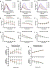
Fig. 3.. Frequency-dependent activation. Impact of frequency on contractile force and kinetics of human NF (n=36), FI (n=9), and FNI (n=23) LV trabeculae.
(A) Twitch contractions of a trabecula from NF heart (#694855) showing a positive FFR. (B) Twitch contractions of a trabecula from FI heart (#214010) showing a blunted/negative FFR. (C) Twitch contractions of a trabecula from FNI heart (#373249) showing a blunted/negative FFR. (D) NF trabeculae exhibited positive FFR, while HF trabeculae exhibited negative FFR. (E) The same as panel (D), but data are presented as a fraction of their corresponding Fdev at 0.5 Hz. NF trabeculae have significantly higher Fdev values when compared to those of HF trabeculae at 1.5 to 3 Hz. (F, G) NF trabeculae showed no significant differences in Fdia or TTP compared to both HF groups. (H, I) NF trabeculae showed significantly shorter RT50 and TT90 compared to those of HF trabeculae at lower frequencies. (J, K) NF trabeculae showed significantly faster dF/dt and -dF/dt at higher frequencies and significantly faster -dF/dt/Fdev at lower frequencies compared to those exhibited by HF trabeculae. Data are presented as means ± SEM; A two-way repeated measures ANOVA followed by Tukey’s or Dunnett’s post hoc tests, where appropriate, to correct for multiple comparisons; *P<0.05 (NF vs. FNI); #P<0.05 (NF vs. FI) at corresponding stimulation frequency. FFR, force frequency relationship; other abbreviations as in Fig. 2.
Fig. 4.
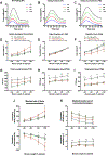
Fig. 4.. Length-dependent activation. Impact of muscle length on contractile force and kinetics of human NF (n=24), FI (n=6), and FNI (n=19) LV trabeculae.
(A-C) Twitch contractions of three trabeculae isolated from NF heart (#442404), FI heart (#214101), and FNI heart (#465645) at 85% (L85), 90% (L90), 95% (L95), and 100% of optimal length (L100). (D) FI trabeculae have significantly higher Fdev at L100 compared to those of NF and FNI trabeculae. (E) The same as panel (D), but data are presented as a fraction of Fdev at L100. (F) FNI trabeculae exhibited a significantly higher Fdia than that of NF trabeculae at L100 and that of FI trabeculae at L95 and L100. (G) FNI trabeculae showed a significantly longer TTP at L85, L90, and L95 compared to those of NF trabeculae. (H, I) FNI trabeculae have significantly longer RT50 and TT90 at all muscle lengths compared to those of NF and FI trabeculae except at L90 where RT50 was longer than that of FI, but not significant. (J) At L100, FI trabeculae showed a significantly faster dF/dt vs. FNI trabeculae and significantly faster -dF/dt vs. NF and FNI trabeculae. (K) NF trabeculae showed faster dF/dt/Fdev at all muscle lengths compared to HF trabeculae, while FNI trabeculae showed a significantly slower -dF/dt/Fdev at all muscle lengths compared to those of NF and FI trabeculae except at L90. Data are presented as means ± SEM; A two-way repeated measures ANOVA followed by Tukey’s or Dunnett’s post hoc tests, where appropriate, to correct for multiple comparisons; *P<0.05 (NF vs. FNI); #P<0.05 (NF vs. FI); P<0.05 (FI vs. FNI) at corresponding muscle length. Abbreviations as in Fig. 2.
Fig. 5.
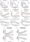
Fig. 5.. Impact of β-adrenergic stimulation (increasing concentration of isoproterenol [1 nM - 1 μM]) on contractile force and kinetics of human NF (n=21), FI (n=5), and FNI (n=16) LV trabeculae.
(A-C) Twitch contractions of three trabeculae isolated from NF heart (#632941), FI heart (#214010), and FNI heart (#478852) at increasing concentrations of isoproterenol. At the highest concentrations of isoproterenol, arrhythmic behavior due to calcium overload started to emerge in some preparations, as can be gleaned from non-exponential late relaxation phase. (D) Fdev was increased in all groups upon addition of isoproterenol in a concentration-dependent manner. (E) The same as panel (D), but data are presented as a fraction of Fdev at baseline. NF trabeculae have significantly higher Fdev values than those of FI and FNI trabeculae at isoproterenol concentrations of 30 nM to 1 μM. (F, G) There were no significant differences in Fdia or TTP between all groups at different isoproterenol concentrations. (H) FNI trabeculae have significantly longer RT50 than those of NF and FI trabeculae at isoproterenol concentrations of 1 nM to 10 nM. (I) FNI trabeculae have a significantly longer TT90 at isoproterenol concentrations of 1 nM to 30 nM compared to those of FI trabeculae and at all concentrations compared to those of NF trabeculae. (J) NF trabeculae have significantly faster dF/dt and -dF/dt than those of FNI trabeculae at isoproterenol concentrations of equal or greater than 30 nM. (K) NF trabeculae showed faster dF/dt/Fdev compared to those of FNI trabeculae at all isoproterenol concentrations, but was only significant at 0.3 μM, while NF trabeculae showed a significantly faster -dF/dt/Fdev at isoproterenol concentrations of equal or greater than 3 nM compared to those of FNI trabeculae. Data are presented as means ± SEM; A two-way repeated measures ANOVA followed by Tukey’s or Dunnett’s post hoc tests, where appropriate, to correct for multiple comparisons; *P<0.05 (NF vs. FNI); #P<0.05 (NF vs. FI); P<0.05 (FI vs. FNI) at corresponding isoproterenol concentration. Abbreviations as in Fig. 2.

References

    1. Yancy CW, Jessup M, Bozkurt B, Butler J, Casey DE, Drazner MH, et al. 2013 ACCF/AHA guideline for the management of heart failure: a report of the American College of Cardiology Foundation/American Heart Association Task Force on Practice Guidelines. J Am Coll Cardiol 2013; 62(16): e147–e239. - PubMed
    1. Orso F, Fabbri G, Maggioni AP. Epidemiology of heart failure. Heart Failure: Springer; 2016. p. 15–33. - PubMed
    1. Virani SS, Alonso A, Benjamin EJ, Bittencourt MS, Callaway CW, Carson AP, et al. Heart disease and stroke statistics—2020 update: a report from the American Heart Association. Circulation 2020; 141(9): E139–E596. - PubMed
    1. Felker GM, Shaw LK, O’Connor CM. A standardized definition of ischemic cardiomyopathy for use in clinical research. J Am Coll Cardiol 2002; 39(2): 210–8. - PubMed
    1. Frank O Zur dynamik des herzmuskels. Z Biol 1895; 32: 370–447.

Publication types

MeSH terms