

Polyploid giant cancer cell characterization: New frontiers in predicting response to chemotherapy in breast cancer
- PMID: 33766651
- PMCID: PMC8672208
- DOI: 10.1016/j.semcancer.2021.03.017
Polyploid giant cancer cell characterization: New frontiers in predicting response to chemotherapy in breast cancer
Abstract
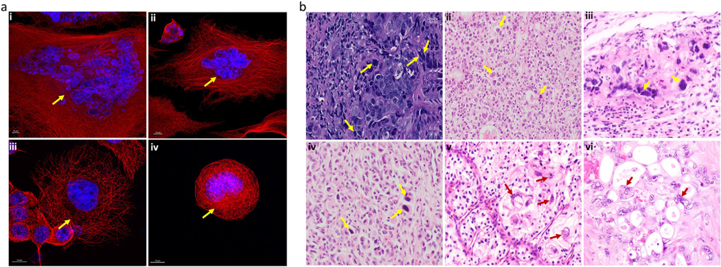
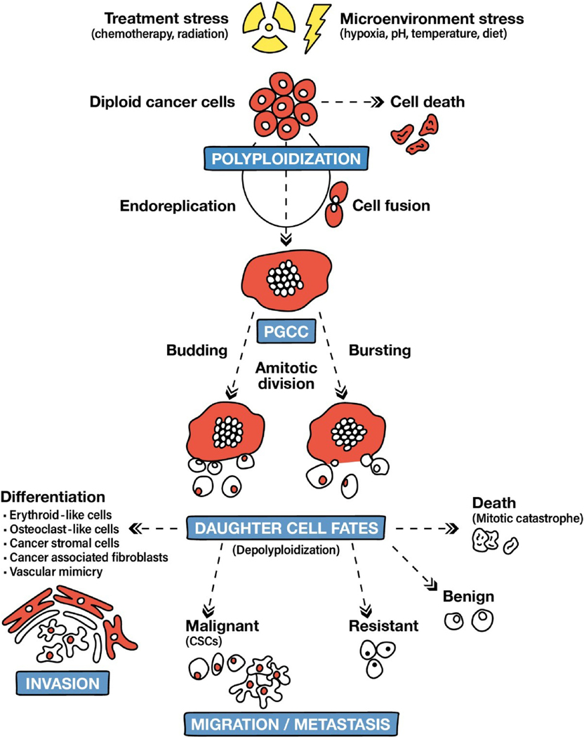
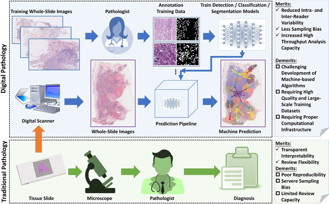
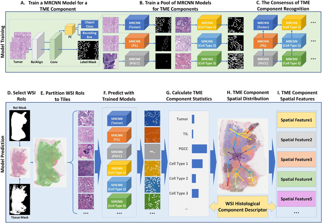
Although polyploid cells were first described nearly two centuries ago, their ability to proliferate has only recently been demonstrated. It also becomes increasingly evident that a subset of tumor cells, polyploid giant cancer cells (PGCCs), play a critical role in the pathophysiology of breast cancer (BC), among other cancer types. In BC, PGCCs can arise in response to therapy-induced stress. Their progeny possess cancer stem cell (CSC) properties and can repopulate the tumor. By modulating the tumor microenvironment (TME), PGCCs promote BC progression, chemoresistance, metastasis, and relapse and ultimately impact the survival of BC patients. Given their pro- tumorigenic roles, PGCCs have been proposed to possess the ability to predict treatment response and patient prognosis in BC. Traditionally, DNA cytometry has been used to detect PGCCs.. The field will further derive benefit from the development of approaches to accurately detect PGCCs and their progeny using robust PGCC biomarkers. In this review, we present the current state of knowledge about the clinical relevance of PGCCs in BC. We also propose to use an artificial intelligence-assisted image analysis pipeline to identify PGCC and map their interactions with other TME components, thereby facilitating the clinical implementation of PGCCs as biomarkers to predict treatment response and survival outcomes in BC patients. Finally, we summarize efforts to therapeutically target PGCCs to prevent chemoresistance and improve clinical outcomes in patients with BC.
Keywords: Artificial intelligence; Breast cancer; Chemo resistance; Patient prognosis; Polyploid giant cancer cell.
Copyright © 2021 Elsevier Ltd. All rights reserved.
Conflict of interest statement
Conflicts of interest
The authors declare no conflicts of interest.
Figures
Similar articles
-
Autophagy modulating therapeutics inhibit ovarian cancer colony generation by polyploid giant cancer cells (PGCCs).BMC Cancer. 2022 Apr 14;22(1):410. doi: 10.1186/s12885-022-09503-6. BMC Cancer. 2022. PMID: 35421971 Free PMC article.
-
Single-cell morphological and transcriptome analysis unveil inhibitors of polyploid giant breast cancer cells in vitro.Commun Biol. 2023 Dec 21;6(1):1301. doi: 10.1038/s42003-023-05674-5. Commun Biol. 2023. PMID: 38129519 Free PMC article.
-
High-Throughput Empirical and Virtual Screening To Discover Novel Inhibitors of Polyploid Giant Cancer Cells in Breast Cancer.Anal Chem. 2025 Mar 18;97(10):5498-5506. doi: 10.1021/acs.analchem.4c05138. Epub 2025 Mar 4. Anal Chem. 2025. PMID: 40040372 Free PMC article.
-
Polyploid giant cancer cells and cancer progression.Front Cell Dev Biol. 2022 Oct 5;10:1017588. doi: 10.3389/fcell.2022.1017588. eCollection 2022. Front Cell Dev Biol. 2022. PMID: 36274852 Free PMC article. Review.
-
Dormant cancer cells and polyploid giant cancer cells: The roots of cancer recurrence and metastasis.Clin Transl Med. 2024 Feb;14(2):e1567. doi: 10.1002/ctm2.1567. Clin Transl Med. 2024. PMID: 38362620 Free PMC article. Review.
Cited by
-
Presence of cells in the polyaneuploid cancer cell (PACC) state predicts the risk of recurrence in prostate cancer.Prostate. 2023 Feb;83(3):277-285. doi: 10.1002/pros.24459. Epub 2022 Nov 13. Prostate. 2023. PMID: 36372998 Free PMC article.
-
Exploration of drug resistance mechanisms in triple negative breast cancer cells using a microfluidic device and patient tissues.Elife. 2024 Mar 27;12:RP88830. doi: 10.7554/eLife.88830. Elife. 2024. PMID: 38536720 Free PMC article.
-
High-Throughput Empirical and Virtual Screening to Discover Novel Inhibitors of Polyploid Giant Cancer Cells in Breast Cancer.bioRxiv [Preprint]. 2024 Sep 24:2024.09.23.614522. doi: 10.1101/2024.09.23.614522. bioRxiv. 2024. Update in: Anal Chem. 2025 Mar 18;97(10):5498-5506. doi: 10.1021/acs.analchem.4c05138. PMID: 39386568 Free PMC article. Updated. Preprint.
-
Nuclear morphology predicts cell survival to cisplatin chemotherapy.Neoplasia. 2023 Aug;42:100906. doi: 10.1016/j.neo.2023.100906. Epub 2023 May 10. Neoplasia. 2023. PMID: 37172462 Free PMC article.
-
Emerging Paradigms in Cancer Metastasis: Ghost Mitochondria, Vasculogenic Mimicry, and Polyploid Giant Cancer Cells.Cancers (Basel). 2024 Oct 19;16(20):3539. doi: 10.3390/cancers16203539. Cancers (Basel). 2024. PMID: 39456632 Free PMC article. Review.
References
-
- Davoli T, de Lange T, The causes and consequences of polyploidy in normal development and cancer, Annual Review of Cell and Developmental Biology. 27 (2011) 585–610. - PubMed
-
- Liu Z, Yue S, Chen X, Kubin T, Braun T, Regulation of cardiomyocyte polyploidy and multinucleation by CyclinG1, Circulation Research. 106 (2010) 1498. - PubMed
-
- Takegahara N, Kim H, Mizuno H, Sakaue-Sawano A, Miyawaki A, Tomura M, Kanagawa O, Ishii M, Choi Y, Involvement of receptor activator of nuclear factor-κB ligand (RANKL)-induced incomplete cytokinesis in the polyploidization of osteoclasts, Journal of Biological Chemistry. 291 (2016) 3439–3454. - PMC - PubMed
Publication types
MeSH terms
Grants and funding
LinkOut - more resources
Full Text Sources
Other Literature Sources
Medical
