A randomized and blinded trial of inhaled nitric oxide in a piglet model of pediatric cardiopulmonary resuscitation
- PMID: 33766668
- PMCID: PMC8096708
- DOI: 10.1016/j.resuscitation.2021.03.004
A randomized and blinded trial of inhaled nitric oxide in a piglet model of pediatric cardiopulmonary resuscitation
Abstract
Aim: Inhaled nitric oxide (iNO) during cardiopulmonary resuscitation (CPR) improved systemic hemodynamics and outcomes in a preclinical model of adult in-hospital cardiac arrest (IHCA) and may also have a neuroprotective role following cardiac arrest. The primary objectives of this study were to determine if iNO during CPR would improve cerebral hemodynamics and mitochondrial function in a pediatric model of lipopolysaccharide-induced shock-associated IHCA.
Methods: After lipopolysaccharide infusion and ventricular fibrillation induction, 20 1-month-old piglets received hemodynamic-directed CPR and were randomized to blinded treatment with or without iNO (80 ppm) during and after CPR. Defibrillation attempts began at 10 min with a 20-min maximum CPR duration. Cerebral tissue from animals surviving 1-h post-arrest underwent high-resolution respirometry to evaluate the mitochondrial electron transport system and immunohistochemical analyses to assess neuropathology.
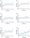
Results: During CPR, the iNO group had higher mean aortic pressure (41.6 ± 2.0 vs. 36.0 ± 1.4 mmHg; p = 0.005); diastolic BP (32.4 ± 2.4 vs. 27.1 ± 1.7 mmHg; p = 0.03); cerebral perfusion pressure (25.0 ± 2.6 vs. 19.1 ± 1.8 mmHg; p = 0.02); and cerebral blood flow relative to baseline (rCBF: 243.2 ± 54.1 vs. 115.5 ± 37.2%; p = 0.02). Among the 8/10 survivors in each group, the iNO group had higher mitochondrial Complex I oxidative phosphorylation in the cerebral cortex (3.60 [3.56, 3.99] vs. 3.23 [2.44, 3.46] pmol O2/s mg; p = 0.01) and hippocampus (4.79 [4.35, 5.18] vs. 3.17 [2.75, 4.58] pmol O2/s mg; p = 0.02). There were no other differences in mitochondrial respiration or brain injury between groups.
Conclusions: Treatment with iNO during CPR resulted in superior systemic hemodynamics, rCBF, and cerebral mitochondrial Complex I respiration in this pediatric cardiac arrest model.
Keywords: Cardiopulmonary resuscitation; Cerebral blood flow; Hemodynamics; In-hospital cardiac arrest; Inhaled nitric oxide; Laboratory; Pediatrics; Physiology; Pulmonary hypertension; Shock.
Copyright © 2021 Elsevier B.V. All rights reserved.
Figures


References
Publication types
MeSH terms
Substances
Grants and funding
LinkOut - more resources
Full Text Sources
Other Literature Sources
Medical

