. 2021 Apr 13;118(15):e2025866118.
doi: 10.1073/pnas.2025866118.

Generation of SARS-CoV-2 reporter replicon for high-throughput antiviral screening and testing

Affiliations

Generation of SARS-CoV-2 reporter replicon for high-throughput antiviral screening and testing

Xi He et al. Proc Natl Acad Sci U S A. .

Abstract

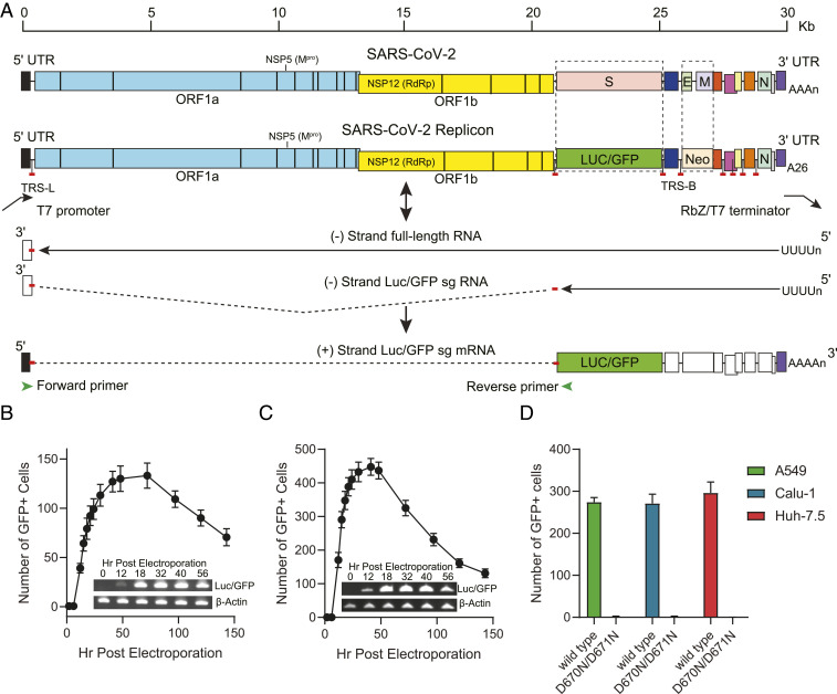
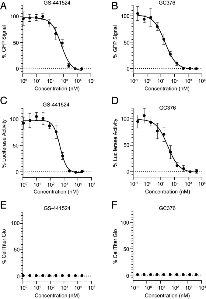
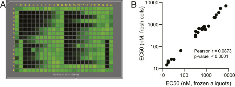
Severe acute respiratory syndrome coronavirus 2 (SARS-CoV-2) research and antiviral discovery are hampered by the lack of a cell-based virus replication system that can be readily adopted without biosafety level 3 (BSL-3) restrictions. Here, the construction of a noninfectious SARS-CoV-2 reporter replicon and its application in deciphering viral replication mechanisms and evaluating SARS-CoV-2 inhibitors are presented. The replicon genome is replication competent but does not produce progeny virions. Its replication can be inhibited by RdRp mutations or by known SARS-CoV-2 antiviral compounds. Using this system, a high-throughput antiviral assay has also been developed. Significant differences in potencies of several SARS-CoV-2 inhibitors in different cell lines were observed, which highlight the challenges of discovering antivirals capable of inhibiting viral replication in vivo and the importance of testing compounds in multiple cell culture models. The generation of a SARS-CoV-2 replicon provides a powerful platform to expand the global research effort to combat COVID-19.

Keywords: COVID-19; SARS-CoV-2; antivirals; high-throughput antiviral screening; replicon.


Conflict of interest statement

Competing interest statement: All authors are employees of Merck and Company, Inc. A provisional patent application on the discoveries of this work has been filed.

Figures

Fig. 1.
Fig. 1.
Construction and transcriptional analysis of SARS-CoV-2 replicon. (A) Schematic diagram of reporter replicon genome structure, replication, and sg mRNA production. The S and E/M ORFs in the wt viral genome were deleted and replaced with Luc/GFP and Neo genes, respectively. The full-length replicon cDNA was flanked by a T7 promoter and a polyA/HDV ribozyme and T7 terminator cassette. The in vitro-transcribed replicon RNA is copied by replicase complex to produce genomic or subgenomic-sized negative stranded RNAs (solid lines). The negative stranded sg RNAs are used as templates to produce the sg mRNAs for expression of reporter and structural proteins. The sg mRNAs consist of the leader at 5′-UTR of the genome and mRNA body sequences joined by a short and conserved sequence motif, the transcription-regulating sequence (TRS). The location of the sequences encoding major antiviral targets NSP5 (Mpro) and NSP12 (RdRp), the untranscribed regions (dashed lines), TRS leader (TRS-L) and TRS body (TRS-B) are also indicated. (B and C) Kinetics of GFP expression post-electroporation and detection of the sg mRNA expressing Luc/GFP reporter products. 293T (B) and A549 (C) cells were electroporated with in vitro-transcribed replicon RNA, and the number of GFP-positive cells in each well containing 1 × 10⁴ cells were counted at indicated time points. Total RNA was also collected and purified at indicated times from electroporated cells. RT-PCR products from mRNAs expressing Luc/GFP or β-actin were confirmed by gel electrophoresis. (D) GFP reporter signals produced by the wt and RdRp mutant replicons. The A549, Calu-1, and Huh-7.5 cells were electroporated with T7 polymerase transcribed wt replicon RNA or the RdRp D760N/D761N double-mutant replicon RNA and transferred to 384-well plates. The number of GFP-positive cells in each well were counted at 30 h post-electroporation.
Fig. 2.
Fig. 2.
Dose-dependent responses of SARS-CoV-2 replicon reporter activity to GS-441524 and GC376. The 293T cells were electroporated with T7 polymerase transcribed replicon RNA and incubated with GS-441524 or GC376 compounds at indicated concentrations. The activities were determined by percentage of GFP or luciferase signals comparing to DMSO-treated cells (A–D). The cytotoxicity of GS-441524 and GC376 to the cells were also examined (E and F).
Fig. 3.
Fig. 3.
Adaptation of the SARS-CoV-2 replicon-based assay to high-throughput platform. (A) The plate view of GFP expression in replicon electroporated cells treated with compounds. The 293T cells were electroporated with T7 polymerase transcribed replicon RNA and incubated with control compounds in 384-well plates for 30 h. The plates were scanned using an Acumen eX3. (B) Correlation between the EC50 values measured using freshly or frozen electroporated cells. A total of 27 control compounds was tested using either fresh or cryopreserved 293T cells electroporated by SARS-CoV-2 replicon RNA as described in Materials and Methods. The strength of correlation is determined by the Pearson test.

Similar articles

Cited by

References

    1. Dong E., Du H., Gardner L., An interactive web-based dashboard to track COVID-19 in real time. Lancet Infect. Dis. 20, 533–534 (2020). - PMC - PubMed
    1. Thorlund K., et al. ., A real-time dashboard of clinical trials for COVID-19. Lancet Digit. Health 2, e286–e287 (2020). - PMC - PubMed
    1. Puelles V. G., et al. ., Multiorgan and renal tropism of SARS-CoV-2. N. Engl. J. Med. 383, 590–592 (2020). - PMC - PubMed
    1. Zhang Y., et al. ., New understanding of the damage of SARS-CoV-2 infection outside the respiratory system. Biomed. Pharmacother. 127, 110195 (2020). - PMC - PubMed
    1. Chu H., et al. ., Comparative tropism, replication kinetics, and cell damage profiling of SARS-CoV-2 and SARS-CoV with implications for clinical manifestations, transmissibility, and laboratory studies of COVID-19: An observational study. Lancet Microbe 1, e14–e23 (2020). - PMC - PubMed

Substances