Editorial
. 2021 Mar 25;372(6549):1413-1418.
doi: 10.1126/science.abg9175. Online ahead of print.

mRNA vaccination boosts cross-variant neutralizing antibodies elicited by SARS-CoV-2 infection

Affiliations
Editorial

mRNA vaccination boosts cross-variant neutralizing antibodies elicited by SARS-CoV-2 infection

Leonidas Stamatatos et al. Science. .

Abstract

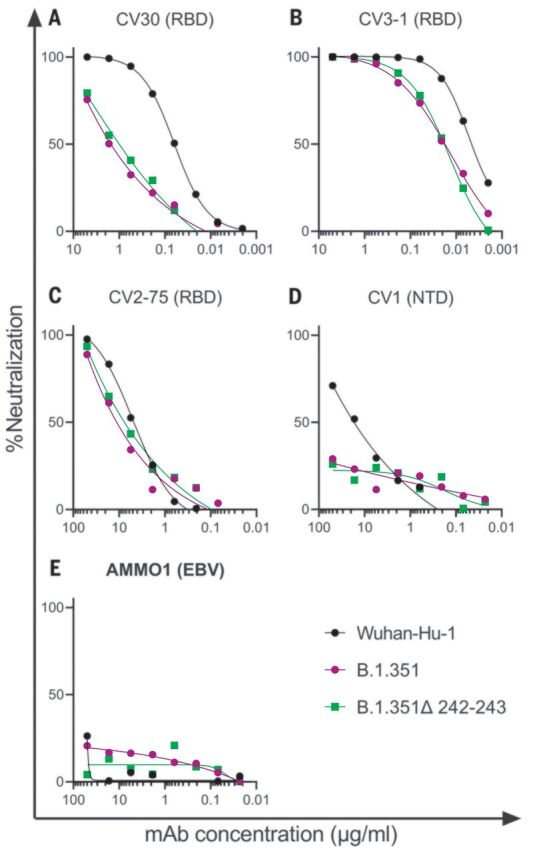
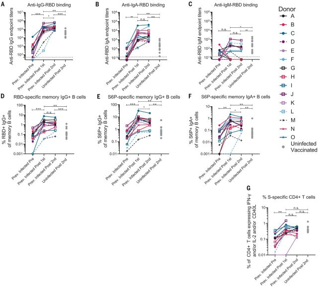
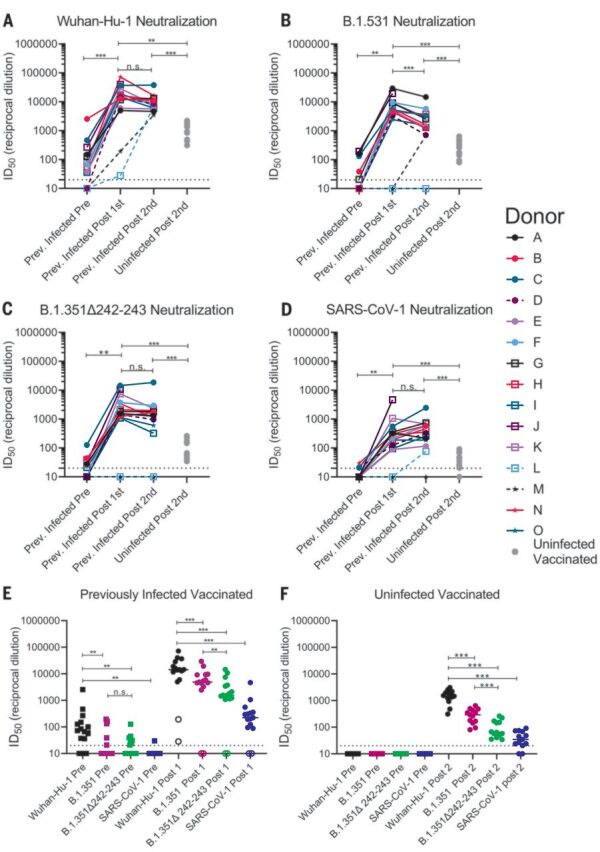
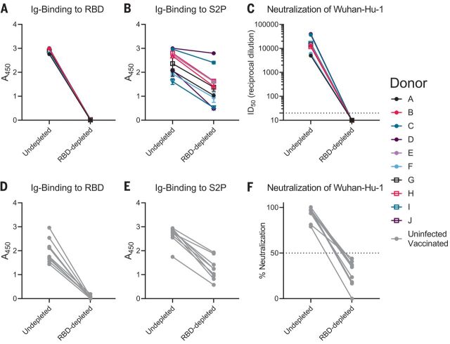
Emerging SARS-CoV-2 variants have raised concerns about resistance to neutralizing antibodies elicited by previous infection or vaccination. We examined whether sera from recovered and naïve donors collected prior to, and following immunizations with existing mRNA vaccines, could neutralize the Wuhan-Hu-1 and B.1.351 variants. Pre-vaccination sera from recovered donors neutralized Wuhan-Hu-1 and sporadically neutralized B.1.351, but a single immunization boosted neutralizing titers against all variants and SARS-CoV-1 by up to 1000-fold. Neutralization was due to antibodies targeting the receptor binding domain and was not boosted by a second immunization. Immunization of naïve donors also elicited cross-neutralizing responses, but at lower titers. Our study highlights the importance of vaccinating both uninfected and previously infected persons to elicit cross-variant neutralizing antibodies.


Figures

Fig. 1
Fig. 1. B.1.351 variants show decreased susceptibility to neutralizing mAbs.
(A to E) The ability of the indicated mAbs to neutralize Wuhan-Hu-1, B.1.351, and B.1.351–Δ242-243 pseudovirus infectivity in 293T-hACE2 cells was measured as indicated. The epitope specificity of each mAb is shown in parentheses. EBV, Epstein-Barr virus. Data points represent the mean of two technical replicates. Data are representative of two independent experiments.
Fig. 2
Fig. 2. A single dose of a spike-derived mRNA vaccine elicits a strong recall response.
(A to C) IgG (A), IgA (B), and IgM (C) end-point antibody titers specific to the RBD of the Wuhan-Hu-1 variant were measured in serum collected from PIDs before and after one or two immunizations with the Pfizer-BioNTech or Moderna mRNA vaccines by ELISA, as indicated. End-point titers measured in sera from NDs after two vaccine doses are shown for comparison (gray dots). (D) Frequency of Wuhan-Hu-1 RBD-specific IgG+ memory B cells (live, IgD, CD19+, CD20+, CD3, CD14, CD56, singlet, and lymphocytes) in peripheral blood mononuclear cells (PBMCs) from PIDs was measured before and after one or two immunizations. (E and F) The frequency of S6P-specific IgG+ (E) and IgA+ (F) memory B cells in PBMCs from PIDs was measured before and after one or two immunizations. The frequencies of memory B cells from NDs after two vaccine doses are shown for comparison in (D) to (F) (gray dots). (G) The frequency of S-specific CD4+ T cells expressing interferon-γ (IFN-γ) and/or interleukin-2 (IL-2) and/or CD40L in PBMCs from PIDs was measured before and after one or two immunizations. The frequencies of S-specific CD4+ T cells in PBMCs from uninfected donors after two vaccine doses are shown for comparison (gray dots). Experiments were performed once. Significant differences in infected donors before or after vaccination [(A) to (G)] were determined using a Wilcoxon signed rank test (n.s., not significant; *P < 0.05; **P < 0.01; and ***P < 0.001). Significant differences between previously infected and uninfected donors [(A) to (G)] were determined using a Wilcoxon rank sum test (*P < 0.05; **P < 0.01; and ***P < 0.001).
Fig. 3
Fig. 3. Preexisting SARS-CoV-2 nAb responses are boosted by a single dose of a spike-derived mRNA vaccine.
(A to D) The serum dilution resulting in 50% neutralization (ID50) of Wuhan-Hu-1 (A), B.1.351 (B), B.1.351–Δ242-243 (C), and SARS-CoV-1 (D) pseudoviruses was measured in PIDs before and after one or two immunizations with the Pfizer-BioNTech or Moderna vaccines and in NDs after two vaccine doses, as indicated. Data points between PIDs who were symptomatic and asymptomatic are connected by solid and dashed lines, respectively, in (A) to (D). (E) Serum dilution resulting in 50% neutralization (ID50) from PIDs before (squares) and after (circles) a single immunization with the Pfizer-BioNTech or Moderna vaccines against Wuhan-Hu-1, B.1.351, B.1.351–Δ242-243, and SARS-CoV-1 pseudoviruses, as indicated. PIDs who were asymptomatic and negative for anti-IgG RBD antibodies and RBD-specific IgG+ memory B cells before vaccination are shown as open circles. (F) Neutralizing potency (ID50) of serum from NDs after two immunizations with the Pfizer-BioNTech or Moderna vaccines against the indicated pseudoviruses. Each data point represents a different donor, and the horizonal bars represent the medians in (E) and (F). The dashed lines demarcate the lowest serum dilutions tested. Experiments were performed once. Significant differences in infected donors before or after vaccination, or from the same time point against different variants, were determined using a Wilcoxon signed rank test (*P < 0.05; **P < 0.01; and ***P < 0.001). Significant differences between previously infected and uninfected donors were determined using a Wilcoxon rank sum test (*P < 0.05; **P < 0.01; and ***P < 0.001).
Fig. 4
Fig. 4. Vaccine-elicited nAbs target the RBD.
RBD-binding antibodies were adsorbed from sera from PIDs after receiving a single vaccine dose or from NDs after receiving two vaccine doses using Wuhan-Hu-1 RBD immobilized to magnetic beads. (A and B) Antibody binding in undepleted or RBD-depleted sera from PIDs was measured to RBD at a 1:500 dilution (A) and S2P at a 1:4500 dilution (B) by ELISA, as indicated. A450, absorbance at 450 nM. (C) The serum dilution resulting in 50% neutralization (ID50) of the Wuhan-Hu-1 pseudovirus was measured in undepleted or RBD-depleted sera from the PIDs in (A) and (B). (D and E) Antibody binding in undepleted and RBD-depleted sera from NDs was measured to RBD at a 1:500 dilution (D) and S2P at a 1:500 dilution (E) by ELISA. (F) The percent neutralization of a 1:120 dilution of undepleted or RBD-depleted sera from the donors in (D) and (E) was measured against the Wuhan-Hu-1 pseudovirus. Experiments were performed once.

Update of

Comment in

References

    1. Dong E., Du H., Gardner L., An interactive web-based dashboard to track COVID-19 in real time. Lancet Infect. Dis. 20, 533–534 (2020). 10.1016/S1473-3099(20)30120-1 - DOI - PMC - PubMed
    1. Zhou P., Yang X.-L., Wang X.-G., Hu B., Zhang L., Zhang W., Si H.-R., Zhu Y., Li B., Huang C.-L., Chen H.-D., Chen J., Luo Y., Guo H., Jiang R.-D., Liu M.-Q., Chen Y., Shen X.-R., Wang X., Zheng X.-S., Zhao K., Chen Q.-J., Deng F., Liu L.-L., Yan B., Zhan F.-X., Wang Y.-Y., Xiao G.-F., Shi Z.-L., A pneumonia outbreak associated with a new coronavirus of probable bat origin. Nature 579, 270–273 (2020). 10.1038/s41586-020-2012-7 - DOI - PMC - PubMed
    1. Zhu N., Zhang D., Wang W., Li X., Yang B., Song J., Zhao X., Huang B., Shi W., Lu R., Niu P., Zhan F., Ma X., Wang D., Xu W., Wu G., Gao G. F., Tan W., China Novel Coronavirus Investigating and Research Team , A Novel Coronavirus from Patients with Pneumonia in China, 2019. N. Engl. J. Med. 382, 727–733 (2020). 10.1056/NEJMoa2001017 - DOI - PMC - PubMed
    1. Walls A. C., Park Y.-J., Tortorici M. A., Wall A., McGuire A. T., Veesler D., Structure, Function, and Antigenicity of the SARS-CoV-2 Spike Glycoprotein. Cell 181, 281–292.e6 (2020). 10.1016/j.cell.2020.02.058 - DOI - PMC - PubMed
    1. Hoffmann M., Kleine-Weber H., Schroeder S., Krüger N., Herrler T., Erichsen S., Schiergens T. S., Herrler G., Wu N.-H., Nitsche A., Müller M. A., Drosten C., Pöhlmann S., SARS-CoV-2 Cell Entry Depends on ACE2 and TMPRSS2 and Is Blocked by a Clinically Proven Protease Inhibitor. Cell 181, 271–280.e8 (2020). 10.1016/j.cell.2020.02.052 - DOI - PMC - PubMed

Publication types