

A Zic2/Runx2/NOLC1 signaling axis mediates tumor growth and metastasis in clear cell renal cell carcinoma
- PMID: 33767130
- PMCID: PMC7994417
- DOI: 10.1038/s41419-021-03617-8
A Zic2/Runx2/NOLC1 signaling axis mediates tumor growth and metastasis in clear cell renal cell carcinoma
Abstract
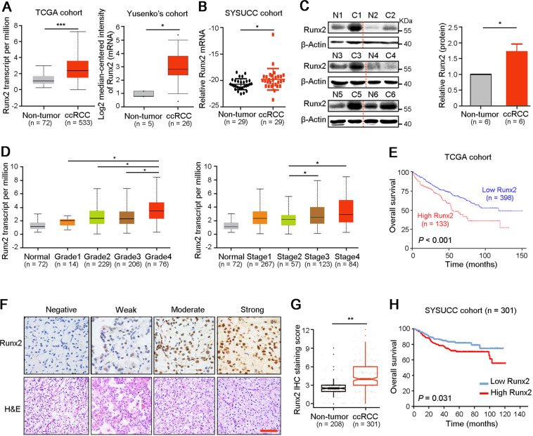
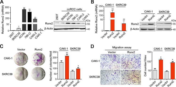
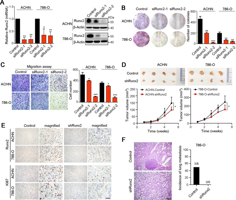
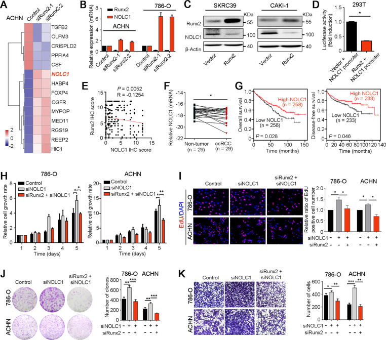
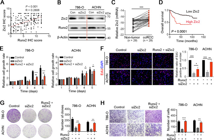
Clear cell renal cell carcinoma (ccRCC) is one of the most common malignancies with rapid growth and high metastasis, but lacks effective therapeutic targets. Here, using public sequencing data analyses, quantitative real-time PCR assay, western blotting, and IHC staining, we characterized that runt-related transcription factor 2 (Runx2) was significantly upregulated in ccRCC tissues than that in normal renal tissues, which was associated with the worse survival of ccRCC patients. Overexpression of Runx2 promoted malignant proliferation and migration of ccRCC cells, and inversely, interfering Runx2 with siRNA attenuates its oncogenic ability. RNA sequencing and functional studies revealed that Runx2 enhanced ccRCC cell growth and metastasis via downregulation of tumor suppressor nucleolar and coiled-body phosphoprotein 1 (NOLC1). Moreover, increased Zic family member 2 (Zic2) was responsible for the upregulation of Runx2 and its oncogenic functions in ccRCC. Kaplan-Meier survival analyses indicated that ccRCC patients with high Zic2/Runx2 and low NOLC1 had the worst outcome. Therefore, our study demonstrates that Zic2/Runx2/NOLC1 signaling axis promotes ccRCC progression, providing a set of potential targets and prognostic indicators for patients with ccRCC.
Conflict of interest statement
The authors declare no competing interests.
Figures
Similar articles
-
FOXM1-regulated ZIC2 promotes the malignant phenotype of renal clear cell carcinoma by activating UBE2C/mTOR signaling pathway.Int J Biol Sci. 2023 Jun 26;19(11):3293-3306. doi: 10.7150/ijbs.84067. eCollection 2023. Int J Biol Sci. 2023. PMID: 37496990 Free PMC article.
-
RUNX2 interacts with SCD1 and activates Wnt/β-catenin signaling pathway to promote the progression of clear cell renal cell carcinoma.Cancer Med. 2023 Mar;12(5):5764-5780. doi: 10.1002/cam4.5326. Epub 2022 Oct 6. Cancer Med. 2023. PMID: 36200301 Free PMC article.
-
Phosphorylated MAPK11 promotes the progression of clear cell renal cell carcinoma by maintaining RUNX2 protein abundance.J Cell Mol Med. 2023 Sep;27(17):2583-2593. doi: 10.1111/jcmm.17870. Epub 2023 Jul 31. J Cell Mol Med. 2023. PMID: 37525479 Free PMC article.
-
VHL mutation-mediated SALL4 overexpression promotes tumorigenesis and vascularization of clear cell renal cell carcinoma via Akt/GSK-3β signaling.J Exp Clin Cancer Res. 2020 Jun 8;39(1):104. doi: 10.1186/s13046-020-01609-8. J Exp Clin Cancer Res. 2020. PMID: 32513235 Free PMC article.
-
Regulation of Runx2 and Its Signaling Pathways by MicroRNAs in Breast Cancer Metastasis.Curr Protein Pept Sci. 2021;22(7):534-547. doi: 10.2174/1389203721666201116115337. Curr Protein Pept Sci. 2021. PMID: 33200704 Review.
Cited by
-
FOXM1-regulated ZIC2 promotes the malignant phenotype of renal clear cell carcinoma by activating UBE2C/mTOR signaling pathway.Int J Biol Sci. 2023 Jun 26;19(11):3293-3306. doi: 10.7150/ijbs.84067. eCollection 2023. Int J Biol Sci. 2023. PMID: 37496990 Free PMC article.
-
FOXA1 activates NOLC1 transcription through NOTCH pathway to promote cell stemness in lung adenocarcinoma.Kaohsiung J Med Sci. 2025 Feb;41(2):e12930. doi: 10.1002/kjm2.12930. Epub 2025 Jan 10. Kaohsiung J Med Sci. 2025. PMID: 39789998 Free PMC article.
-
Ginsenoside compound K alleviates osteoarthritis by inhibiting NLRP3‑mediated pyroptosis.Exp Ther Med. 2023 Jul 10;26(2):406. doi: 10.3892/etm.2023.12105. eCollection 2023 Aug. Exp Ther Med. 2023. PMID: 37522058 Free PMC article.
-
Role of RUNX2 in breast cancer development and drug resistance (Review).Oncol Lett. 2023 Mar 15;25(5):176. doi: 10.3892/ol.2023.13762. eCollection 2023 May. Oncol Lett. 2023. PMID: 37033103 Free PMC article. Review.
-
Expression of lymphoid structure-associated cytokine/chemokine gene transcripts in tumor and protein in serum are prognostic of melanoma patient outcomes.Front Immunol. 2023 Jun 22;14:1171978. doi: 10.3389/fimmu.2023.1171978. eCollection 2023. Front Immunol. 2023. PMID: 37435077 Free PMC article.
References
Publication types
MeSH terms
Substances
LinkOut - more resources
Full Text Sources
Other Literature Sources
Medical
