. 2021 Mar 25;12(1):1890.
doi: 10.1038/s41467-021-22128-w.

Scaffolding proteins guide the evolution of algal light harvesting antennas

Affiliations

Scaffolding proteins guide the evolution of algal light harvesting antennas

Harry W Rathbone et al. Nat Commun. .

Abstract

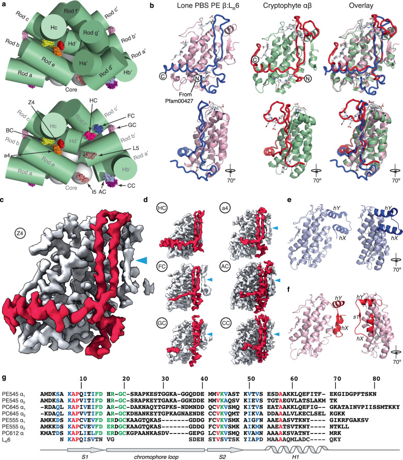
Photosynthetic organisms have developed diverse antennas composed of chromophorylated proteins to increase photon capture. Cryptophyte algae acquired their photosynthetic organelles (plastids) from a red alga by secondary endosymbiosis. Cryptophytes lost the primary red algal antenna, the red algal phycobilisome, replacing it with a unique antenna composed of αβ protomers, where the β subunit originates from the red algal phycobilisome. The origin of the cryptophyte antenna, particularly the unique α subunit, is unknown. Here we show that the cryptophyte antenna evolved from a complex between a red algal scaffolding protein and phycoerythrin β. Published cryo-EM maps for two red algal phycobilisomes contain clusters of unmodelled density homologous to the cryptophyte-αβ protomer. We modelled these densities, identifying a new family of scaffolding proteins related to red algal phycobilisome linker proteins that possess multiple copies of a cryptophyte-α-like domain. These domains bind to, and stabilise, a conserved hydrophobic surface on phycoerythrin β, which is the same binding site for its primary partner in the red algal phycobilisome, phycoerythrin α. We propose that after endosymbiosis these scaffolding proteins outcompeted the primary binding partner of phycoerythrin β, resulting in the demise of the red algal phycobilisome and emergence of the cryptophyte antenna.


Conflict of interest statement

Authors declare no competing interests.

Figures

Fig. 1
Fig. 1. CALM domain proteins stabilise ‘lone’ PE β subunits in the red algal PBS.
a ‘lone’ PE β subunits (coloured protein surface rendering) peripherally associated to red algal PBS rods (green cylinders) observed in the cryo-EM structure of the red algal PBS from P. purpureum (PDB 6KGX). Lower panel: Rod d’, Rod g’ and Ha’ removed. Labelling is according to Ma et al.. b revised model of the C-terminal domain of LR6 with its cognate PE β subunit is structurally homologous to the cryptophyte-αβ protomer. c, d sculpted EM map density showing cryptophyte-like protomers from the P. purpureum red algal PBS (CALM domains in red and PE β in grey) labelled according to. Cyan triangle indicates the β strand in PE β. e, f comparison of metamorphic states of PE β in two rotations with secondary structure labelled: e the red algal PBS hexamer form and f the cryptophyte-like form. g structure-based sequence alignment of cryptophyte α subunits (PDB 1XG0 (PE545), 4LMS (PC645), 4LMX (PE555), and 4LM6 (PC612)) and the C-terminal CALM domain of LR6. Red = identity, blue = conserved, and green = chromophore interacting.
Fig. 2
Fig. 2. The CaRSPs of P. purpureum.
ac sculpted EM map density for LR6, CaRSP2, and CaRSP1, respectively (red), with their associated PE β subunits (coloured purple-blue-teal by their order in the cluster). Both the density and the peptide trace are shown for each CaRSP. A reference to the positions of each cluster is given in the central panel. d the domain organization of the three P. purpureum CaRSPs (left) and the structure-based alignment of the modelled CALM domains (right). Residue numbers of the beginning and end of each domain are shown (left). Structure and sequence motifs are shown (right; Ω = Y/F/W and ɸ = hydrophobic with orange being the aromatic motif, and red and blue signifying identity and similarity, respectively).
Fig. 3
Fig. 3. The binding surface of PE β is hydrophobic and strictly conserved.
CALM domains (a LR6 CALM1), cryptophyte α subunits (b PE555 PDB 4LMX), and red algal PBS PE α subunits (c PE hexamer PDB 3V57) bind to conserved hydrophobic surfaces of PE β as shown in CPK representation (hydrophobic surfaces appear white). Partner proteins (top panels) are peeled off their respective PE β subunits (lower panels) 180° around the fold line with a black silhouette left in their place. Colour of the residue labels corresponds to the cartoon just to left of each CALM. d, e, and f are mappings of sequence variability (Shannon entropy) onto the molecular surfaces of the PE β structures (white = identity; red = 97% identity or lower; where d, e and f correspond, respectively, to structures in ab and c above). The conserved and hydrophobic surfaces are congruent. d and f show the variability for red algal sequences only while e includes cryptophyte sequences. g, h, and i are cartoon representations of PE β subunits (grey) with their partner proteins (red).
Fig. 4
Fig. 4. Model for the evolution of red algal and cryptophyte antennas.
Top row: Red algae acquire linker protein LR1, which couples red algal PBS ring structures using its Pfam00427 and Pfam01383 domains (inset). Second row: the gene expressing LR1 was transferred to the nucleus, where it expanded and diversified. LR2, “Linker 3”, LR3 and LR6 retain C-terminal domains with some similarity to Pfam01383. CALM domains appeared in the red algal PBS linker family (LR6) or, alternatively, in an ancestral CaRSP. Third row: CaRSP proteins lose Pfam00427 domain, expand and diversify in the red algal nucleus. Bottom row: a single CALM protein appeared in the ancestral cryptophyte. It acquired a chromophore plus a plastid targeting sequence. It formed a stable cryptophyte-αβ protomer with a PE β subunit which then dimerised, creating the cryptophyte antenna.

References

    1. Rathbone HW, et al. Coherent phenomena in photosynthetic light harvesting: part two-observations in biological systems. Biophys. Rev. 2018;10:1443–1463. doi: 10.1007/s12551-018-0456-x. - DOI - PMC - PubMed
    1. Bryant DA, Canniffe DP. How nature designs light-harvesting antenna systems: design principles and functional realization in chlorophototrophic prokaryotes. J. Phys. B: At. Mol. Optical Phys. 2018;51:033001. doi: 10.1088/1361-6455/aa9c3c. - DOI
    1. Scholes GD, Mirkovic T, Turner DB, Fassioli F, Buchleitner A. Solar light harvesting by energy transfer: from ecology to coherence. Energy Environ. Sci. 2012;5:9374–9393. doi: 10.1039/c2ee23013e. - DOI
    1. Cenci U, et al. Nuclear genome sequence of the plastid-lacking cryptomonad Goniomonas avonlea provides insights into the evolution of secondary plastids. BMC Biol. 2018;16:137. doi: 10.1186/s12915-018-0593-5. - DOI - PMC - PubMed
    1. Kim JI, et al. Evolutionary dynamics of cryptophyte plastid genomes. Genome Biol. Evol. 2017;9:1859–1872. doi: 10.1093/gbe/evx123. - DOI - PMC - PubMed

Publication types

LinkOut - more resources