. 2021 Mar 25;12(4):316.
doi: 10.1038/s41419-021-03572-4.

Pharmacological targeting of the novel β-catenin chromatin-associated kinase p38α in colorectal cancer stem cell tumorspheres and organoids

Affiliations

Pharmacological targeting of the novel β-catenin chromatin-associated kinase p38α in colorectal cancer stem cell tumorspheres and organoids

Martina Lepore Signorile et al. Cell Death Dis. .

Abstract

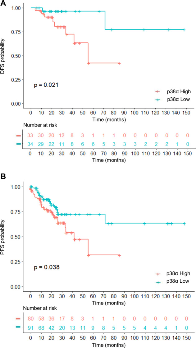
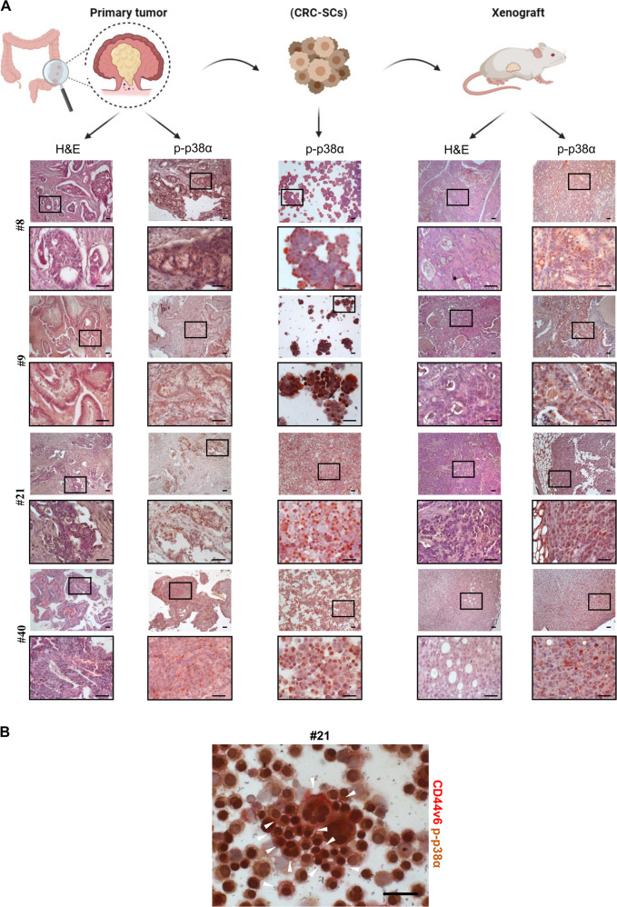
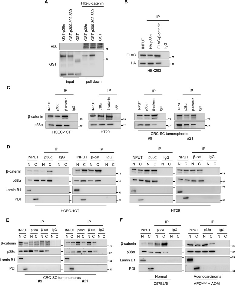
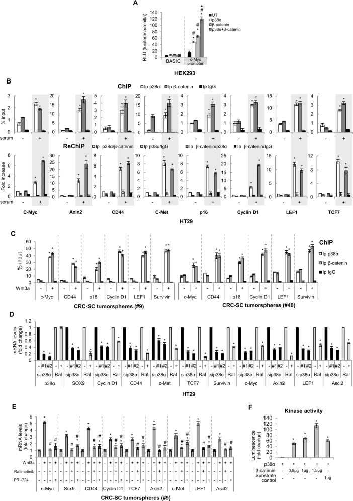
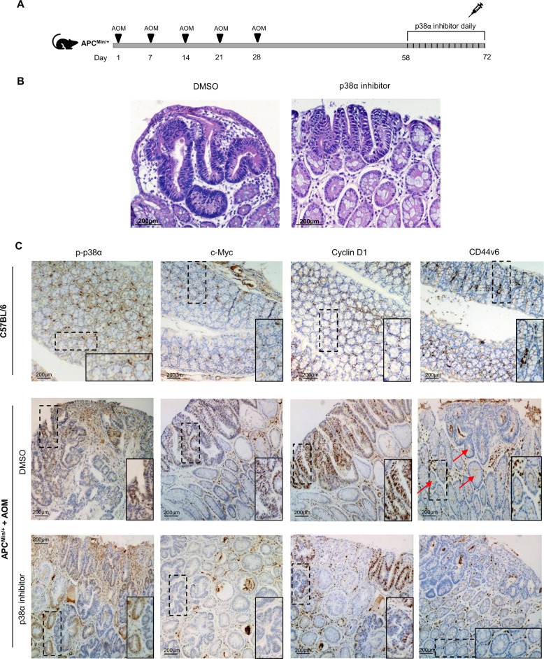
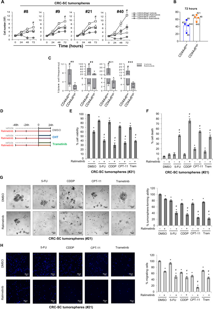
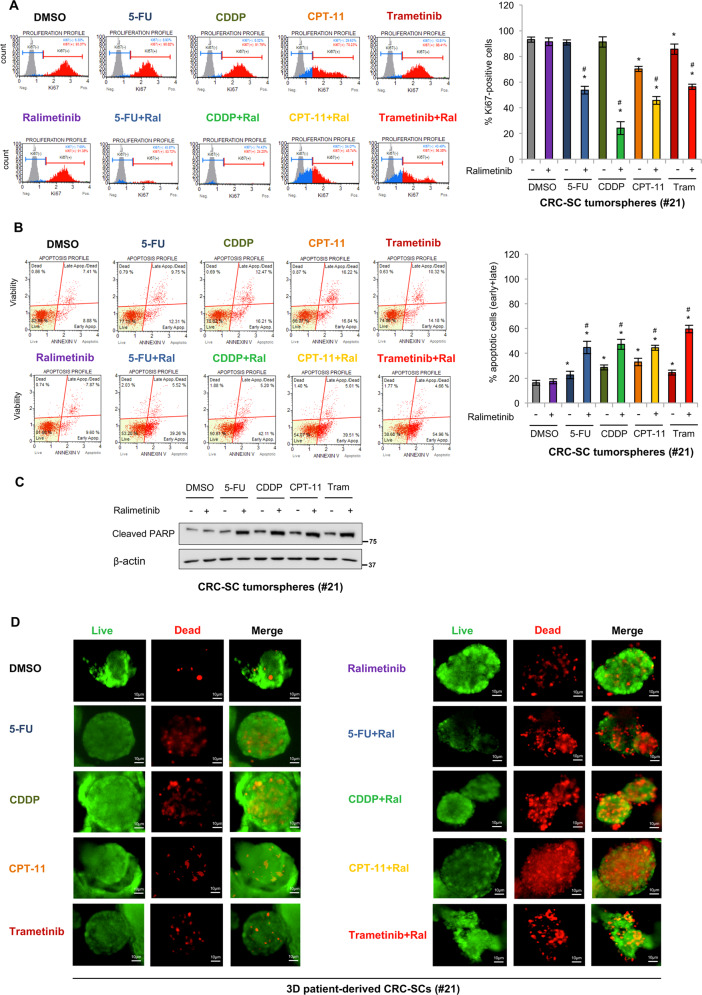
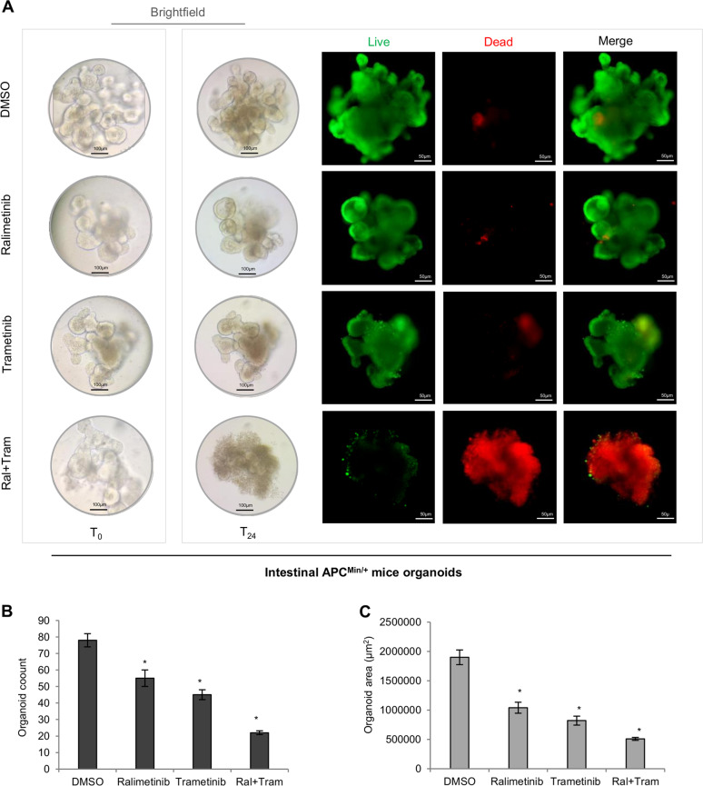
The prognosis of locally advanced colorectal cancer (CRC) is currently unsatisfactory. This is mainly due to drug resistance, recurrence, and subsequent metastatic dissemination, which are sustained by the cancer stem cell (CSC) population. The main driver of the CSC gene expression program is Wnt signaling, and previous reports indicate that Wnt3a can activate p38 MAPK. Besides, p38 was shown to feed into the canonical Wnt/β-catenin pathway. Here we show that patient-derived locally advanced CRC stem cells (CRC-SCs) are characterized by increased expression of p38α and are "addicted" to its kinase activity. Of note, we found that stage III CRC patients with high p38α levels display reduced disease-free and progression-free survival. Extensive molecular analysis in patient-derived CRC-SC tumorspheres and APCMin/+ mice intestinal organoids revealed that p38α acts as a β-catenin chromatin-associated kinase required for the regulation of a signaling platform involved in tumor proliferation, metastatic dissemination, and chemoresistance in these CRC model systems. In particular, the p38α kinase inhibitor ralimetinib, which has already entered clinical trials, promoted sensitization of patient-derived CRC-SCs to chemotherapeutic agents commonly used for CRC treatment and showed a synthetic lethality effect when used in combination with the MEK1 inhibitor trametinib. Taken together, these results suggest that p38α may be targeted in CSCs to devise new personalized CRC treatment strategies.


Conflict of interest statement

The authors declare no competing interests.

Figures

Fig. 1
Fig. 1. p38α is a potential marker of therapy efficacy in stage III CRC patients.
A, B Kaplan–Meier curve of disease-free survival (DFS) (A) and progression-free survival (PFS) (B) in stage III CRC patients as a function of p38α levels.
Fig. 2
Fig. 2. CRC-SCs recapitulate parental tumor features.
A Representative hematoxylin and eosin staining and immunohistochemical analysis of p-p38α in primary CRC, isolated CRC-SCs, and CRC-SC-derived xenografts. Scale bars, 100 µm. B Representative immunocytochemical analysis of CD44v6 (red) and p-p38α (brown) in CRC-SCs #21. White arrow-heads indicate CD44v6+/p-p38α+ CRC-SCs. Scale bar, 100 µm.
Fig. 3
Fig. 3. Functional interaction between p38α and β-catenin in in vitro models of CRC.
A In vitro binding assay between GST-p38α fusion protein and HIS-β-catenin. Bound proteins were analyzed by immunoblotting using anti-GST and anti-HIS antibodies. B Co-immunoprecipitation with anti-HA and anti-FLAG antibodies in HEK293 cells overexpressing HA-p38α or FLAG-β-catenin. C Co-immunoprecipitation of endogenous p38α and β-catenin in the indicated cells. D and E Co-immunoprecipitation of endogenous p38α and β-catenin in nuclear and cytoplasmic fractions of the indicated cells. F Co-immunoprecipitation of endogenous p38α and β-catenin in nuclear and cytoplasmic fractions from C57BL/6 mice normal colon tissue and AOM-treated APCMin/+ mice adenocarcinoma tissue. Input corresponds to 10% of the lysate. Anti-IgGs were used as negative controls. Lamin B1: nuclear loading control; PDI: cytoplasmic loading control; N = nucleus, C = cytoplasm, β-cat = β-catenin.
Fig. 4
Fig. 4. p38α is a novel β-catenin chromatin-associated kinase.
A Luciferase assay for c-Myc promoter activity. HEK293 cells were serum-starved for 24 h and transfected with either the empty vector (pcDNA) or pcDNA3.1-HAHA-p38α and/or pcDNA-β-catenin expression constructs. B Chromatin immunoprecipitation (ChIP) and re-ChIP assays in HT29 cells. Cells were serum-starved for 48 h and then switched to a serum-containing medium for 4 h. In ChIP assays (upper panels), chromatin was pulled down with anti-p38α and anti-β-catenin antibodies. In re-ChIP assays (lower panels), chromatin was pulled down with anti-p38α antibodies and then re-immunoprecipitated with anti-β-catenin antibodies and vice versa. Anti-IgGs were used as negative controls. C ChIP with anti-p38α and anti-β-catenin antibodies. CRC-SC tumorspheres were treated or not with Wnt3a (50 ng/ml) for 4 h. B, C Quantification was done using the % input method. D Real-time PCR analysis of β-catenin target genes in HT29 cells treated with ralimetinib (10 µM) or two p38α-specific siRNAs (sip38α #1 and #2) for 48 h. E Real-time PCR analysis of β-catenin target genes in CRC-SC tumorspheres treated with Wnt3a (50 ng/ml) for 4 h with or without ralimetinib (10 µM) or PRI-724 (25 nM) for 20 h. D, E Data are presented as mRNA fold change vs. control. F In vitro kinase assay showing β-catenin phosphorylation by p38α. UT = untransfected. A *P < 0.05 vs. BASIC, #P < 0.05 vs. untransfected cells, P < 0.05 vs. p38α-transfected or β-catenin-transfected cells. B *P < 0.05 vs. serum-starved cells. C, E *P < 0.05 vs. untreated cells, and #P < 0.05 vs. Wnt3a-treated cells. D *P < 0.05 vs. control (DMSO or control siRNA). F *P < 0.05 vs. active p38α.
Fig. 5
Fig. 5. p38α inhibition downregulates CRC-SC markers in an in vivo model.
A Mice treatment scheme. B Hematoxylin and eosin staining of AOM-treated APCMin/+ mice injected with the p38α inhibitor SB202190 or DMSO. Original magnification: 200x. C Immunohistochemistry analysis of colon tissue sections from C57BL/6 and AOM-treated APCMin/+ mice injected with the p38α inhibitor SB202190 or DMSO. Original magnification: 100x.
Fig. 6
Fig. 6. Targeting p38α in patient-derived stage III CRC-SCs to circumvent chemoresistance.
A Growth kinetics of CD44v6low-enriched and CD44v6high-enriched CRC-SCs treated with ralimetinib (10 μM) or the vehicle for up to 72 h. B Viable cell number variation in CD44v6low- and CD44v6high-enriched CRC-SCs treated with ralimetinib (10 μM) for 72 h. Values were normalized against those of vehicle-treated cells. C Limiting dilution assay performed on CD44v6low-enriched and CD44v6high-enriched CRC-SCs. The graph shows the clonogenic capacity of each cell subset. AC CD44v6low and CD44v6high represent cell samples enriched for the top 20% cells with the lowest and highest expression of CD44v6, respectively. A, C *P < 0.05: CD44v6high treated with ralimetinib vs. CD44v6high treated with the vehicle; and #P < 0.05: CD44v6low treated with ralimetinib vs. CD44v6low treated with the vehicle. D Treatment scheme: CRC-SCs were pre-treated with ralimetinib (10 μM) for 48 h and then treated with 5-FU (2 μM), CDDP (30 μM), CPT-11 (30 μM), or trametinib (1 nM) for another 24 h in the presence of ralimetinib. E Quantification of cell viability by Cell Titer Glo in CRC-SCs #21 treated as described in (D). F Quantification of cell death by trypan blue staining in CRC-SCs #21 treated as described in (D). G Colony-forming ability of CRC-SCs #21 seeded onto double-layer soft agar and treated as described in (D). Data represent the percentage of colonies relative to DMSO-treated cells. Original magnification: 100x. H Migratory ability of growth factor-starved CRC-SCs #21 placed in the inner chamber of transwell plates and treated with the indicated compounds for 16 h. Migrating cells were fixed and counted under a fluorescence microscope. Original magnification: 100x. Tram = trametinib. *P < 0.05: treatment vs. control (DMSO); and #P < 0.05: combined treatment vs. corresponding single treatments.
Fig. 7
Fig. 7. Combined treatment with ralimetinib and chemotherapeutics or trametinib has a synergistic cytotoxic effect.
A Flow cytometry analysis of Ki67 expression in CRC-SCs #21 treated as described in Fig. 6D. Populations were gated identically using the unstained background populations shown in gray behind the Ki67-negative (blue) and Ki67-positive (red) populations. The graph on the right summarizes the percentage of Ki67-positive cells. B Flow cytometry analysis of annexin V staining in CRC-SCs #21 treated as described in Fig. 6D. The graph on the right summarizes the percentage of apoptotic cells (early + late). C Immunoblot analysis of cleaved PARP levels in CRC-SCs #21 treated as described in Fig. 6D. β-actin was used as a loading control. D Live/dead staining of CRC-SCs #21 grown as 3D cultures and treated as described in Fig. 6D. Tram = trametinib. Green: live cells; red: dead cells. *P < 0.05: treatment vs. control (DMSO); and #P < 0.05: combined treatment vs. corresponding single treatments.
Fig. 8
Fig. 8. Targeting p38α as part of a synthetic lethality approach in APCMin/+ mice intestinal organoids.
A Brightfield imaging of organoids formation from single adenoma intestinal crypts isolated from APCMin/+ mice at T0 and after 24 h treatment with ralimetinib (10 μM) and/or trametinib (1 nM). Treated organoids were also subjected to live/dead staining. Green: live cells; red: dead cells. B Quantification of APCMin/+ mice intestinal organoids after 24 h treatment with ralimetinib (10 μM) and/or trametinib (1 nM). C Average area of APCMin/+ mice intestinal organoids after 24 h treatment with ralimetinib (10 μM) and/or trametinib (1 nM), as measured using ImageJ software. *P < 0.05: treatment vs. control (DMSO).

References

    1. Keum N, Giovannucci E. Global burden of colorectal cancer: emerging trends, risk factors and prevention strategies. Nat. Rev. Gastroenterol. Hepatol. 2019;16:713–732. doi: 10.1038/s41575-019-0189-8. - DOI - PubMed
    1. Lieu C, et al. Duration of oxaliplatin-containing adjuvant therapy for Stage III colon cancer: ASCO Clinical Practice Guideline. J. Clin. Oncol. 2019;37:1436–1447. doi: 10.1200/JCO.19.00281. - DOI - PubMed
    1. Li C, et al. Survival nomograms for stage III colorectal cancer. Medicine. 2018;97:e13239. doi: 10.1097/MD.0000000000013239. - DOI - PMC - PubMed
    1. Sheth KR, Clary BM. Management of hepatic metastases from colorectal cancer. Clin. Colon Rectal Surg. 2005;18:215–223. doi: 10.1055/s-2005-916282. - DOI - PMC - PubMed
    1. Zhou Y, et al. Cancer stem cells in progression of colorectal cancer. Oncotarget. 2018;9:33403–33415. doi: 10.18632/oncotarget.23607. - DOI - PMC - PubMed

Publication types

MeSH terms