. 2021 Mar 25;11(1):6849.
doi: 10.1038/s41598-021-86211-4.

In silico studies reveal structural deviations of mutant profilin-1 and interaction with riluzole and edaravone in amyotrophic lateral sclerosis

Affiliations

In silico studies reveal structural deviations of mutant profilin-1 and interaction with riluzole and edaravone in amyotrophic lateral sclerosis

Ahmad Shahir Sadr et al. Sci Rep. .

Abstract

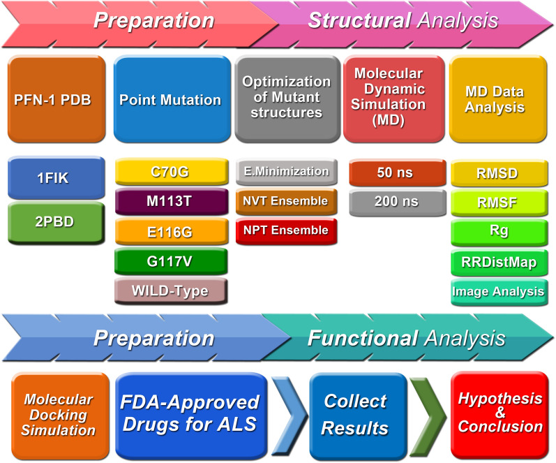
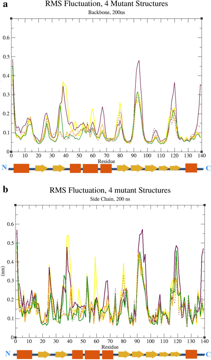
This study aimed to investigate four of the eight PFN-1 mutations that are located near the actin-binding domain and determine the structural changes due to each mutant and unravel how these mutations alter protein structural behavior. Swapaa's command in UCSF chimera for generating mutations, FTMAP were employed and the data was analyzed by RMSD, RMSF graphs, Rg, hydrogen bonding analysis, and RRdisMaps utilizing Autodock4 and GROMACS. The functional changes and virtual screening, structural dynamics, and chemical bonding behavior changes, molecular docking simulation with two current FDA-approved drugs for ALS were investigated. The highest reduction and increase in Rg were found to exist in the G117V and M113T mutants, respectively. The RMSF data consistently shows changes nearby to this site. The in silico data described indicate that each of the mutations is capable of altering the structure of PFN-1 in vivo. The potential effect of riluzole and edaravone two FDA approved drugs for ALS, impacting the structural deviations and stabilization of the mutant PFN-1 is evaluated using in silico tools. Overall, the analysis of data collected reveals structural changes of mutant PFN-1 protein that may explain the neurotoxicity and the reason(s) for possible loss and gain of function of PFN-1 in the neurotoxic model of ALS.


Conflict of interest statement

M.K. is the founder, president, and CEO of RockGen Therapeutics, LLC.

Figures

Figure 1
Figure 1
A diagrammatic summary of the preparation, structural and functional analysis of PFN-1. The PDB file of PFN-1 obtained from the protein data-bank, 4 point mutations under the investigation added one at a time, optimized, performed molecular dynamic simulation, and data analysis carried out as outlined in the cartoon format. The lower part of the diagram demonstrates the outline of our work with respect to the effect of the two FDA approved drugs for ALS.
Figure 2
Figure 2
Two-dimensional and three-dimensional view of ALS FDA-approved drugs (riluzole and edaravone) before and after B3LYP protocol optimization of spatial energy of the Gaussian program. The structure of the (A) Riluzole and (B) Edaravone were obtained from the Drug bank (www.drugbank.ca).
Figure 3
Figure 3
The four amino acid residues are located in the actin-binding domain of PFN-1. The Cys 70 to Gly is a polar amino acid with uncharged R groups changed to a nonpolar amino acid with the smallest R group. The Met 113 to Thr mutation is a nonpolar amino acid with an aliphatic R group changed to a polar amino acid with an uncharged R group. The Glu 116 to Gly is a negatively charged R group changed to a nonpolar amino acid with the smallest R group. The Gly 117 to Val is a nonpolar aliphatic residue that is substituted to Val, with the same nonpolar aliphatic R group.
Figure 4
Figure 4
The position of the mutated 4 amino acids in PFN-1 associated with pathogenic injuries in ALS. (A) Ribbon view showing the location of the reported mutated 4 amino acids. (B) Surface views of the structure and sphere views of the actin-binding domain showing the amino acids E116, G117 located on the surface. Amino acids C70 and M113 are located deep inside the structure. (C) Secondary structure of the protein sequence and the position of the mutated amino acids in the polypeptide chain. The C70 and M113 are located on the beta-strand domain and E116 and G117 are on the turn.
Figure 5
Figure 5
The RMSD data generated from a triplicate runs for 200 ns. (A) RMSD of structures after 200 ns relative to the structure in a decent energy-minimized, equilibrated system. (B) Compared to RMSD values for 200 ns.
Figure 6
Figure 6
The Radius of gyration for wild-type and all mutant structures After 200 ns MD simulation. (A) Changes in the radius of gyration of the wild-type structure and all mutant structures in an outline, each marked with different colors. (B) Rate of variation of Radius of gyration.
Figure 7
Figure 7
Root Mean Square Fluctuations (RMSF) of backbone and side-chain atoms versus residue number of the WT, and all mutant structures. (A) RMSF structures based on the backbone fluctuations of all structures. (B) RMSF structures are based on side-chain fluctuations of all structures.
Figure 8
Figure 8
Identified hydrogen bonding and hydrophobic interacting amino acid residues in the actin-binding domain. The amino acid residues Arg 74, Gly 120, Lys 125 and Thr 89, Val 60, Ser 71, Lys 90, and Lys 69 were found to participate in hydrogen bonding (formula image 
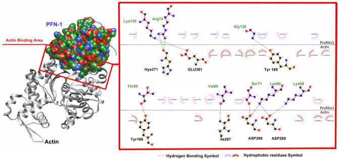
). On the actin side, residues are His 371, Glu 361, Tyr 169, Tyr 166, Ile 287, Asp 286, and 288. The amino acid residues involved in the hydrophobic interaction (formula image ) are identified and noted in the figure.
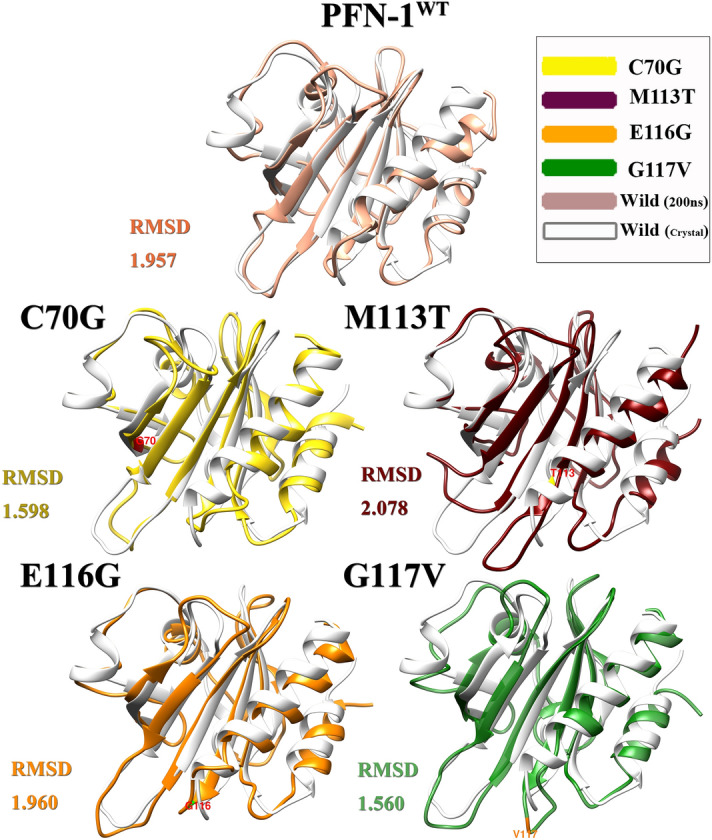
Figure 9
Figure 9
Superimposition of PFN-1WT (white) over the mutant structures (the predefined colors) after 200 ns MD simulation. In this case with RMSD 1.957 which is related to the wild structure of PFN-1 before and after 200 ns MD simulation, The RMSD of M113T mutations with a value of 2.078 is larger than the PFN-1WT structure and the C70G and G117V mutations with a value of 1.560 and 1.598 respectively, are less than the PFN-1WT, while RMSD value for E116G is almost equal to the PFN-1WT.
Figure 10
Figure 10
Difference RRDistMaps (for viewing and comparing protein distance) representing the difference between the PFN-1WT and mutant forms of PFN-1. The difference between the Wild and mutant structures at the end of 50 ns MD is 20.27 for the E116G mutation, which is 11 for the C70G mutation, 13.4, and 15.21 for the G117V and M113T mutations, respectively.
Figure 11
Figure 11
Binding energies (Kcal/Mol) of mutant forms of PFN-1 and PFN1WT vs the two FDA-approved drugs for ALS.
Figure 12
Figure 12
Possible Interaction of FDA-Approved Drugs for ALS; Edaravone, Riluzole with mutant PFN-1 at C70G, M113, and G117 residues determined with molecular docking simulation are shown in ribbon, sticks, and molecular view. (a) Riluzole and Edaravone interacting with C70G mutant PFN-1 (b). Riluzole and Edaravone interacting with M113T mutant PFN-1. (c) Riluzole and Edaravone interacting with G117V mutant PFN-1.

References

    1. Longo, D.L. Robert H. Brown, D. Phil., MD, and Ammar AlChalabi, Ph. D., FRCP, Dip. Stat. N. Engl. J. Med.377, 162–172 (2017).
    1. Dervishi I, Ozdinler PH. Incorporating upper motor neuron health in ALS drug discovery. Drug Discov. Today. 2018;23(3):696–703. doi: 10.1016/j.drudis.2018.01.027. - DOI - PMC - PubMed
    1. Nguyen HP, Van Broeckhoven C, van der Zee J. ALS genes in the genomic era and their implications for FTD. Trends Genet. 2018;34(6):404–423. doi: 10.1016/j.tig.2018.03.001. - DOI - PubMed
    1. Wu C-H, Fallini C, Ticozzi N, Keagle PJ, Sapp PC, Piotrowska K, et al. Mutations in the profilin 1 gene cause familial amyotrophic lateral sclerosis. Nature. 2012;488(7412):499–503. doi: 10.1038/nature11280. - DOI - PMC - PubMed
    1. Alkam D, Feldman EZ, Singh A, Kiaei M. Profilin1 biology and its mutation, actin (g) in disease. Cell. Mol. Life Sci. 2017;74(6):967–981. doi: 10.1007/s00018-016-2372-1. - DOI - PMC - PubMed

Publication types

MeSH terms