

A 3D analysis of growth trajectory and integration during early human prenatal facial growth
- PMID: 33767268
- PMCID: PMC7994314
- DOI: 10.1038/s41598-021-85543-5
A 3D analysis of growth trajectory and integration during early human prenatal facial growth
Abstract
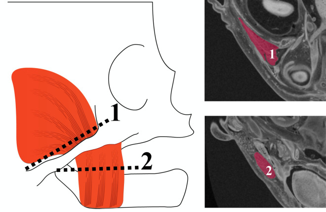
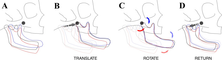
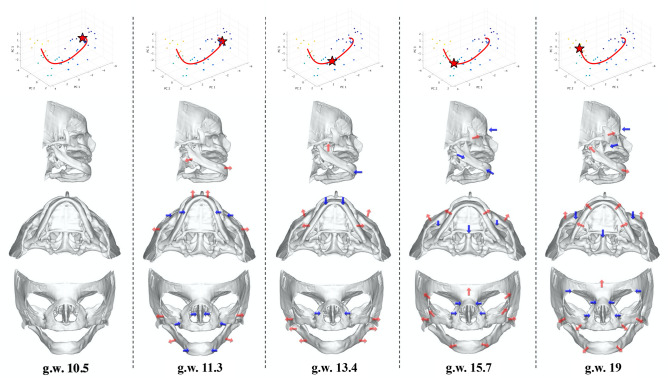
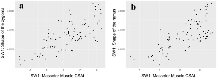
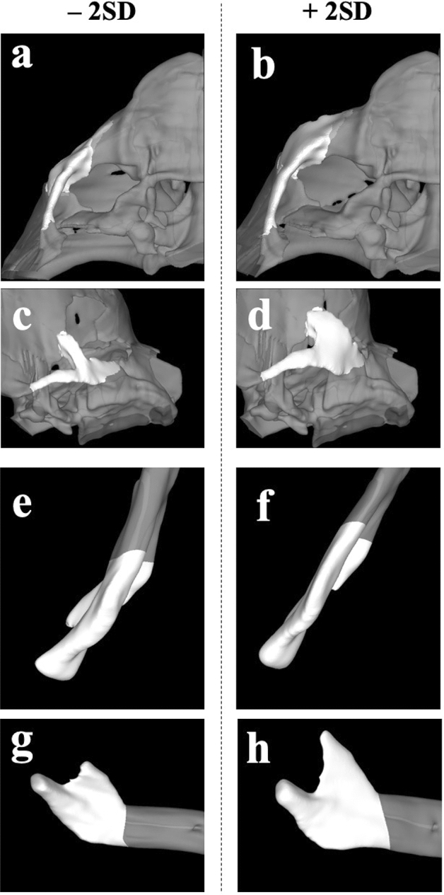
Significant shape changes in the human facial skeleton occur in the early prenatal period, and understanding this process is critical for studying a myriad of congenital facial anomalies. However, quantifying and visualizing human fetal facial growth has been challenging. Here, we applied quantitative geometric morphometrics (GM) to high-resolution magnetic resonance images of human embryo and fetuses, to comprehensively analyze facial growth. We utilized non-linear growth estimation and GM methods to assess integrated epigenetic growth between masticatory muscles and associated bones. Our results show that the growth trajectory of the human face in the early prenatal period follows a curved line with three flexion points. Significant antero-posterior development occurs early, resulting in a shift from a mandibular prognathic to relatively orthognathic appearance, followed by expansion in the lateral direction. Furthermore, during this time, the development of the zygoma and the mandibular ramus is closely integrated with the masseter muscle.
Conflict of interest statement
The authors declare no competing interests.
Figures
Similar articles
-
Growth of the masseter muscle in rhesus monkeys (Macaca mulatta).Am J Phys Anthropol. 1983 Mar;60(3):401-10. doi: 10.1002/ajpa.1330600310. Am J Phys Anthropol. 1983. PMID: 6846512
-
Effects of masticatory muscle function and bite-raising on mandibular morphology in the growing rat.Swed Dent J Suppl. 2001;(150):1-49. Swed Dent J Suppl. 2001. PMID: 11803646
-
[Comparative study of masticatory muscles in the suncus and the mouse].Kaibogaku Zasshi. 2001 Dec;76(6):523-32. Kaibogaku Zasshi. 2001. PMID: 11806145 Japanese.
-
The Face – A Musculoskeletal Perspective. A literature review.Swiss Dent J. 2018 Sep 10;128(9):678-688. doi: 10.61872/sdj-2018-09-442. Swiss Dent J. 2018. PMID: 30056693 Review.
-
[Functional structure of the human temporal-masseter muscle complex in the fetus and the adult].Orthod Fr. 1987;58 Pt 2:549-65. Orthod Fr. 1987. PMID: 3334688 Review. French. No abstract available.
Cited by
-
A Geometric Morphometric Study on Sexual Dimorphism in Viscerocranium.Biology (Basel). 2022 Sep 9;11(9):1333. doi: 10.3390/biology11091333. Biology (Basel). 2022. PMID: 36138812 Free PMC article.
-
The first 3D analysis of the sphenoid morphogenesis during the human embryonic period.Sci Rep. 2022 Mar 28;12(1):5259. doi: 10.1038/s41598-022-08972-w. Sci Rep. 2022. PMID: 35347174 Free PMC article.
-
Morphometric analysis of secondary palate development in human embryos.J Anat. 2022 Dec;241(6):1287-1302. doi: 10.1111/joa.13745. Epub 2022 Aug 19. J Anat. 2022. PMID: 35983845 Free PMC article.
-
3D anthropometry of the nasolabial region in children aged 3 to 9 months as reference database for clinical assessment.Sci Rep. 2025 Jul 28;15(1):27443. doi: 10.1038/s41598-025-11024-8. Sci Rep. 2025. PMID: 40721459 Free PMC article.
-
3D atlas of the human fetal chondrocranium in the middle trimester.Sci Data. 2024 Jun 13;11(1):626. doi: 10.1038/s41597-024-03455-1. Sci Data. 2024. PMID: 38871782 Free PMC article.
References
-
- Carlson DS. Theories of craniofacial growth in the postgenomic era. Semin. Orthod. 2005;11:172–183. doi: 10.1053/j.sodo.2005.07.002. - DOI
-
- DeMyer W, Zeman W, Palmer C. The face predicts the brain: Diagnostic significance of median facial anomalies for holoprosencephaly (arhinencephaly) Pediatrics. 1964;2:256–256. - PubMed
Publication types
MeSH terms
LinkOut - more resources
Full Text Sources
Other Literature Sources
