. 2021 Mar 25;11(1):6927.
doi: 10.1038/s41598-021-86380-2.

In silico investigation of critical binding pattern in SARS-CoV-2 spike protein with angiotensin-converting enzyme 2

Affiliations

In silico investigation of critical binding pattern in SARS-CoV-2 spike protein with angiotensin-converting enzyme 2

Farzaneh Jafary et al. Sci Rep. .

Abstract

Severe acute respiratory syndrome coronavirus-2 (SARS-CoV-2) is a newly-discovered coronavirus and responsible for the spread of coronavirus disease 2019 (COVID-19). SARS-CoV-2 infected millions of people in the world and immediately became a pandemic in March 2020. SARS-CoV-2 belongs to the beta-coronavirus genus of the large family of Coronaviridae. It is now known that its surface spike glycoprotein binds to the angiotensin-converting enzyme-2 (ACE2), which is expressed on the lung epithelial cells, mediates the fusion of the cellular and viral membranes, and facilitates the entry of viral genome to the host cell. Therefore, blocking the virus-cell interaction could be a potential target for the prevention of viral infection. The binding of SARS-CoV-2 to ACE2 is a protein-protein interaction, and so, analyzing the structure of the spike glycoprotein of SARS-CoV-2 and its underlying mechanism to bind the host cell receptor would be useful for the management and treatment of COVID-19. In this study, we performed comparative in silico studies to deeply understand the structural and functional details of the interaction between the spike glycoprotein of SARS-CoV-2 and its cognate cellular receptor ACE2. According to our results, the affinity of the ACE2 receptor for SARS-CoV-2 was higher than SARS-CoV. According to the free energy decomposition of the spike glycoprotein-ACE2 complex, we found critical points in three areas which are responsible for the increased binding affinity of SARS-CoV-2 compared with SARS-CoV. These mutations occurred at the receptor-binding domain of the spike glycoprotein that play an essential role in the increasing the affinity of coronavirus to ACE2. For instance, mutations Pro462Ala and Leu472Phe resulted in the altered binding energy from - 2 kcal mol-1 in SARS-COV to - 6 kcal mol-1 in SARS-COV-2. The results demonstrated that some mutations in the receptor-binding motif could be considered as a hot-point for designing potential drugs to inhibit the interaction between the spike glycoprotein and ACE2.


Conflict of interest statement

The authors declare no competing interests.

Figures
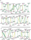
Figure 1
Figure 1
Two-dimensional interaction schemes for the spike-ACE2 complex: SARS-CoV (A), chimeric structure (B), and SARS-CoV-2 (C). ACE2 and spike proteins residues denoted by A and E in parenthesis respectively. Also, hydrogen bonds and hydrophobic interactions are colored in yellow and green lines respectively. The images have been obtained by LigPlot+ v.1.4.5 (https://www.ebi.ac.uk/thornton-srv/software/LigPlus/).
Figure 2
Figure 2
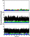
The fractional H-bonds of the receptor-spike protein complex during the simulation: hydrogen bonds between four regions of the ACE2 receptor (19–33, 35–54, 325–331, and 334–339) and receptor-binding motif in SARS-CoV (A), chimeric structure (B) and SARS-CoV-2 (C). Black, red, green and blue lines in each graph are related to residues 19–33, 35–54, 353–358 and 325–331 of the ACE2 receptor, respectively. The fractional H-bond graphs have been plotted using xmgrace from Grace plotting tool (https://plasma-gate.weizmann.ac.il/Grace/).
Figure 3
Figure 3
The interaction network analysis of ACE2-spike protein complex during the simulation: (A) in SARS-CoV (2ajf) and (B) SARS-CoV-2 (6m0j) structures. The PDB structures obtained from the initial (A1 and A2 for SARS-CoV, and B1 and B2 for SARS-CoV-2) and final (A3 and A4 for SARS-CoV, and B3 and B4 for SARS-CoV-2) 500 frames of the simulation that were analyzed by NAPS. The red points in A1, A3, B1 and B3 indicate critical amino acids that participated in the spike-ACE2 interaction The images have been obtained by UCSF Chimera 1.13.1 (http://www.rbvi.ucsf.edu/chimera/) and NAPS server (http://bioinf.iiit.ac.in/NAPS/).
Figure 4
Figure 4
Free energy decomposition of the spike protein residues in the spike-ACE2 complex: (A) SARS-COV and (B) SARS-COV-2. The red graph shows binding free energy for each residue in the receptor-binding motif of the spike protein.
Figure 5
Figure 5
Free energy decomposition of the ACE2 residues in the spike-ACE2 complex: (A) SARS-COV and (B) SARS-COV-2. The red graph shows binding free energy for each residue in ACE2 receptor.
Figure 6
Figure 6
Native contact pattern for the SARS-CoV and SARS-CoV2 in interaction with ACE2: The native contact pattern between the receptor-binding motif of the spike protein and ACE2 in SARS-CoV (A) and SARS-CoV-2 (B) during the simulation. Residues with high values of the native contact (based on R-factor) involved in the interactions are designated as green, yellow, and red which identified in part A2 and B2. A3 and B3 are the region located in the middle of the receptor-binding motif in SARS-CoV and SARS-CoV-2 respectively that acts as a clamp for the binding of the virus to ACE2. The images have been obtained by Swiss-PDB viewer 4.0.1 (https://spdbv.vital-it.ch/).

Similar articles

Cited by

References

    1. Payne S. Family coronaviridae. Viruses. 2017 doi: 10.1016/B978-0-12-803109-4.00017-9. - DOI
    1. Sahin AR, et al. Novel coronavirus (COVID-19) outbreak: a review of the current literature. EJMO. 2020;4:1–7.
    1. Zhong NS, et al. Epidemiology and cause of severe acute respiratory syndrome (SARS) in Guangdong, People's Republic of China, in February, 2003. Lancet. 2003;362:1353–1358. doi: 10.1016/S0140-6736(03)14630-2. - DOI - PMC - PubMed
    1. De Wit E, van Doremalen N, Falzarano D, Munster VJ. SARS and MERS: recent insights into emerging coronaviruses. Nat. Rev. Microbiol. 2016;14:523–534. doi: 10.1038/nrmicro.2016.81. - DOI - PMC - PubMed
    1. Zaki AM, van Boheemen S, Bestebroer TM, Osterhaus AD, Fouchier RA. Isolation of a novel coronavirus from a man with pneumonia in Saudi Arabia. N. Engl. J. Med. 2012;367:1814–1820. doi: 10.1056/NEJMoa1211721. - DOI - PubMed

Substances

LinkOut - more resources