. 2021 Mar 25;4(1):408.
doi: 10.1038/s42003-021-01956-y.

Bacteria-specific pro-photosensitizer kills multidrug-resistant Staphylococcus aureus and Pseudomonas aeruginosa

Affiliations

Bacteria-specific pro-photosensitizer kills multidrug-resistant Staphylococcus aureus and Pseudomonas aeruginosa

Min Lu et al. Commun Biol. .

Abstract

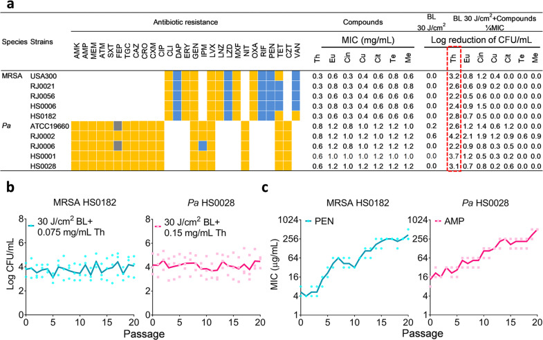
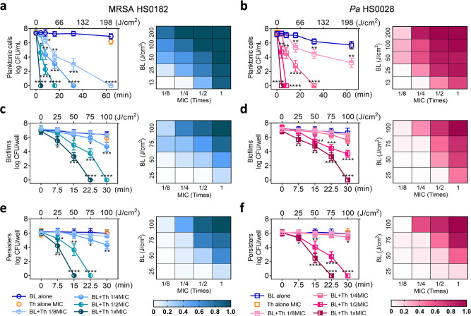
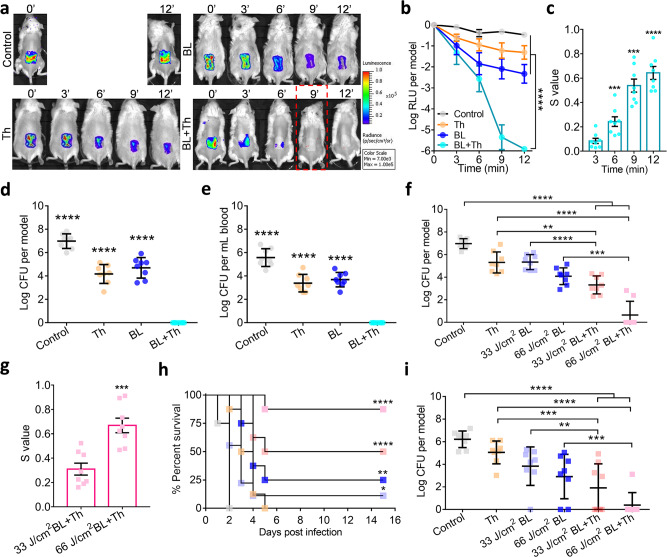
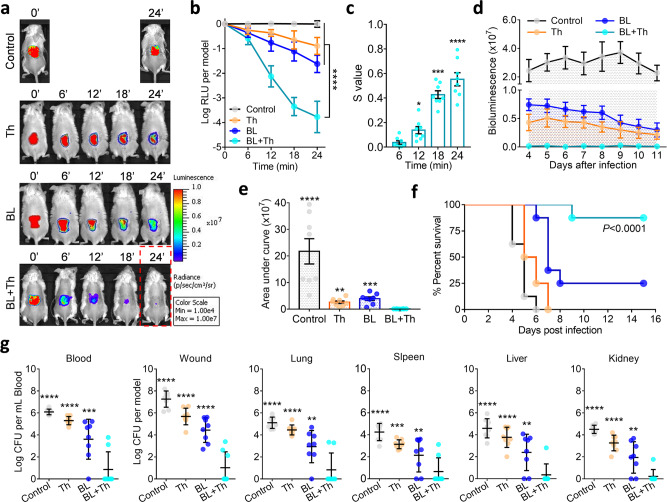
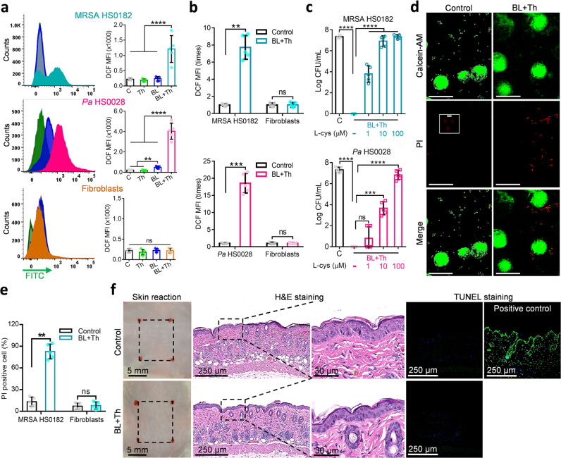
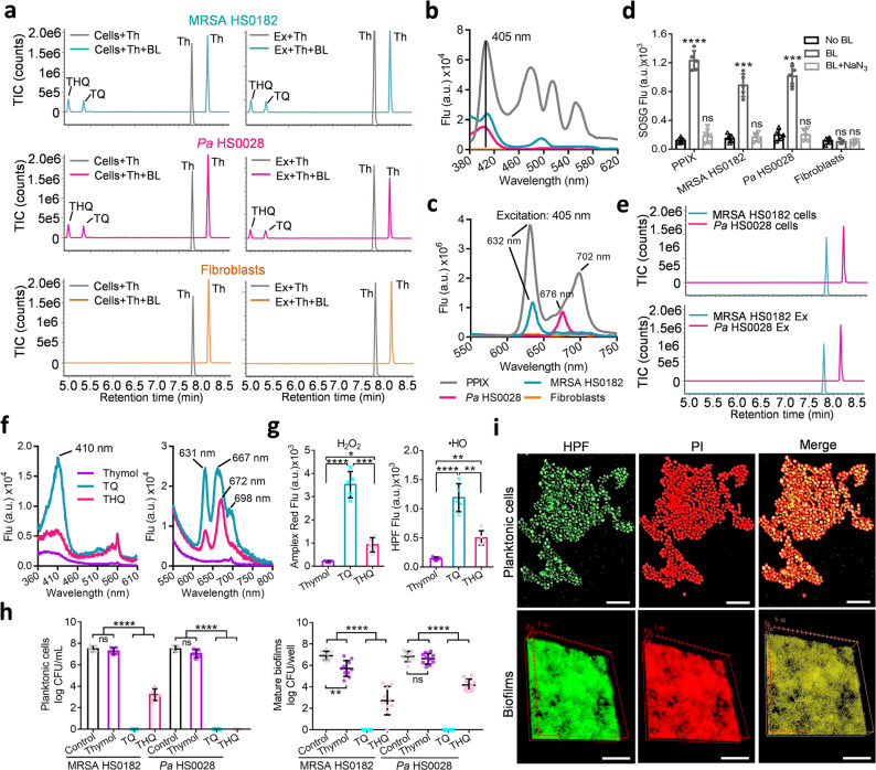
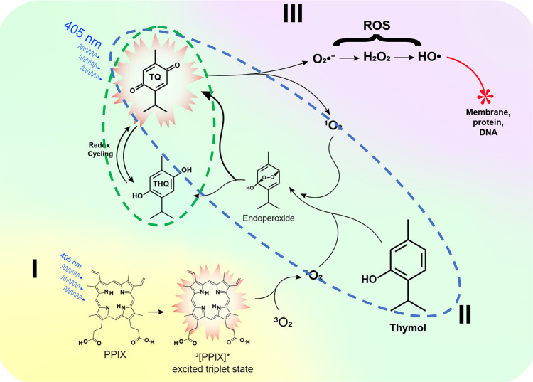
The emergence of multidrug-resistant bacteria has become a real threat and we are fast running out of treatment options. A combinatory strategy is explored here to eradicate multidrug-resistant Staphlococcus aureus and Pseudomonas aeruginosa including planktonic cells, established biofilms, and persisters as high as 7.5 log bacteria in less than 30 min. Blue-laser and thymol together rapidly sterilized acute infected or biofilm-associated wounds and successfully prevented systematic dissemination in mice. Mechanistically, blue-laser and thymol instigated oxidative bursts exclusively in bacteria owing to abundant proporphyrin-like compounds produced in bacteria over mammalian cells, which transformed harmless thymol into blue-laser sensitizers, thymoquinone and thymohydroquinone. Photo-excitations of thymoquinone and thymohydroquinone augmented reactive oxygen species production and initiated a torrent of cytotoxic events in bacteria while completely sparing the host tissue. The investigation unravels a previously unappreciated property of thymol as a pro-photosensitizer analogous to a prodrug that is activated only in bacteria.


Conflict of interest statement

The authors declare no competing interests.

Figures

Fig. 1
Fig. 1. Screening bactericidal activity of BL combined with essential oil compounds.
a Bacteria of 5 MRSA and 5 Pa strains were tested for their susceptibility to various antibiotics: susceptible (blue boxes), intermediate (gray boxes), or resistant (yellow boxes) and MIC values to Th (thymol), Eu (eugenol), Cin (cinnamaldehyde), Cu (cuminaldehyde), Cit (citral-a), Te (terpinen-4-ol), and Me (menthol). Bactericidal efficacies of 30 J/cm2 BL in the absence and presence of an indicated compound at 1/4 MICs were obtained against the 10 MDR strains. b and c Resistance development of MRSA HS0182 and Pa HS0028 to the sub-lethal doses of BL plus thymol (b), penicillin (PEN) alone (c, left), or ampicillin (AMP) alone (c, right), respectively. Each value represents the mean of triplicate assays. Antibiotics in a: AMK, amikacin; MEM, meropenem; ATM, aztreonam; SXT, trimethoprim-sulfamethoxazole; FEP, cefepime; TGC, tigecycline; CAZ, ceftazidime; CRO, ceftriaxone; CXM, cefuroxime; CIP, ciprofloxacin; CLI, clindamycin; DAP, daptomycin; ERY, erythromycin; GEN, gentamicin; IPM, imipenem; LVX, levofloxacin; LNZ, linezolid; LZD, linezolid; MXF, moxifloxacin; NIT, nitrofurantoin; OXA, oxacillin; RIF, rifampin; TET, tetracycline; CZT, ceftizoxime; and VAN, vancomycin.
Fig. 2
Fig. 2. BL and thymol synergistically inactivate planktonic cells, established biofilms, and bacterial persisters.
Killing curves (left) and synergy evalution heat map (right) of MRSA HS0182 (a, c, and e) and Pa HS0028 (b, d, and f) in planktonic cells (a and b), established biofilms (c and d), and bacterial persisters (e and f) treated with an increasing BL fluence alone, thymol at 1x MIC alone, or BL combined with an increasing concentration of thymol (a fraction of MIC). Checkerboards in the corresponding right panels show S-values for different combinations of BL and thymol as assessed by the Bliss Independence model according to the following formula: S-value = (logCFU/mLBL/logCFU/mLControl)(logCFU/mLTh/logCFU/mLControl) − (logCFU/mLBL+Th/logCFU/mLControl). LogCFU/mLBL, logCFU/mLTh, logCFU/mLBL+Th, and logCFU/mLcontrol are the number of viable bacteria remaining after treatment with BL alone, thymol alone, combination of BL and thymol, or sham light, respectively. 0 < S < 1 indicates a synergistic interaction, whereas S < 0 indicates an antagonistic interaction. Results are presented as mean ± SD of four to six replicates from five independent experiments. ****P < 0.0001; ***P < 0.001; **P < 0.01; *P < 0.05; and ns, no significance.
Fig. 3
Fig. 3. Synergistic disinfections of murine burns by topical application of BL and thymol.
Full-thickness 3rd-burn-wounds were infected with USA300 (ae) or Pa HS0028 (fi) at 5 × 106 CFU in 50 μL of PBS for 30 min as acute infection. ae The USA300-infected wounds were exposed to sham (control), 50 µL of thymol at 2 mg/mL (Th), indicated times of BL exposure (BL), or both (BL + Th). a Bacterial luminescence images of representative wounds were acquired at indicated times after various treatments. b Mean luminescence was presented as logarithmic relative luminescence units (log RLU) per model relative to time zero. c S-values are calculated by the Bliss Independence model as Fig. 2. d and e Bacterial burdens in the wounds (d) and blood (e) were assayed 7 days after acute infection shown as log CFU per model. fi The Pa HS0028-infected burns were treated with sham (control), 50 µL of thymol at 10 mg/mL (Th), BL exposure at 33 J/cm2 or 66 J/cm2, or both and log CFU per wound were determined on day 1 after the indicated treatments. g S-values confirm the synergy between BL and thymol against acute Pa HS0028 infection. h Kaplan–Meier survival curves of Pa HS0028-infected mice. i Bacterial loads in the burns were quantified either prior death or at the end of the experiments. All results are presented as mean ± SD of eight mice. Zero in d, e, f, and i was below the detection limit (40 CFU per wound and 20 CFU per mL blood). ****P < 0.0001; ***P < 0.001; **P < 0.01; and ns, no significance.
Fig. 4
Fig. 4. BL together with thymol rescues mice from lethal USA300 biofilm-associated infection.
Murine 3rd-burn-wounds were infected with USA300 at 5 × 107 CFU in 50 µL of PBS for 72 h to form mature biofilms. The infected wounds were treated with sham (control), 50 µL of thymol at 10 mg/mL (Th), indicated times of BL (BL), or both (BL + Th). a Bacterial luminescence images of representative wounds were acquired at indicated times. b Mean luminescence was acquired over time and presented as log RLU per model. c, S-values are calculated by the Bliss Independence model as Fig. 2. d and e Mean luminescence was acquired from days 4 to 11 after an indicated treatment and the mean areas under the luminescence curves were summarized in e. f Kaplan–Meier survival curves of USA300 biofilm-associated mice in response to an indicated treatment. g Bacterial loads in the blood, wounds, lungs, spleens, livers, and kidneys were quantified just prior death or on day 15 after bacterial inoculation. All results are presented as mean ± SD of eight biological replicates. Zero in g was below the detection limit (40 CFU per model for murine wounds or organs and 20 CFU per mL blood). ****P < 0.0001; ***P < 0.001; **P < 0.01; and ns, no significance.
Fig. 5
Fig. 5. No adverse effects of BL combined with thymol treatment on fibroblast and murine skin.
a Flow cytometric analyses of intracellular ROS in MRSA HS0182 (upper), Pa HS0028 (medium), and fibroblasts (bottom) treated with sham light (C), thymol at 1/2 MIC (Th), an indicated BL fluence (BL), or both (BL + Th) by DCFH-DA fluorescent staining. MRSA HS0182 was exposed to thymol at 0.15 mg/mL, BL at 50 J/cm2, or both, and Pa HS0028 to thymol at 0.3 mg/mL, BL at 25 J/cm2, or both. Alternatively, fibroblasts alone (a, bottom) or the cells co-cultured with MRSA HS0182 (b, upper) or Pa HS0028 (c, bottom) were treated with thymol at 0.5 mg/mL, BL at 50 J/cm2, or both. DCF mean florescence intensity (MFI) on gate of bacteria or fibroblasts was presented in a and fold changes of DCF MFI relative to sham-treated controls were shown in b. c Dose-dependent effects of antioxidant L-cys on the bactericidal activity of the combined therapy: 50 J/cm2 BL and 0.15 mg/mL thymol for MRSA HS0182 and 25 J/cm2 BL and 0.3 mg/mL thymol for Pa HS0028. d Representative fluorescence images of co-culture of MRSA HS0182 and fibroblasts were shown, in which the dead and viable cells were visualized by PI (red) and calcein-AM staining (green), respectively. Scale bars, 20 µm. An area in the middle panel (control PI) was enlarged to show a few PI-stained bacteria (Scale bars, 2 µm). e PI+ MRSA HS0182 and PI+ fibroblasts in co-cultures were also counted manually and presented as percentages relative to a total of cells. f No adverse effect of topical application of BL and thymol in murine skin. The dorsal skin was topically treated with sham (control) or 100 J/cm2 BL and 50 µL of thymol at 20 mg/mL (BL + Th) once a day for 5 consecutive days. On day 6, the skins were processed by H&E histological examination and TUNEL assay. DNase I-treated skins were TUNEL stained in parallel as positive-staining controls. All results are presented as mean ± SD of at least five biological replicates. Images in d and f are representative of five independent experiments. ****P < 0.0001; ***P < 0.001; **P < 0.01; and ns, no significance.
Fig. 6
Fig. 6. Photo-oxidation of thymol to generate photosensitizers TQ and THQ exclusively in bacteria.
a UPLC-VION-IMS-QTOF-MS/MS analyses of thymol oxidation in cells (left) or extracts (right) of MRSA HS0182 (upper), Pa HS0028 (medium), or fibroblasts (bottom) treated with 0.5 mg/mL thymol in the absence or presence of 50 J/cm2 BL exposure. b and c Exciation (b) and emission (c) spectra of an indicated cellular extract in comparison with those of PPIX at 10 µM. d 1O2 is notably generated by 50 J/cm2 BL in MRSA HS0182, Pa HS0028, and PPIX solution but not in fibroblasts. 1O2 generation was blunted by NaN3 (BL + NaN3), a 1O2-specific quencher. e UPLC-VION-IMS-QTOF-MS/MS analyses of photo-oxidized thymol in the presence of NaN3 at 10 µM. BL illumination at 50 J/cm2, thymol at 0.5 mg/mL. f Exciation (left) and emission (right) spectra of thymol, TQ, and THQ each at 0.5 mg/mL. g H2O2 and •HO were generated by thymol, TQ, and THQ each at 0.2 mg/mL in combination with 50 J/cm2 BL. h Bactericidal activities of thymol, TQ, and THQ against planktonic cells (left) and established biofilms (right) of MRSA HS0182 and Pa HS0028 in the presence of 20 J/cm2 BL. The concentrations of three compounds were same at 0.05 mg/mL for planktonic cells and 0.1 mg/mL for established biofilms. i Representative fluorescence images of planktonic cells (upper) and established biofilms (bottom) of MRSA HS0182 after a lethal dose of the duo treatment. Bacterial viability and intracellular •HO were evaluated by PI and HPF staining, respectively. Scale bars: 2 µm in upper panel and 50 µm in bottom panel. All images in a, b, c, e, f, and i are representative of five independent experiments. Results in d, g, and h are presented as mean ± SD of at least five independent experiments. ****P < 0.0001; ***P < 0.001; **P < 0.01; *P < 0.05; and ns, no significance.
Fig. 7
Fig. 7. A schematic of phototoxic cascade reactions in the presence of BL and thymol.
Endogenous proporphyrins-like compounds absorb BL to attain an excited triplet-state from an electronic ground state and react with a ground state molecular oxygen (3O2) to generate singlet oxygen (1O2) (I). The 1O2 oxidizes thymol to form TQ and/or THQ via an endoperoxide intermediate (II). The resultant TQ and THQ, particually, TQ act as a photosensitizer and generates more O2 and 1O2 that can in turn oxidize thymol, continuously replenishing the TQ pool, which forms the first autoxidation cycle (blue-dashed outline). The second autoxidation cycle comprises THQ oxidation into TQ that can be then photo-hydrolyzed into THQ (green dashed outline). O2 undergoes dismutation and forms H2O2 that is converted into the most detrimental •HO via a Fenton reaction or photolysis (III). The deleterious •HO initiates a chain reaction that oxidatively damages the bacteria.

Similar articles

Cited by

References

    1. World Health Organization. 2019 Antibacterial Agents in Clinical Development: an Analysis of the Antibacterial Clinical Development Pipeline. (WHO, 2019).
    1. World Health Organization. Prioritization of Pathogens to Guide Discovery, Research and Development of New Antibiotics for Drug-resistant Bacterial Infections, Including Tuberculosis. (WHO, 2017).
    1. Olsen I. Biofilm-specific antibiotic tolerance and resistance. Eur. J. Clin. Microbiol. Infect. Dis. 2015;34:877–886. doi: 10.1007/s10096-015-2323-z. - DOI - PubMed
    1. Lewis K. Persister cells, dormancy and infectious disease. Nat. Rev. Microbiol. 2007;5:48–56. doi: 10.1038/nrmicro1557. - DOI - PubMed
    1. Kim W, et al. A new class of synthetic retinoid antibiotics effective against bacterial persisters. Nature. 2018;556:103–107. doi: 10.1038/nature26157. - DOI - PMC - PubMed

Publication types

MeSH terms