

Kinetic changes in the spinal cord occupation rate of dural sac in cervical spondylotic myelopathy
- PMID: 33767534
- PMCID: PMC7973381
- DOI: 10.1016/j.jor.2021.03.005
Kinetic changes in the spinal cord occupation rate of dural sac in cervical spondylotic myelopathy
Abstract
Objective: The study aimed to establish the spinal cord occupation rate of the dural sac during flexion and extension.
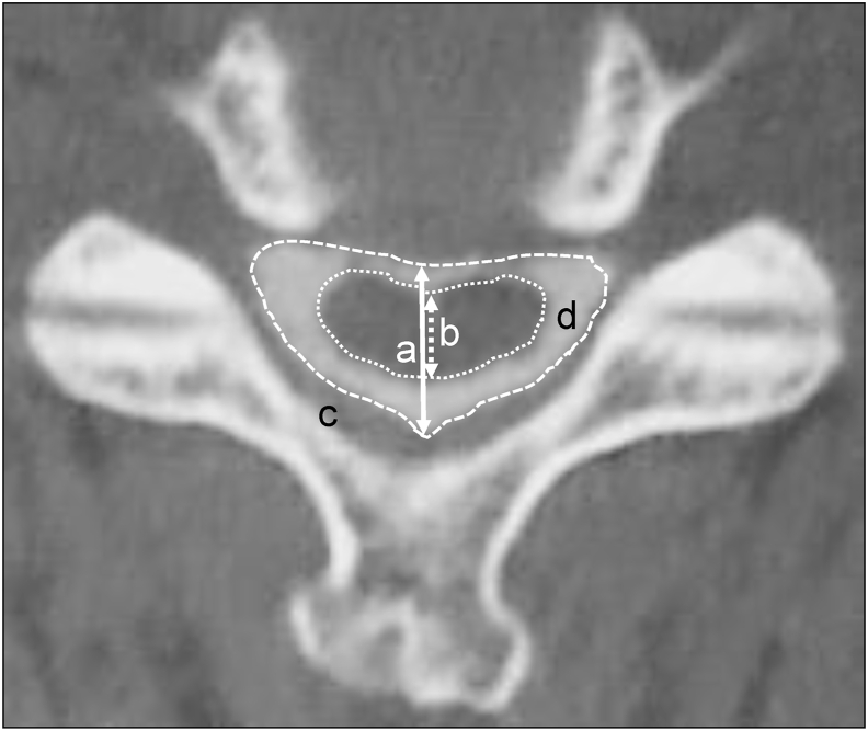
Methods: We measured the cross-sectional area of the dural sac and the spinal cord between C2/C3 and C7/T1 disc levels in 100 patients with cervical spondylotic myelopathy and 1211 asymptomatic subjects.
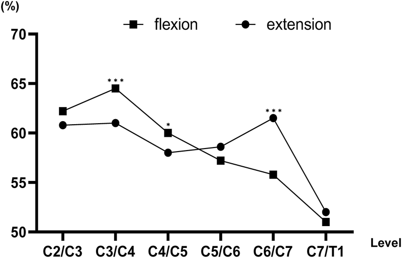
Results: The spinal cord occupation rate of the dural sac in the cross-sectional area was higher on extension than on flexion at the mid-lower cervical spine.
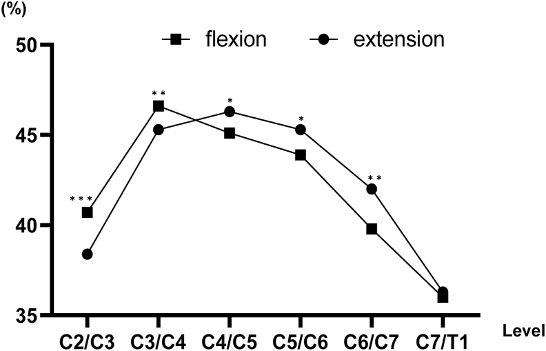
Conclusions: The spinal cord occupation rate of the dural sac in the cross-sectional area was highest at the C4/C5 and C5/C6 levels on extension.
Keywords: Asymptomatic subjects; Cervical spondylotic myelopathy; Dural sac; Kinetic change; Occupation rate; Spinal cord.
© 2021 Professor P K Surendran Memorial Education Foundation. Published by Elsevier B.V. All rights reserved.
Conflict of interest statement
The authors have no financial conflicts of interest.
Figures
Similar articles
-
Dynamic changes in longitudinal stretching of the spinal cord in thoracic spine: Focus on the spinal cord occupation rate of dural sac.Clin Neurol Neurosurg. 2020 Nov;198:106225. doi: 10.1016/j.clineuro.2020.106225. Epub 2020 Sep 8. Clin Neurol Neurosurg. 2020. PMID: 32942132
-
Pathoanatomic investigation of cervical spondylotic myelopathy.Fukushima J Med Sci. 2004 Dec;50(2):47-54. doi: 10.5387/fms.50.47. Fukushima J Med Sci. 2004. PMID: 15779570
-
Pathological changes of cervical spinal canal in cervical spondylotic myelopathy: A retrospective study on 39 cases.Clin Neurol Neurosurg. 2019 Jun;181:133-137. doi: 10.1016/j.clineuro.2019.04.016. Epub 2019 Apr 15. Clin Neurol Neurosurg. 2019. PMID: 31039495
-
Influence of developmental cervical stenosis on dural sac space.Chin Med J (Engl). 2014;127(22):3857-61. Chin Med J (Engl). 2014. PMID: 25421180
-
Venous hypertensive myelopathy associated with cervical spondylosis.Spine J. 2016 Nov;16(11):e751-e754. doi: 10.1016/j.spinee.2016.06.003. Epub 2016 Jun 9. Spine J. 2016. PMID: 27293119 Review.
Cited by
-
Radiological and Clinical Significance of Cervical Dynamic Magnetic Resonance Imaging for Cervical Spondylotic Myelopathy.Neurospine. 2024 Jun;21(2):443-454. doi: 10.14245/ns.2448166.083. Epub 2024 Jun 30. Neurospine. 2024. PMID: 38955522 Free PMC article.
References
-
- Karadimas S.K., Erwin W.M., Ely C.G., Dettori J.R., Fehlings M.G. Pathophysiology and natural history of cervical spondylotic myelopathy. Spine (Phila Pa 1976) 2013 Oct 15;38(22 Suppl 1):S21–S36. - PubMed
-
- Nouri A., Tetreault L., Singh A., Karadimas S.K., Fehlings M.G. Degenerative cervical myelopathy: epidemiology, genetics and pathogenesis. Spine (Phila Pa 1976) 2015 Jun 15;40(12):E675–E693. - PubMed
-
- Yu M., Tang Y., Liu Z., Sun Y., Liu X. The morphological and clinical significance of developmental cervical stenosis. Eur Spine J. 2015 Aug;24(8):1583–1589. - PubMed
-
- Kadanka Z., Kerkovsky M., Bednarik J., Jarkovsky J. Cross-sectional transverse area and hyperintensities on magnetic resonance imaging in relation to the clinical picture in cervical spondylotic myelopathy. Spine (Phila Pa 1976) 2007 Nov 1;32(23):2573–2577. - PubMed
-
- Takeshima T., Omokawa S., Takaoka T., Araki M., Ueda Y., Takakura Y. Sagittal alignment of cervical flexion and extension: lateral radiographic analysis. Spine (Phila Pa 1976) 2002 Aug 1;27(15):E348–E355. - PubMed
LinkOut - more resources
Full Text Sources
Other Literature Sources
Miscellaneous
