. 2021 Mar 9:12:632336.
doi: 10.3389/fneur.2021.632336. eCollection 2021.

A Novel Pathogenic HSPG2 Mutation in Schwartz-Jampel Syndrome

Affiliations

A Novel Pathogenic HSPG2 Mutation in Schwartz-Jampel Syndrome

Po-Yu Lin et al. Front Neurol. .

Abstract

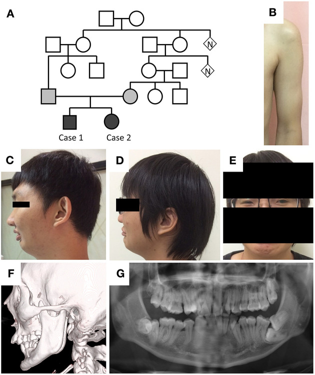
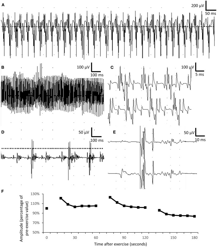
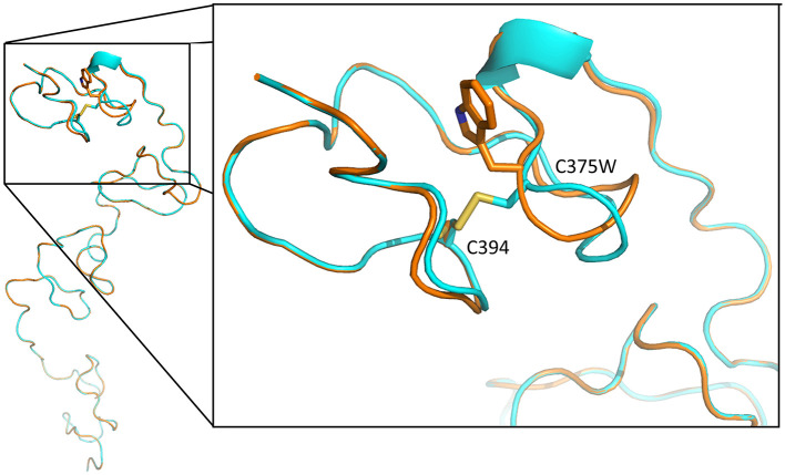
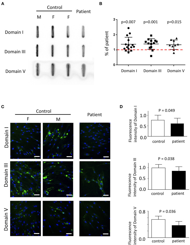
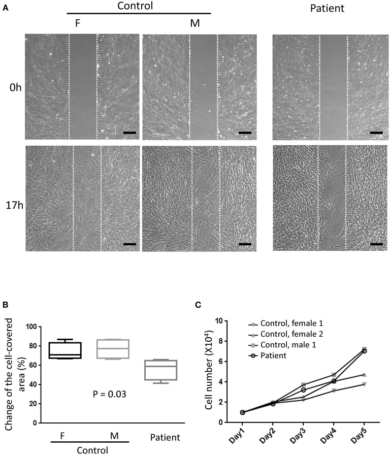
Schwartz-Jampel syndrome is a rare autosomal recessive disease caused by mutation in the heparan sulfate proteoglycan 2 (HSPG2) gene. Its cardinal symptoms are skeletal dysplasia and neuromuscular hyperactivity. Herein, we identified a new pathogenic mutation site (NM_005529.6:c.1125C>G; p.Cys375Trp) of HSPG2 leading to Schwartz-Jampel syndrome by whole-exome sequencing. This mutation carried by the asymptomatic parents was previously registered in a single-nucleotide polymorphism database of the National Institutes of Health as a coding sequence variant rs543805444. The pathogenic nature of this missense mutation was demonstrated by in silico pathogenicity assessment, clinical presentations, and cellular function of primary fibroblast derived from patients. Various in silico software applications predicted the mutation to be pathogenic [Sorting Intolerant From Tolerant (SIFT), 0; Polyphen-2, 1; CADD (Combined Annotation Dependent Depletion), 23.7; MutationTaster, 1; DANN (deleterious annotation of genetic variants using neural networks); 0.9]. Needle electromyography revealed extensive complex repetitive discharges and multiple polyphasic motor unit action potentials in axial and limb muscles at rest. Short exercise test for myotonia showed Fournier pattern I. At cellular levels, mutant primary fibroblasts had reduced levels of secreted perlecan and impaired migration ability but normal capability of proliferation. Patients with this mutation showed more neuromuscular instability and relatively mild skeletal abnormality comparing with previously reported cases.

Keywords: Schwartz–Jampel syndrome; heparan sulfate proteoglycan 2; perlecan; primary fibroblast; short exercise test.


Conflict of interest statement

The authors declare that the research was conducted in the absence of any commercial or financial relationships that could be construed as a potential conflict of interest.

Figures

Figure 1
Figure 1
Phenotypes of the cases. (A) Pedigree of the two cases. (B) Hypertrophic pectoralis major and biceps brachii with thin subcutaneous tissue in case 1. (C,D) Retruded mandible, pursed lip, and supraorbital ridge in both cases. (E) Blepharophimosis in case 2. (F,G) Elongation of the bilateral coronoid processes and malocclusion in case 1.
Figure 2
Figure 2
Electrophysiological studies. (A–C) Complex repetitive discharges were recorded in the biceps brachii. (D,E) Long-duration, polyphasic motor unit action potential suggesting neuropathic changes in the biceps muscle. The duration of motor unit action potential was 5–15 ms in normal muscle, comparing with more than 50-ms duration recorded in the patient. (F) Short exercise test for myotonia in the abductor digiti minimi showed progressively mild decremental change in motor unit action potential in three consecutive phases, compatible with Fournier pattern I.
Figure 3
Figure 3
Tertiary structure of LDL-receptor class A 4 domain of perlecan core protein, predicted with SWISS-MODEL. The blue backbone was wild-type tertiary structure, and the orange one is structure with p.Cys375Trp mutation. The mutation leads to loss a disulfide bond between Cys375 and Cys394 (yellow), resulting in loop conformational change and the disruption of this domain structure.
Figure 4
Figure 4
The expression of perlecan in primary fibroblasts. (A) Quantitative dot blotting of fibroblast conditioned medium from one patient and three normal subjects (one male and two female subjects). Representative pictures from at least three independent experiments. (B) Quantified intensity of each band was normalized to the final cell counts. Dot intensity of all subjects was normalized to the patient. The ratio was compared to one, shown as a dashed red line (one-sample t-test). (C) Representative pictures of immunofluorescence from one patient and two normal subjects (one male and one female subjects) from six independent experiments (green, perlecan; blue, Hoechst; scale bar, 50 μm; F, female; M, male). (A,C) Perlecan was detected by antibody against domain I (top), domain III (middle), or domain V (bottom). (D) Comparisons of quantified expression levels of perlecan between control subjects and patients. Data, mean ± SD.
Figure 5
Figure 5
Migration assay. (A) Representative photographs from six independent experiments showing cell density at the beginning of removing culture insert and 17 h later. The area between the two parallel white dot lines indicated where the culture insert was (F, female; M, male; scale bar, 200 μm). (B) Comparisons of the change of the cell-covered area over time between patients and control subjects. One-way ANOVA, p = 0.03. (C) The proliferation assays showed no difference between primary fibroblasts from the patient and control subjects. Two-way ANOVA, six independent experiments.

Similar articles

Cited by

References

    1. Xu Z, Ichikawa N, Kosaki K, Yamada Y, Sasaki T, Sakai LY, et al. . Perlecan deficiency causes muscle hypertrophy, a decrease in myostatin expression, and changes in muscle fiber composition. Matrix Biol. (2010) 29:461–70. 10.1016/j.matbio.2010.06.001 - DOI - PMC - PubMed
    1. Ning L, Xu Z, Furuya N, Nonaka R, Yamada Y, Arikawa-Hirasawa E. Perlecan inhibits autophagy to maintain muscle homeostasis in mouse soleus muscle. Matrix Biol. (2015) 48:26–35. 10.1016/j.matbio.2015.08.002 - DOI - PubMed
    1. Arikawa-Hirasawa E, Rossi SG, Rotundo RL, Yamada Y. Absence of acetylcholinesterase at the neuromuscular junctions of perlecan-null mice. Nat Neurosci. (2002) 5:119–23. 10.1038/nn801 - DOI - PubMed
    1. Bauche S, Boerio D, Davoine CS, Bernard V, Stum M, Bureau C, et al. . Peripheral nerve hyperexcitability with preterminal nerve and neuromuscular junction remodeling is a hallmark of Schwartz-Jampel syndrome. Neuromuscul Disord. (2013) 23:998–1009. 10.1016/j.nmd.2013.07.005 - DOI - PubMed
    1. Nicole S, Davoine CS, Topaloglu H, Cattolico L, Barral D, Beighton P, et al. . Perlecan, the major proteoglycan of basement membranes, is altered in patients with Schwartz-Jampel syndrome (chondrodystrophic myotonia). Nat Genet. (2000) 26:480–3. 10.1038/82638 - DOI - PubMed

LinkOut - more resources