

Anatomical Variations in the Circulus Arteriosus Cerebri with Clinical Importance - Results of an Magnetic Resonance Angiography Study and Review of Literature
- PMID: 33767900
- PMCID: PMC7981936
- DOI: 10.25259/JCIS_100_2020
Anatomical Variations in the Circulus Arteriosus Cerebri with Clinical Importance - Results of an Magnetic Resonance Angiography Study and Review of Literature
Abstract
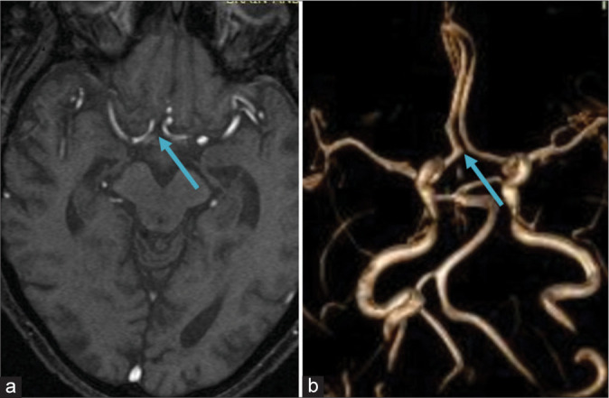
Objectives: Anatomy of circulus arteriosus cerebri (CAC) shows wide variation in different individuals, and population groups and has vital clinical significance in causation and presentation of clinical disease. The literature revealed a connection between the variations of CAC and cerebrovascular disease, ischemia, stroke, aneurysms, and atherosclerosis.
Material and methods: In this study, 513 patients without clinical manifestation in regard to cerebrovascular diseases, who are considered healthy on CAC anatomy, are included. Patients were instructed by clinicians for head imagery with magnetic resonance angiography examination during 2016-2017 periods.
Results: After statistical analysis, 43.27% were male while 56.72% female, 39% were younger than 40 years old. Age interval lies from 11 to 84 years old, mean age 46. The most common variations or 9.74% is when communicant anterior artery absence and absence of both posterior communicant arteries (Type G*/E) more rarely is H*/G (0.2%), G*/D (1.75%), G*/G (0.6%), H*/D (0.4%), H*/E (3.39%), H*/H (0.4%), J*/E (0.6%), while combination J*/D, J*/G, J*/H, G*/H not found. The most often combination is absence of anterior communicant artery and absence of both posterior communicant artery (Type G*/E), more in male 10.36% than female 9.6%.
Conclusion: The CAC is considered to play a critical role in preventing future stroke events in patients with absent of any of the arteries. Knowledge on variations in arteries forming the CAC is with clinical significance, as it is one of the components of CAC which stabilizes cerebral blood flow when principle conduits fail. Knowing the structure of arteries provide clinical knowledge to the surgeons before planning neurovascular surgeries.
Keywords: Cerebral artery; Circulus arteriosus cerebri; Clinical importance; Interventional of cerebral artery; Variations of circulus arteriosus cerebri.
© 2020 Published by Scientific Scholar on behalf of Journal of Clinical Imaging Science.
Conflict of interest statement
There are no conflicts of interest.
Figures


Similar articles
-
The anatomical variation of the circulus arteriosus cerebri in a cadaver cohort representing the population dynamics of the Western Cape.Br J Neurosurg. 2018 Feb;32(1):61-67. doi: 10.1080/02688697.2017.1374348. Epub 2017 Sep 5. Br J Neurosurg. 2018. PMID: 28870111
-
Human cerebral blood supply via circulus arteriosus cerebri: A scoping review on its variations and clinical implications.Heliyon. 2024 Jun 8;10(12):e32648. doi: 10.1016/j.heliyon.2024.e32648. eCollection 2024 Jun 30. Heliyon. 2024. PMID: 38975214 Free PMC article.
-
Morphological examination of the circulus arteriosus cerebri humani (circle of Willis). I. Anterior and posterior communicating arteries.Kaibogaku Zasshi. 1989 Oct;64(5):481-9. Kaibogaku Zasshi. 1989. PMID: 2618572
-
[The correlation of the parameters of the circulus arteriosus cerebri and of the main cerebral arteries and their variability in circulus arteriosus anomalies].Zh Vopr Neirokhir Im N N Burdenko. 1992 Jul-Oct;(4-5):29-33. Zh Vopr Neirokhir Im N N Burdenko. 1992. PMID: 1334310 Russian.
-
Review of the Anatomy of the Distal Anterior Cerebral Artery and Its Anomalies.Turk Neurosurg. 2016;26(5):653-61. doi: 10.5137/1019-5149.JTN.14294-15.1. Turk Neurosurg. 2016. PMID: 27337235 Review.
References
-
- Caplan LR. United Kingdom: Blackwell; 1996. Posterior Circulation Cerebrovascular Syndromes-UpToDate. Available from: https://www.uptodate.com/contents/posterior-circulation-cerebrovascular-... [Last accessed on 2018 Mar 02]
-
- Tanaka H, Fujita N, Enoki T, Matsumoto K, Watanabe Y, Murase K, et al. Relationship between variations in the circle of Willis and flow rates in internal carotid and basilar arteries determined by means of magnetic resonance imaging with semiautomated lumen segmentation: Reference data from 125 healthy volunteers. AJNR Am J Neuroradiol. 2006;27:1770–5. - PMC - PubMed
LinkOut - more resources
Full Text Sources
Other Literature Sources