Case Reports
. 2021 Feb 25:11:11.
doi: 10.25259/JCIS_165_2020. eCollection 2021.

Percutaneous Transhepatic Cholangioscopy and Stone Extraction in a Patient with Recurrent Cholangitis Following Liver Trauma

Affiliations
Case Reports

Percutaneous Transhepatic Cholangioscopy and Stone Extraction in a Patient with Recurrent Cholangitis Following Liver Trauma

Lee K Rousslang et al. J Clin Imaging Sci. .

Abstract

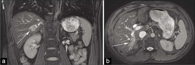
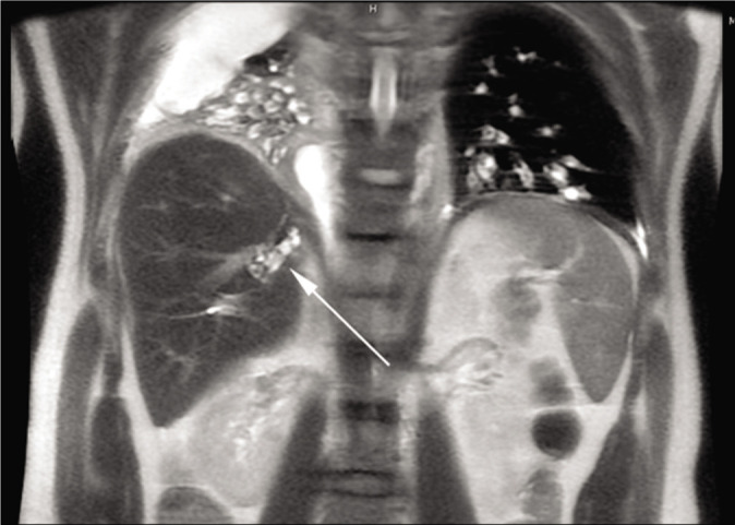
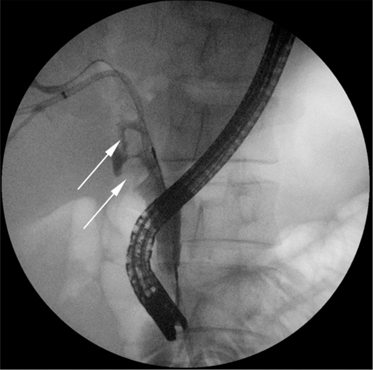
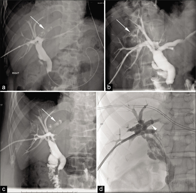
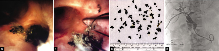
Percutaneous transhepatic cholangioscopy (PTCS) is a safe and effective treatment for obstructive biliary stones, when endoscopic retrograde cholangiopancreatography (ERCP) is unsuccessful or unavailable. Once percutaneous access is gained into the biliary tree by an interventional radiologist, the biliary ducts can be directly visualized and any biliary stones can be managed with lithotripsy, mechanical fragmentation, and/or percutaneous extraction. We report a case of a 45-year-old man who sustained a traumatic liver laceration and associated bile duct injury, complicated by bile duct ectasia and intrahepatic biliary stone formation. Despite undergoing a cholecystectomy, multiple ERCPs, and percutaneous transhepatic cholangiogram with drain placement, the underlying problem was not corrected leading to recurrent bouts of gallstone pancreatitis and cholangitis. He was ultimately referred to an interventional radiologist who extracted the impacted intrahepatic biliary stones that were thought to be causing his recurrent infections through cholangioscopy. This is the first case of PTCS with biliary stone extraction in the setting of recurrent biliary obstruction and cholangitis due to traumatic bile duct injury.

Keywords: Biliary endoscopy; Biliary lithotripsy; Choledochoscopy; Hepatolithiasis; Recurrent cholangitis.


Conflict of interest statement

There are no conflicts of interest.

Figures

Figure 1:
Figure 1:
A 37-year-old man with a remote history of a liver laceration presented with the right upper quadrant abdominal pain, nausea, and vomiting. Coronal (a) and axial (b) fluid-sensitive MR images demonstrate bile duct dilatation in the right lobe of the liver (arrows).
Figure 2:
Figure 2:
A 45-year-old man with recurrent right upper quadrant abdominal pain. Coronal magnetic resonance cholangiopancreatography demonstrates retained intrahepatic stones (arrow) in the right posterior liver.
Figure 3:
Figure 3:
A 45-year-old man with recurrent ascending cholangitis. Endoscopic retrograde cholangiopancreatography demonstrates multiple filling defects in the common bile duct (arrows), but the isolated segment of intrahepatic stones could not be localized.
Figure 4:
Figure 4:
A 45-year-old man with recurrent ascending cholangitis secondary to intrahepatic biliary stones that were not identified at endoscopic retrograde cholangiopancreatography. Intraprocedural fluoroscopic cholangiogram (a-d) demonstrates intrahepatic biliary stones (arrows). A flexible ureteroscope (arrowhead in d) is seen entering into the right hepatic duct.
Figure 5:
Figure 5:
A 45-year-old man with recurrent ascending cholangitis secondary to intrahepatic biliary stones that were not identified at endoscopic retrograde cholangiopancreatography. Endoscopic images reveal the impacted biliary stones (a), with subsequent percutaneous extraction of the stones (b and c). Percutaneous cholangiogram after stone retrieval (d) demonstrates clearing of the filling defects.

Similar articles

References

    1. Neuhaus H. Endoscopic and percutaneous treatment of difficult bile duct stones. Endoscopy. 2003;35:S31–4. doi: 10.1055/s-2003-41534. - DOI - PubMed
    1. Huibregtse K. Complications of endoscopic sphincterotomy and their prevention. N Engl J Med. 1996;335:961. doi: 10.1056/NEJM199609263351309. - DOI - PubMed
    1. Ray AA, Davies ET, Denstedt JD, Razvi H, Denstedt JD. The management of treatment-resistant biliary calculi using percutaneous endourologic techniques. Can J Surg. 2009;52:407–12. - PMC - PubMed
    1. Ponchon T, Genin G, Mitchell R, Henry L, Bory RM, Bodnar D, et al. Methods, indications and results of percutaneous choledochoscopy: A series of 161 procedures. Ann Surg. 1996;1:26–36. doi: 10.1097/00000658-199601000-00005. - DOI - PMC - PubMed
    1. Franzini T, Cardarelli-Leite L, De Moura EG, Morita F, Domingos FU, Carnevale FC, et al. SpyGlass percutaneous transhepatic cholangioscopy-guided lithotripsy of a large intrahepatic stone. Endoscopy. 2017;49:E292–3. doi: 10.1055/s-0043-117943. - DOI - PubMed

Publication types

LinkOut - more resources