Review
. 2021 Feb 19;13(2):e13448.
doi: 10.7759/cureus.13448.

Accessory Vertebral Artery: An Embryological Review With Translation from Adachi

Affiliations
Review

Accessory Vertebral Artery: An Embryological Review With Translation from Adachi

Stephen J Bordes et al. Cureus. .

Abstract

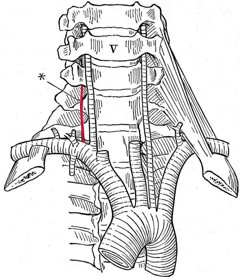
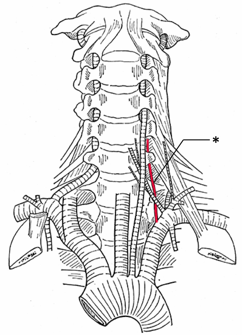
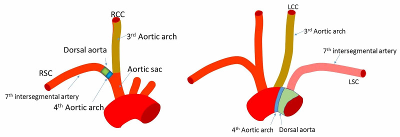
The vertebral arteries arise from the posterior superior aspect of the bilateral subclavian arteries and course superiorly through the transverse foramina of C1-C6 vertebrae before joining one another along the anterior surface of the pons. Developmental variations during the fourth to sixth weeks of embryonic development may result in the formation of accessory vertebral arteries, i.e., ipsilateral vertebral arteries of dual origin. This anatomical variation is distinct from and often confused with vertebral artery duplications and fenestrations. This article reviews the anatomy and embryology of the accessory vertebral artery with excerpts from Buntaro Adachi's classic text on vascular anatomical variations. Knowledge of accessory vertebral vessels is important during vascular and spinal procedures of the head and neck. Furthermore, these variations have been associated with cerebrovascular pathologies, such as stroke, dissection, and other hemodynamic anomalies.

Keywords: accessory vertebral artery; buntaro adachi; embryology; vascular surgery.


Conflict of interest statement

The authors have declared that no competing interests exist.

Figures

Figure 1
Figure 1. Coronal CTA of two patients with accessory vertebral arteries (right arrows)
Both patients also have an aortic arch origin of the left vertebral artery (left arrows). CTA: CT Angiogram
Figure 2
Figure 2. Arteriogram of patient with accessory vertebral artery (left arrow) and left aortic origin of the left vertebral artery (right arrow)
Note that both arteries feed into the intracranial blood supply indicating an anastomosis between the two vessels.
Figure 3
Figure 3. Cadaveric specimen noting a normal right vertebral artery (RVA) and a left accessory vertebral artery (LAVA) with aortic arch origin of the left vertebral artery (LVA)
Figure 4
Figure 4. CTA noting a duplicated vertebral artery (arrows) with normal vertebral artery (asterisks)
CTA: CT Angiogram
Figure 5
Figure 5. Arteriogram noting a fenestrated left vertebral artery (arrow) at the vertebrobasilar junction
The left vertebral artery is noted with asterisks. Also, note the basilar artery (BA) and right vertebral artery (RVA)
Figure 6
Figure 6. Left accessory vertebral artery (asterisk) and left-sided aortic arch origin of the left vertebral artery (After Adachi)
Figure 7
Figure 7. Right accessory vertebral artery (asterisk) with left-sided aortic arch origin of the left vertebral artery (After Adachi)
Figure 8
Figure 8. Left accessory vertebral artery (asterisk) and left-sided aortic arch origin of the left vertebral artery (After Adachi)
Figure 9
Figure 9. Left accessory vertebral artery (red) and left-sided aortic arch origin of the left vertebral artery. Note the relationship between the accessory vertebral artery and thoracic duct colored in black (After Adachi)
Figure 10
Figure 10. Development of the vertebral artery
Left: During the fetal period, along with the development of the pharyngeal arches (not shown), six aortic arches connect the aortic sac and right and left dorsal aortae to supply each pharyngeal arch. Right: The first and second aortic arches regress and almost disappear. The fifth aortic arch becomes a remnant and the sixth aortic arch becomes the pulmonary artery
Figure 11
Figure 11. The third aortic arch becomes the right and left common and internal carotid (not shown) arteries
The left fourth aortic arch becomes the aortic arch, and the right becomes a part of the subclavian artery. The blue green area (Left) would be the origin of the subclavian artery where the right vertebral artery arises. The blue green area (Right) would be the origin of an aortic arch origin of the left vertebral artery. The left normal vertebral or left accessory vertebral artery would arise from the left subclavian artery here shown in pink as derived from the 7th intersegmental artery. RSC, right subclavian artery; RCC, right common carotid artery; LCC, left common carotid artery; LSC, left subclavian artery
Figure 12
Figure 12. The dorsal aortae (DA) gives rise to dorsal (DB) and ventral (VB) branches
Dorsal branches are called intersegmental arteries (30 pairs) and the seventh intersegmental artery becomes the subclavian artery. Intersegmental arteries in the neck usually regress and the upper and lower intersegmental arteries anastomose longitudinally to form new vessels. The vertebral artery is formed by a connection between the postcostal (PostC) anastomosis formed by dorsal branches of the first to sixth intersegmental arteries and seventh intersegmental artery that becomes the subclavian artery. In a similar manner, the thyrocervical trunk and costocervical trunk are formed by precostal (PreC) and posttransverse (PostT) anastomoses, respectively
Figure 13
Figure 13. Table from Adachi’s work illustrating, in schematic form, the various accessory vertebral arteries that he documented (After Adachi)

Similar articles

Cited by

References

    1. Aortic arch origin of the left vertebral artery: an anatomical and radiological study with significance for avoiding complications with anterior approaches to the cervical spine. Tardieu G, Edwards B, Alonso F, et al. Clin Anat. 2017;30:811–816. - PubMed
    1. A case of duplicated right vertebral artery. Motomura M, Watanabe K, Tabira Y, et al. Kurume Med J. 2018;64:69–73. - PubMed
    1. Aberrant origin of vertebral artery and its clinical implications. Yuan SM. Braz J Cardiovasc Surg. 2016;31:52–59. - PMC - PubMed
    1. Vertebral artery anatomical variations as they relate to cervical transforaminal epidural steroid injections. Gitkind AI, Olson TR, Downie SA. Pain Med. 2014;15:1109–1114. - PubMed
    1. Variations of transverse foramina in cervical vertebrae: what happens to the vertebral artery? Zibis A, Mitrousias V, Galanakis N, Chalampalaki N, Arvanitis D, Karantanas A. Eur Spine J. 2018;27:1278–1285. - PubMed

LinkOut - more resources