Dermatoscopy of Cutaneous Granulomatous Disorders
- PMID: 33768021
- PMCID: PMC7982032
- DOI: 10.4103/idoj.IDOJ_543_20
Dermatoscopy of Cutaneous Granulomatous Disorders
Abstract
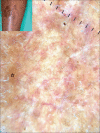
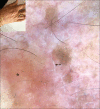
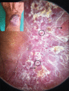
Cutaneous granulomatous disorders represent diseases with underlying granulomas on histology and are broadly divided into infectious and noninfectious disorders. Although histology is sine qua non in diagnosis of granulomatous disorders, lately dermoscopy has come up as a useful tool assisting in diagnosis of granulomatous disorder. Dermoscopy of granulomatous disorder is characterized by localized or diffuse, structureless yellowish-orange areas, along with vessels. Dermoscopic features of granulomatous disorders can be overlapping among various disorders, but detailed accurate assessment of various findings and their pattern may be useful in differentiating among them. In addition to this, peculiar dermatoscopic findings seen can also prove useful in distinguishing between various disorders. Hereby, we discuss dermatoscopic findings of various granulomatous disorders.
Keywords: Cutaneous granulomatous disorders; dermoscopy; granulomatous disorder.
Copyright: © 2021 Indian Dermatology Online Journal.
Conflict of interest statement
There are no conflicts of interest.
Figures






Similar articles
-
Dermoscopy of cutaneous granulomatous disorders: A study of 107 cases.Skin Res Technol. 2023 Jan;29(1):e13273. doi: 10.1111/srt.13273. Skin Res Technol. 2023. PMID: 36704887 Free PMC article.
-
Dermatoscopy of Granulomatous Disorders.Dermatol Clin. 2018 Oct;36(4):369-375. doi: 10.1016/j.det.2018.05.004. Epub 2018 Aug 2. Dermatol Clin. 2018. PMID: 30201146 Review.
-
Dermoscopy of Granuloma Annulare: A Clinical and Histological Correlation Study.Dermatology. 2017;233(1):74-79. doi: 10.1159/000454857. Epub 2017 Jan 19. Dermatology. 2017. PMID: 28099955
-
Dermatoscopic vascular patterns in cutaneous Merkel cell carcinoma.J Am Acad Dermatol. 2012 Jun;66(6):923-7. doi: 10.1016/j.jaad.2011.06.020. Epub 2011 Oct 5. J Am Acad Dermatol. 2012. PMID: 21978573
-
Clinical and Dermoscopic Features Associated With Difficult-to-Recognize Variants of Cutaneous Melanoma: A Systematic Review.JAMA Dermatol. 2020 Apr 1;156(4):430-439. doi: 10.1001/jamadermatol.2019.4912. JAMA Dermatol. 2020. PMID: 32101255
Cited by
-
Type I bare lymphocyte syndrome with novel TAP1 and TAP2 pathogenic variants.JAAD Case Rep. 2024 Jun 25;51:22-25. doi: 10.1016/j.jdcr.2024.05.042. eCollection 2024 Sep. JAAD Case Rep. 2024. PMID: 39345282 Free PMC article. No abstract available.
-
Dermoscopy of the Diverse Spectrum of Cutaneous Tuberculosis in the Skin of Color.Dermatol Pract Concept. 2022 Oct 1;12(4):e2022203. doi: 10.5826/dpc.1204a203. eCollection 2022 Nov. Dermatol Pract Concept. 2022. PMID: 36534549 Free PMC article.
-
Dermoscopy of Granuloma Faciale in the Skin of Color.Indian Dermatol Online J. 2022 Sep 5;13(5):686-687. doi: 10.4103/idoj.idoj_532_21. eCollection 2022 Sep-Oct. Indian Dermatol Online J. 2022. PMID: 36304640 Free PMC article. No abstract available.
-
Primary Cutaneous Histoplasmosis in an Immunocompetent Individual: A Rare Disease from a Dermoscopic Perspective.Indian Dermatol Online J. 2023 Nov 24;15(1):181-182. doi: 10.4103/idoj.idoj_290_23. eCollection 2024 Jan-Feb. Indian Dermatol Online J. 2023. PMID: 38282988 Free PMC article. No abstract available.
-
Dermoscopy in the diagnosis and assessment of treatment response in granulomatous cheilitis.BMJ Case Rep. 2022 Jun 24;15(6):e251200. doi: 10.1136/bcr-2022-251200. BMJ Case Rep. 2022. PMID: 35750429 Free PMC article. No abstract available.
References
-
- Tronnier M, Mitteldorf C. Histologic features of granulomatous skin diseases. Part 1: Non-infectious granulomatous disorders. J Dtsch Dermatol Ges. 2015;13:211–6. - PubMed
-
- Errichetti E, Stinco G. Dermatoscopy of Granulomatous Disorders. Dermatol Clin. 2018;36:369–75. - PubMed
-
- Errichetti E, Stinco G. The practical usefulness of dermoscopy in general dermatology. G Ital Dermatol Venereol. 2015;150:533–46. - PubMed
-
- Lallas A, Errichetti E, Ioannides D. Dermoscopy in General Dermatology. Boca Raton: CRC Press; 2019. [Last Accessed on 2020 Jul 09]. Available from: https://doi.org/10.1201/9781315201733 .
LinkOut - more resources
Full Text Sources

