. 2021 Feb 27:21:28-41.
doi: 10.1016/j.omtm.2021.02.019. eCollection 2021 Jun 11.

Cell-penetrating peptides enhance the transduction of adeno-associated virus serotype 9 in the central nervous system

Affiliations

Cell-penetrating peptides enhance the transduction of adeno-associated virus serotype 9 in the central nervous system

Yuan Meng et al. Mol Ther Methods Clin Dev. .

Erratum in

Abstract

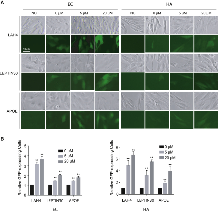
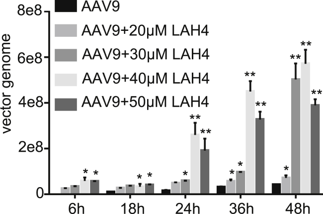
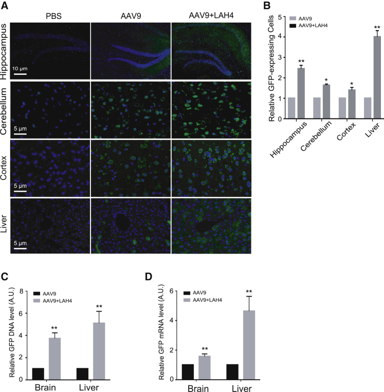
Recombinant adeno-associated viruses (rAAVs) have been widely used in the gene therapy field for decades. However, because of the challenge of effectively delivering rAAV vectors through the blood-brain barrier (BBB), their applications for treatment of central nervous system (CNS) diseases are quite limited. In this study, we found that several cell-penetrating peptides (CPPs) can significantly enhance the in vitro transduction efficiency of AAV serotype 9 (AAV9), a promising AAV vector for treatment of CNS diseases, the best of which was the LAH4 peptide. The enhancement of AAV9 transduction by LAH4 relied on binding of the AAV9 capsid to the peptide. Furthermore, we demonstrated that the LAH4 peptide increased the AAV9 transduction in the CNS in vitro and in vivo after systemic administration. Taken together, our results suggest that CPP peptides can interact directly with AAV9 and increase the ability of this AAV vector to cross the BBB, which further induces higher expression of target genes in the brain. Our study will help to improve the applications of AAV gene delivery vectors for the treatment of CNS diseases.

Keywords: adeno-associated virus serotype 9; blood-brain barrier; cell-penetrating peptides; central nervous system.


Conflict of interest statement

The authors declare no competing interests.

Figures

None
Graphical abstract
Figure 1
Figure 1
CPPs improve AAV9 transduction in HEK293T cells (A) Peptides of LAH4, LEPTIN30, and APOE increased AAV9 transduction to HEK293T cells. HEK293T cells were infected with AAV9/GFP (multiplicity of infection (MOI) of 1,000) alone or precomplexed without or with 5 or 20 μM LAH4, LEPTIN30, or APOE. Images were taken by an inverted fluorescence microscope (Nikon). (B) Quantification of green fluorescent protein (GFP) fluorescence level in (A). Error bars represent the standard error of the mean (SEM) from triplicate experiments. Data are means ± SEM (n = 3; >100 cells per experiment). ∗p < 0.05, ∗∗p < 0.01, by one-way ANOVA followed by a Turkey’s test.
Figure 2
Figure 2
CPPs improve AAV9 transduction in ECs and HAs (A) Peptides of LAH4, LEPTIN30, and APOE increased AAV9 transduction to ECs and HAs. ECs or HAs were infected with AAV9/GFP (MOI of 1,000) alone or precomplexed without or with 5 or 20 μM LAH4, LEPTIN30, or APOE. Images were taken by an inverted fluorescence microscope (Nikon). (B) Quantification of GFP-expressing cells in (A). Error bars represent the SEM from triplicate experiments. Data are means ± SEM (n = 3; >100 cells per experiment). ∗∗p < 0.01, by one-way ANOVA followed by a Turkey’s test.
Figure 3
Figure 3
CPPs improve the target gene expression in HEK293T cells, ECs, and HAs (A) Peptides of LAH4, LEPTIN30, and APOE increased GFP mRNA level in cells. HEK293 cells, ECs, or HAs were infected with AAV9-GFP (MOI of 1,000) alone or pre-complexed without or with 5 or 20 μM LAH4, LEPTIN30, or APOE. Total RNA was isolated from cells, and mRNA levels of GFP were assessed by quantitative polymerase chain reaction (qPCR). The 2−ΔΔCt method was used to calculate the relative GFP mRNA level. All of the data were then normalized by the mean percentage of GFP expression in AAV9 only cell. All error bars represent the SEM from triplicate experiments. Data are means ± SEM (n = 3). ∗p < 0.05, ∗∗p < 0.01, by one-way ANOVA followed by a Turkey’s test. (B) Peptides of LAH4, LEPTIN30, and APOE increased GFP protein level in cells. HEK293 cells, ECs, or HAs were infected with AAV9/GFP (MOI of 1,000) alone or precomplexed without or with 5 or 20 μM LAH4, LEPTIN30, or APOE. Total protein was extracted and GFP expression levels in three cell lines were determined by western blots. Ponceau S-stained total protein was used as a loading control. (C) Quantification of the level of GFP protein in (B). Error bars represent the SEM from triplicate experiments. Data are means ± SEM (n = 3). ∗p < 0.05, ∗∗p < 0.01, by one-way ANOVA followed by a Turkey’s test.
Figure 4
Figure 4
Colocalization of the LAH4 peptides and AAV9 particles in cells (A) AAV9 particles were incubated with FITC-CPP (green) and subsequently probed with a mouse monoclonal antibody specific for intact AAV (red). The nucleus is in blue and a single cell is shown. Overlapping green and red signals appear as yellow in the merged image. The image was taken by a Nikon confocal microscope. Arrows indicate colocalized AAV9 particles and LAH4 peptides. Scale bar represents 10 μm. (B) The AAV9-LAH4-FITC mixture was incubated without or with mouse serum and subsequently probed with a monoclonal antibody specific for intact AAV (red). Overlapping green and red signals appear as yellow in the merged image. The image was taken by Nikon confocal microscope. Scale bar represents 20 μm. (C) Quantification the colocalization of each cell in (B). n = 3; >50 cells per experiment.
Figure 5
Figure 5
Optimization of AAV9-LAH4 complex formation (A) Effect of incubation time on AAV9 transduction efficiency. Scale bar represents 40 μm. (B) Total RNA was isolated from cells in (A), and mRNA levels of GFP were assessed by qPCR. Data are means ± SEM (n = 3; one-way ANOVA followed by t test). ∗∗p < 0.01. (C) GFP protein level in ECs transduced with the AAV9-LAH4 complex formed after 0, 30, and 120 min. Total protein was extracted, and western blots were performed using anti-GFP antibody. The Ponceau S-stained total protein was used as a loading control. (D) Quantification of the protein level in (C). Data are means ± SEM (n = 3; one-way ANOVA followed by a t test). ∗∗p < 0.01. (E) Effect of incubation temperature on AAV9 transduction efficiency. Scale bar represents 40 μm. (F) Total RNA was isolated from cells incubated at 4°C, 25°C, or 37°C in (E), and mRNA levels of GFP were assessed by quantitative polymerase chain reaction. Data are means ± SEM (n = 3; one-way ANOVA followed by a t test). ∗p < 0.05, ∗∗p < 0.01. (G) GFP protein level in ECs transduced with the AAV9-LAH4 complex formed at 4°C, 25°C, or 37°C. Total protein was extracted and western blots were performed using anti-GFP antibody. The Ponceau S-stained total protein was used as a loading control. (H) Quantification of the protein level in (G). Data are means ± SEM (n = 3; one-way ANOVA followed by t test). ∗p < 0.05, ∗∗p < 0.01.
Figure 6
Figure 6
LAH4 increased the penetration of AAV9 through the BBB model in vitro hCMEC/D3 cells were cultured in a monolayer and incubated without or with LAH4 at concentration gradients of 20, 30, 40, and 50 μM. The media in the basal chamber were collected at different time points and the viral titer was analyzed by qPCR. All treatments were performed in triplicate. Data are means ± SEM (n = 3; one-way ANOVA followed by a t test). ∗p < 0.05, ∗∗p < 0.01, compared to cells with the AAV9 treatment.
Figure 7
Figure 7
The LAH4 peptide facilitates gene delivery of AAV9 in mouse brain and liver (A) The transgene expression in tissues from C57BL/6 mice injected with AAV9 or AAV9-LAH4 complexes. Tissues of the liver and brain (hippocampus, cerebellum, cortex) were taken at day 21 after tail vein injection and immunohistochemically stained with antibodies against GFP (green) and liver, respectively. Scale bars represent 5 or 10 μm. (B) Quantification of the relative number of positive cells in (A). Data are means ± SEM (n = 3; one-way ANOVA followed by a t test). ∗p < 0.05, ∗∗p < 0.01. (C) Genomic DNA was isolated from tissues of the liver and brain and GFP DNA was assessed by qPCR. The 2−ΔΔCt method was used to calculate the relative DNA level. Data are means ± SEM (n = 3; one-way ANOVA followed by a t test). ∗∗p < 0.01. (D) Total RNA was isolated from tissues of the liver and brain and mRNA levels of GFP were assessed by qPCR. The 2−ΔΔCt method was used to calculate the relative mRNA level. Data are means ± SEM (n = 3; one-way ANOVA followed by a t test). ∗∗p < 0.01.
Figure 8
Figure 8
AAV9-LAH4 complex leads to increased transduction in neurons The transgene expression in neuron cells from C57BL/6 mice injected with PBS, AAV9, or AAV9-LAH4 complexes was examined. (A and B) Tissues of the brain were taken at day 21 day after tail vein injection and immunohistochemically stained with antibodies against GFP (green color) and NeuN (red color) (A) or GFAP (red color) (B), respectively. Representative images of the GFP expression (green) of AAV9, NeuN (red), or GFAP (red) (B) were merged with DAPI (blue). Scale bars represent 10 μm.
Figure 9
Figure 9
The LAH4 peptide did not increase the immune response in the brain (A) Total RNA was isolated from tissues of the liver and brain, and mRNA levels of IL-1β and TNF-α were assessed by qPCR. The 2−ΔΔCt method was used to calculate the relative expression of IL-1β and TNF-α. All of the data were then normalized by the mean percentage of cytokine expression in mice injected with AAV9 only. Data are means ± SEM (n = 3; one-way ANOVA followed by a t test). ∗p < 0.05. (B) Analysis of T lymphocyte infiltration in brain tissues from C57BL/6 mice injected with AAV9 or AAV9-LAH4 complexes at 21 days. Tissues were immunohistochemically stained with antibodies against CD4 and CD8 (red). Scale bar represents 5 μm. (C) Quantification of the relative number of positive cells in (B). Data are means ± SEM (n = 3; one-way ANOVA followed by a t test). ∗p < 0.05.

References

    1. Berns K.I., Muzyczka N. AAV: an overview of unanswered questions. Hum. Gene Ther. 2017;28:308–313. - PMC - PubMed
    1. Wang D., Tai P.W.L., Gao G. Adeno-associated virus vector as a platform for gene therapy delivery. Nat. Rev. Drug Discov. 2019;18:358–378. - PMC - PubMed
    1. Gaudet D., Méthot J., Déry S., Brisson D., Essiembre C., Tremblay G., Tremblay K., de Wal J., Twisk J., van den Bulk N. Efficacy and long-term safety of alipogene tiparvovec (AAV1-LPLS447X) gene therapy for lipoprotein lipase deficiency: an open-label trial. Gene Ther. 2013;20:361–369. - PMC - PubMed
    1. Mingozzi F., High K.A. Therapeutic in vivo gene transfer for genetic disease using AAV: progress and challenges. Nat. Rev. Genet. 2011;12:341–355. - PubMed
    1. Büning H. Gene therapy enters the pharma market: the short story of a long journey. EMBO Mol. Med. 2013;5:1–3. - PMC - PubMed

LinkOut - more resources