. 2020;2(3):86-99.
doi: 10.33696/diabetes.1.024.

Improved Wound Closure Rates and Mechanical Properties Resembling Native Skin in Murine Diabetic Wounds Treated with a Tropoelastin and Collagen Wound Healing Device

Affiliations

Improved Wound Closure Rates and Mechanical Properties Resembling Native Skin in Murine Diabetic Wounds Treated with a Tropoelastin and Collagen Wound Healing Device

Robert S Kellar et al. J Diabetes Clin Res. 2020.

Abstract

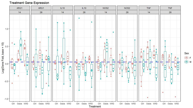
Chronic wounds in patients suffering from type II diabetes mellitus (DMII) where wounds remain open with a complicated pathophysiology, healing, and recovery process is a public health concern. Normal wound healing plays a critical role in wound closure, restoration of mechanical properties, and the biochemical characteristics of the remodeled tissue. Biological scaffolds provide a tissue substitute to help facilitate wound healing by mimicking the extracellular matrix (ECM) of the dermis. In the current study an electrospun biomimetic scaffold, wound healing device (WHD), containing tropoelastin (TE) and collagen was synthesized to mimic the biochemical and mechanical characteristics of healthy human skin. The WHD was compared to a commercially available porcine small intestinal submucosa (SIS) matrix that has been used in both partial and full-thickness wounds, Oasis® Wound Matrix. Using a diabetic murine model C57BKS.Cg-m+/+Leprdb/J mice (db/db) wound closure rates, histochemistry (CD31 and CD163), qPCR (GAPDH, TNF-α, NOS2, ARG1 and IL10), and mechanical testing of treated wound sites were evaluated. The WHD in a splinted, full thickness, diabetic murine wound healing model demonstrated skin organ regeneration, an enhanced rate of wound closure, decreased tissue inflammation, and a stronger and more durable remodeled tissue that more closely mimics native unwounded skin compared to the control device.

Keywords: Diabetic wound; Elastin; Electrospinning; Tropoelastin; Wound healing device.


Figures

Figure 1:
Figure 1:
Left Uniaxial Testing Device (UTTD). Right, Assembled device and graphic user interface.
Figure 2:
Figure 2:
Polynomial regression analysis among treatment groups over 28 days. Percent wound closure was standardized to day 0 for each succeeding measurement. Shown values are means ± SEM.
Figure 3:
Figure 3:
Gross images of full thickness splinted wounds in a diabetic murine model. Days 6–16 of healing demonstrate accelerated wound closure in the WHD treatment group compared to the Oasis Matrix Scale: each line = 1 mm.
Figure 4:
Figure 4:
Hematoxylin and Eosin stained samples from WHD and Oasis Wound Matrix-treated wound sites after 28 days of remodeling. WHD-treated wound sites demonstrated wound regeneration where the skin organ and tissue had evidence of functional restoration with the presence of hair follicles (yellow arrows). Scale bars = 600 μm.
Figure 5:
Figure 5:
Analysis of elastin positive staining percentage between day 14 and 28. Asterisk represents significantly different group means where p=0.0003. Comparison was conducted using a two-sided t-test with equal variance (Levenne’s Test; p=0.08). Means are illustrated by crosses.
Figure 6:
Figure 6:
Microvessel density (CD31) faceted by day, treatment, and sex.
Figure 7:
Figure 7:
CD31 reacted tissues used to quantify the presence of microvasculature in the wounded tissue sites. Both treatments had a large amount of microvasculature present at 14 days (Left). While there was no significant difference in the amount of microvasculature measured per square micron, the area measured in the WHD was smaller due to the progression of wound healing in the 28-day samples (Right). Macro-image scale bars = 1 mm, micro-image scale bars = 200 μm.
Figure 8:
Figure 8:
CD163 positive stain count compared across treatments. Compact letter display illustrates significant differences between treatments when p-value <0.05. WHD treatment had significantly less macrophage presence during healing compared to Oasis.
Figure 9.
Figure 9.
Phase I Immunohistochemistry (CD163) highlighting tissue macrophage presence in remodeled wounds at 14 days (left) and 28 days (right). The WHD demonstrated lower levels of tissue macrophage presence compared to Oasis.
Figure 10:
Figure 10:
Treatment differences in gene expression faceted by sex, treatment, day, and gene. No significant differences were noted.
Figure 11:
Figure 11:
Stress vs. Strain analysis of excised murine skin for each treatment. Lines represent spline regression models with 95% confidence intervals. Data was divided into 3 intervals x<0.2, 0.2 ≤ x ≤ 0.3, and x>0.3 applying a cubic prediction model to each section. Although not statistically significant, due to the amount of variation introduced as strain increases, WHD withstood more than 0.4 MPa of stress before yielding compared to control. NW group: Non-wounded skin.

References

    1. Frykberg RG, Banks J. Challenges in the treatment of chronic wounds. Advances in Wound Care. 2015. September 1;4(9):560–82. - PMC - PubMed
    1. Sen CK. Human wounds and its burden: an updated compendium of estimates. Advances in Wound Care. 2019. February 14;8(2):39–48. - PMC - PubMed
    1. Järbrink K, Ni G, Sönnergren H, Schmidtchen A, Pang C, Bajpai R, et al. The humanistic and economic burden of chronic wounds: a protocol for a systematic review. Systematic Reviews. 2017. December 1;6(1):15. - PMC - PubMed
    1. Falanga V Wound healing and its impairment in the diabetic foot. The Lancet. 2005. November 12;366(9498):1736–43. - PubMed
    1. New CD. report: more than 100 million Americans have diabetes or prediabetes. Centers for Disease Control and Prevention; 2017. Available from: https://www.cdc.gov/media/releases/2017/p0718-diabetes-report.html. 2017.

LinkOut - more resources