. 2021 Jul 19;33(6):1961-1979.
doi: 10.1093/plcell/koab091.

Arabidopsis cryptochrome 1 controls photomorphogenesis through regulation of H2A.Z deposition

Affiliations

Arabidopsis cryptochrome 1 controls photomorphogenesis through regulation of H2A.Z deposition

Zhilei Mao et al. Plant Cell. .

Abstract

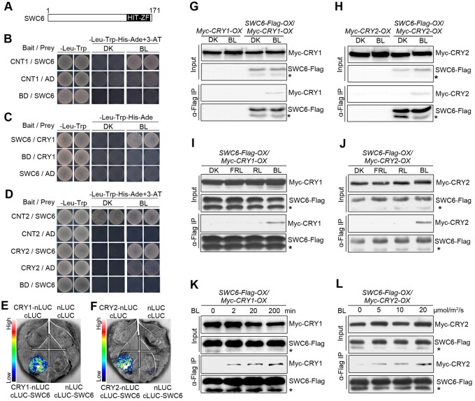
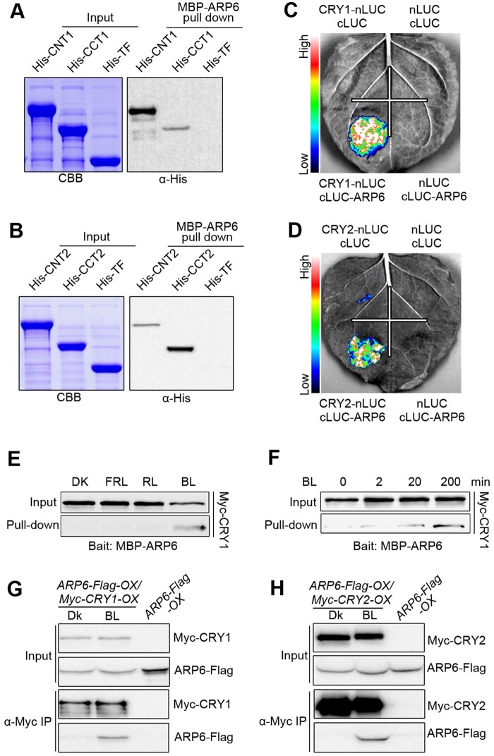
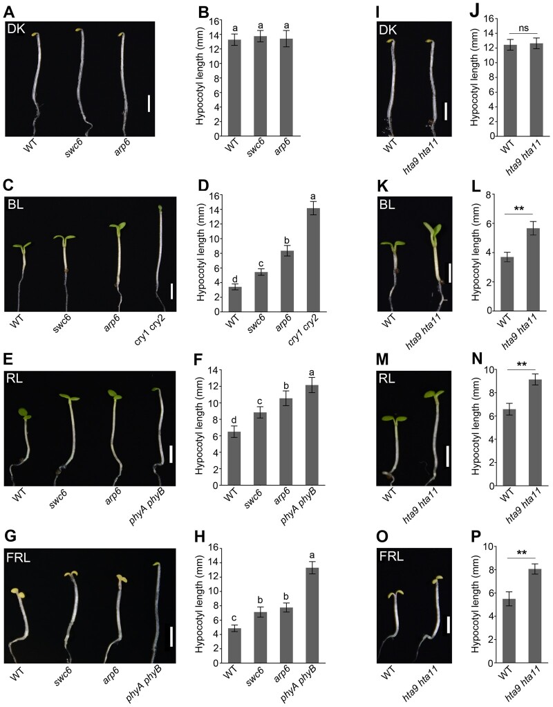
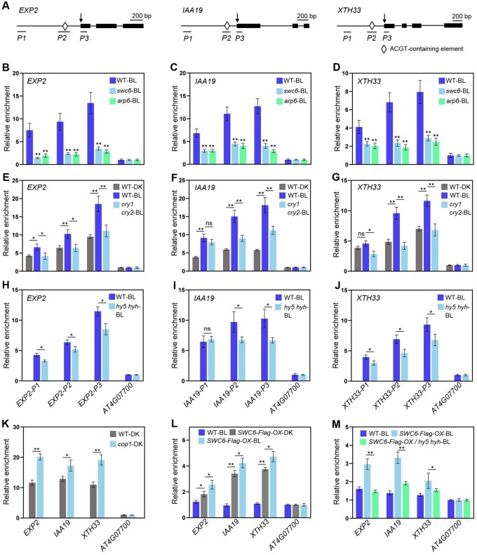
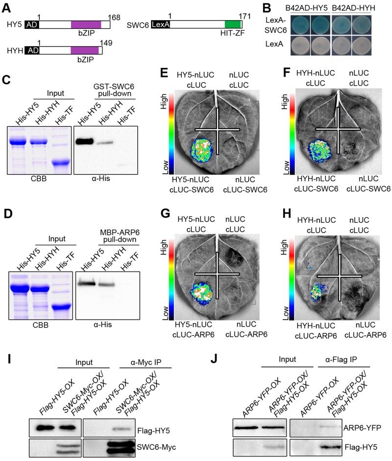
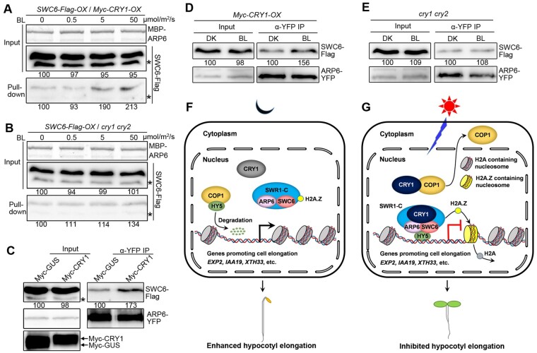
Light is a key environmental cue that fundamentally regulates plant growth and development, which is mediated by the multiple photoreceptors including the blue light (BL) photoreceptor cryptochrome 1 (CRY1). The signaling mechanism of Arabidopsis thaliana CRY1 involves direct interactions with CONSTITUTIVE PHOTOMORPHOGENIC 1 (COP1)/SUPPRESSOR OF PHYA-105 1 and stabilization of COP1 substrate ELONGATED HYPOCOTYL 5 (HY5). H2A.Z is an evolutionarily conserved histone variant, which plays a critical role in transcriptional regulation through its deposition in chromatin catalyzed by SWR1 complex. Here we show that CRY1 physically interacts with SWC6 and ARP6, the SWR1 complex core subunits that are essential for mediating H2A.Z deposition, in a BL-dependent manner, and that BL-activated CRY1 enhances the interaction of SWC6 with ARP6. Moreover, HY5 physically interacts with SWC6 and ARP6 to direct the recruitment of SWR1 complex to HY5 target loci. Based on previous studies and our findings, we propose that CRY1 promotes H2A.Z deposition to regulate HY5 target gene expression and photomorphogenesis in BL through the enhancement of both SWR1 complex activity and HY5 recruitment of SWR1 complex to HY5 target loci, which is likely mediated by interactions of CRY1 with SWC6 and ARP6, and CRY1 stabilization of HY5, respectively.


Figures

Figure 1
Figure 1
CRY1 and CRY2 interact with SWC6 in a BL-dependent manner. A, Schematic diagram depicting the HIT-ZF domain of SWC6 protein. B, Yeast two-hybrid assays showing the interaction of CNT1 with SWC6. Yeast cells coexpressing the indicated combinations of constructs were grown on SD –Trp–Leu or SD–Trp–Leu–His–Ade medium with 10 mM 3-AT in continuous DK or BL (30 μmol/m2/s). BD, GAL4 DNA-binding domain; AD, GAL4 DNA-activation domain . C and D, Yeast two-hybrid assays showing the interactions of CRY1 with SWC6 (C), and CNT2 and CRY2 with SWC6 (D). E and F, Split-LUC assays indicating the interactions of CRY1 and CRY2 with SWC6 in N. benthamiana cells. G and H, Co-IP assays showing BL-induced interactions of CRY1 and CRY2 with SWC6 in Arabidopsis. Myc-CRY1-OX or Myc-CRY2-OX or SWC6-Flag-OX/Myc-CRY1-OX or SWC6-Flag-OX/Myc-CRY2-OX seedlings were adapted in DK for 2 d and then exposed to BL (30 μmol/m2/s for CRY1 and 20 μmol/m2/s for CRY2) or still adapted in DK for 1 h, followed by IP with an anti-Flag antibody. The IP (SWC6) and co-IP signals (CRY1 and CRY2) were detected by immunoblots probed with anti-Flag and -Myc antibodies. Asterisks denote truncated SWC6-Flag protein. I and J, Co-IP assay showing BL-specific interactions of CRY1 and CRY2 with SWC6 in Arabidopsis. SWC6-Flag-OX/Myc-CRY1-OX or SWC6-Flag-OX/Myc-CRY2-OX seedlings were adapted in DK for 2 d and then exposed to BL (20 μmol/m2/s) or RL (20 μmol/m2/s) or FRL (5 μmol/m2/s) light or still adapted in DK for 1 h, followed by IP with anti-Flag antibody. K, Co-IP assay showing that CRY1–SWC6 interaction is enhanced in response to increasing exposure times of BL. SWC6-Flag-OX/Myc-CRY1-OX seedlings were adapted in DK for 2 d and then exposed to BL (30 μmol/m2/s) for different times. L, Co-IP assay showing that CRY2–SWC6 interaction is enhanced in response to increasing intensity of BL. SWC6-Flag-OX/Myc-CRY2-OX seedlings were adapted in DK for 2 d and then exposed to different fluence rates of BL for 1 h.
Figure 2
Figure 2
CRY1 and CRY2 interact with ARP6 in a BL-dependent manner. A and B, In vitro pull-down assays showing the interactions of CNT1 and CCT1 with ARP6 (A), and CNT2 and CCT2 with ARP6 (B). MBP-ARP6 protein served as bait. His-TF, His-TF-CNT1, -CCT1, -CNT2, and -CCT2 served as prey. His-TF was used as negative control. The pulled-down proteins were detected with anti-His antibody. C and D, Split-LUC assays indicating the interactions of CRY1 (C) and CRY2 (D) with ARP6 in N. benthamiana cells. E, Semi-in vivo pull-down assay showing BL-specific interaction of CRY1 with ARP6. Preys were protein extracts from Myc-CRY1-OX seedlings that were DK-adapted for 2 d and then exposed to BL (30 μmol/m2/s) or RL (30 μmol/m2/s) or FRL (5 μmol/m2/s) light or still adapted in DK for 1 h. F, Semi-in vivo pull-down assay showing that CRY1–ARP6 interaction is enhanced in response to increasing exposure times of BL. Preys were protein extracts from Myc-CRY1-OX seedlings that were DK-adapted for 2 d and then exposed to BL (30 μmol/m2/s) for different times. G and H, Co-IP assays showing BL-induced interactions of CRY1 and CRY2 with ARP6 in Arabidopsis. Myc-CRY1-OX Myc-CRY2-OX or ARP6-Flag-OX/Myc-CRY1-OX or ARP6-Flag-OX/Myc-CRY2-OX seedlings were adapted in DK for 2 d and then exposed to BL (30 μmol/m2/s) or still adapted in DK for 1 h, followed by IP with anti-Flag antibody. The IP (ARP6) and co-IP signals (CRY1 and CRY2) were detected by immunoblots probed with anti-Flag and -Myc antibodies.
Figure 3
Figure 3
SWC6, ARP6, and H2A.Z act to inhibit hypocotyl elongation in BL, RL, and FRL. A–H, The swc6, and arp6 mutants show enhanced hypocotyl elongation in BL, RL, and FRL. Seedlings of the indicated genotypes were grown in DK (A), BL (30 μmol/m2/s) (C), RL (50 μmol/m2/s) (E), and FRL (1 μmol/m2/s) (G) for 5 d, and hypocotyl lengths were measured (B, D, F, and H). Bars = 2.5 mm. Letters “a” to “d” indicate statistically significant differences between means for hypocotyl lengths of the indicated genotypes, as determined by ANOVA, followed by LSD test (P < 0.05). I–P, The hta9 hta11 mutant shows enhanced hypocotyl elongation in BL, RL, and FRL. The growth conditions are the same as those in (A–H). Bars = 2.5 mm. Asterisks indicate significant differences between WT and hta9 hta11 seedlings (Student’s t-test: **P < 0.01), and ns denotes no significant differences.
Figure 4
Figure 4
CRYs, ARP6, and HY5/HYH coregulate a large number of genes in the same direction. A, Venn diagrams showing the overlapping genes up- and down-regulated by CRYs and ARP6, which were obtained by analyses of differential expression (FC > 1.5 and P-value < 0.05) from samples of BL-grown WT and cry1 cry2 mutant, and BL-grown WT and arp6 mutant, respectively. B, Hierarchical clustering analyses of the overlapping genes shown in A. Scale bar denotes the log2 value of FC. C, GO analysis showing coordinate regulation of the 1,879 overlapping genes in A by CRYs and ARP6. The numbers on each column denote the percentage of genes in each GO category. Total denotes all Arabidopsis genes. D, Venn diagrams showing the overlapping genes up- and down-regulated by ARP6 and HY5/HYH, which were obtained by analyses of differential expression (FC > 1.5 and P-value < 0.05) from samples of BL-grown WT and arp6 mutant, and BL-grown WT and hy5 hyh mutant, respectively. E, Hierarchical clustering analyses of the overlapping genes shown in D. F, GO analysis showing coordinate regulation of the 2,051 overlapping genes in D by ARP6 and HY5/HYH. The numbers on each column denote the percentage of genes in each GO category.
Figure 5
Figure 5
ChIP-qPCR analyses showing the roles of CRYs, ARP6, SWC6, and HY5/HYH in H2A.Z deposition at HY5 target genes. A, Schematic diagram of EXP2, IAA19, and XTH33 genes with exons indicated as black boxes. Arrowheads denote the transcription start sites. P1–P3 denote the corresponding amplicons for qPCR. B–D, ARP6 and SWC6 are necessary for H2A.Z deposition at +1 nucleosomes of EXP2 (B), IAA19 (C) and XTH33 (D) loci. E–G, CRYs mediate BL-enhanced H2A.Z deposition at the HY5 Target Genes in BL. H–J, HY5 and HYH promote H2A.Z deposition at their target genes in BL. K, H2A.Z deposition at +1 nucleosomes of HY5 target loci is enhanced in cop1-4 mutant. L and M, SWC6 binding to the HY5 target genes promoting cell elongation is dependent on HY5 and HYH in BL. In B–J, L, and M, 5-d-old white light-grown seedlings were adapted in DK for 2 d, and then either exposed to BL (50 μmol/m2/s) or still adapted in DK for 6 h. In K, 5-d-old white light-grown WT and cop1 seedlings were adapted in DK for 2 d, and then harvested and cross-linked. The +1 nucleosome fragments of EXP2, IAA19, XTH33, and AT4G07700 (negative control) were immunoprecipitated by anti-HTA9 or anti-Flag antibody, and then qPCR was performed to quantify the enrichment of the indicated genes, which was normalized to that of AT4G07700. Error bars, SDs of three biological replicates. Asterisks indicate significant differences between different genotypes of plants subjected to different treatments (Student’s t-test: **P < 0.01, *P < 0.05).
Figure 6
Figure 6
SWC6 and ARP6 interact with HY5 and HYH. A, Schematic diagram of HY5, HYH, and SWC6 proteins tested in LexA yeast two-hybrid assays. bZIP denotes basic leucine zipper domain. B, LexA yeast two-hybrid assays showing interactions of SWC6 with HY5 and HYH. Yeast cells coexpressing the indicated combinations of constructs were grown on SD –Trp–His–Ura with X-gal. The blue precipitates on the plates represent the GUS activities. C and D, In vitro pull-down assays showing the interactions of HY5 and HYH with SWC6 (C) and ARP6 (D). GST-SWC6 and MBP-ARP6 protein served as baits. His-TF, His-TF-HY5, and -HYH served as preys. Input images show CBB staining. The pulled-down proteins were detected with anti-His antibody. E–H, Split-LUC assays indicating the interactions of SWC6 with HY5 (E) and HYH (F), and the interactions of ARP6 with HY5 (G) and HYH (H). I and J, Co-IP assays showing the interactions of HY5 with SWC6 (I) and ARP6 (J) in Arabidopsis. Flag-HY5-OX and SWC6-Myc-OX/Flag-HY5-OX seedlings were grown in BL (30 μmol/m2/s) for 7 d, followed by IP with anti-Myc antibody. The IP (SWC6) and co-IP signals (HY5) were detected by immunoblots probed with anti-Myc and -Flag antibodies (I). ARP6-YFP-OX and ARP6-YFP-OX/Flag-HY5-OX seedlings were grown in BL (30 μmol/m2/s) for 7 d, followed by IP with anti-Flag antibody. The IP (HY5) and co-IP signals (ARP6) were detected by immunoblots probed with anti-Flag and -YFP antibodies (J).
Figure 7
Figure 7
CRY1 likely enhances the association of SWC6 with ARP6. A and B, Semi-in vivo pull-down assays showing that CRY1 may promote SWC6–ARP6 interaction in a BL fluence rate-dependent manner. MBP-ARP6 protein served as bait. SWC6-Flag protein extracts prepared from SWC6-Flag-OX/Myc-CRY1-OX and SWC6-Flag-OX/cry1 cry2 seedlings adapted in DK for 2 d and then exposed to different fluence rates of BL for 1 h served as preys. C, Co-IP assays showing that CRY1 may promote the interaction of SWC6 with ARP6 in N. benthamiana. SWC6-Flag and ARP6-YFP were coexpressed with Myc-GUS or Myc-CRY1 in N. benthamiana leaves, respectively. ARP6-YFP served as bait, and SWC6-Flag served as prey. Myc-GUS and Myc-CRY1 were detected with anti-Myc antibody. D and E, Co-IP assays showing CRY1 promotion of the association of SWC6 with ARP6 by BL in Arabidopsis protoplasts. SWC6-Flag and ARP6-YFP were coexpressed in Myc-CRY1-OX (D) and cry1 cry2 protoplasts (E). The transformed protoplasts were DK-adapted for 16 h, and then exposed to BL (30 μmol/m2/s) or still adapted to DK for 1 h. SWC6-Flag served as prey and ARP6-YFP served as bait. F and G, A model illustrating how CRY1 mediates regulation of H2A.Z deposition at HY5 target loci. In DK, CRY1 is inactive and unable to interact with COP1 and SWC6/ARP6 to regulate their activities. COP1 is fully functional, while SWR1 complex is not. HY5 undergoes ubiquitination and degradation, and SWR1 complex can hardly be recruited to HY5 target loci to mediate H2A.Z deposition, leading to active expression of HY5 target genes promoting cell elongation and enhanced hypocotyl elongation (F). Upon BL irradiation, CRY1 is activated, and interacts with ARP6 and SWC6 to enhance ARP6–SWC6 interaction, leading to enhanced SWR1 complex activity. At the same time, CRY1 interacts with COP1 to inhibit its activity and promote its translocation from nucleus to cytoplasm, leading to promotion of HY5 accumulation and enhanced recruitment of SWR1 complex to HY5 target loci. These two pathways work together to enhance SWR1 complex-mediated H2A.Z deposition at HY5 target genes to regulate their expression and mediate CRY1 inhibition of hypocotyl elongation (G).

References

    1. Ahmad M, Cashmore AR (1993) HY4 gene of A. thaliana encodes a protein with characteristics of a blue-light photoreceptor. Nature 366: 162–166 - PubMed
    1. Ang LH, Chattopadhyay S, Wei N, Oyama T, Okada K, Batschauer A, Deng XW (1998) Molecular interaction between COP1 and HY5 defines a regulatory switch for light control of Arabidopsis development. Mol Cell 1: 213–222 - PubMed
    1. Briggs WR, Christie JM (2002) Phototropins 1 and 2: versatile plant blue-light receptors. Trends Plant Sci 7: 204–210 - PubMed
    1. Bruggemann E, Handwerger K, Essex C, Storz G (1996) Analysis of fast neutron-generated mutants at the Arabidopsis thaliana HY4 locus. Plant J 10: 755–760 - PubMed
    1. Burko Y, Seluzicki A, Zander M, Pedmale UV, Ecker JR, Chory J (2020) Chimeric activators and repressors define HY5 activity and reveal a light-regulated feedback mechanism. Plant Cell 32: 967–983 - PMC - PubMed

Publication types

MeSH terms