. 2021 Oct 22;144(9):2863-2878.
doi: 10.1093/brain/awab141.

Kcnq2/Kv7.2 controls the threshold and bi-hemispheric symmetry of cortical spreading depolarization

Affiliations

Kcnq2/Kv7.2 controls the threshold and bi-hemispheric symmetry of cortical spreading depolarization

Isamu Aiba et al. Brain. .

Abstract

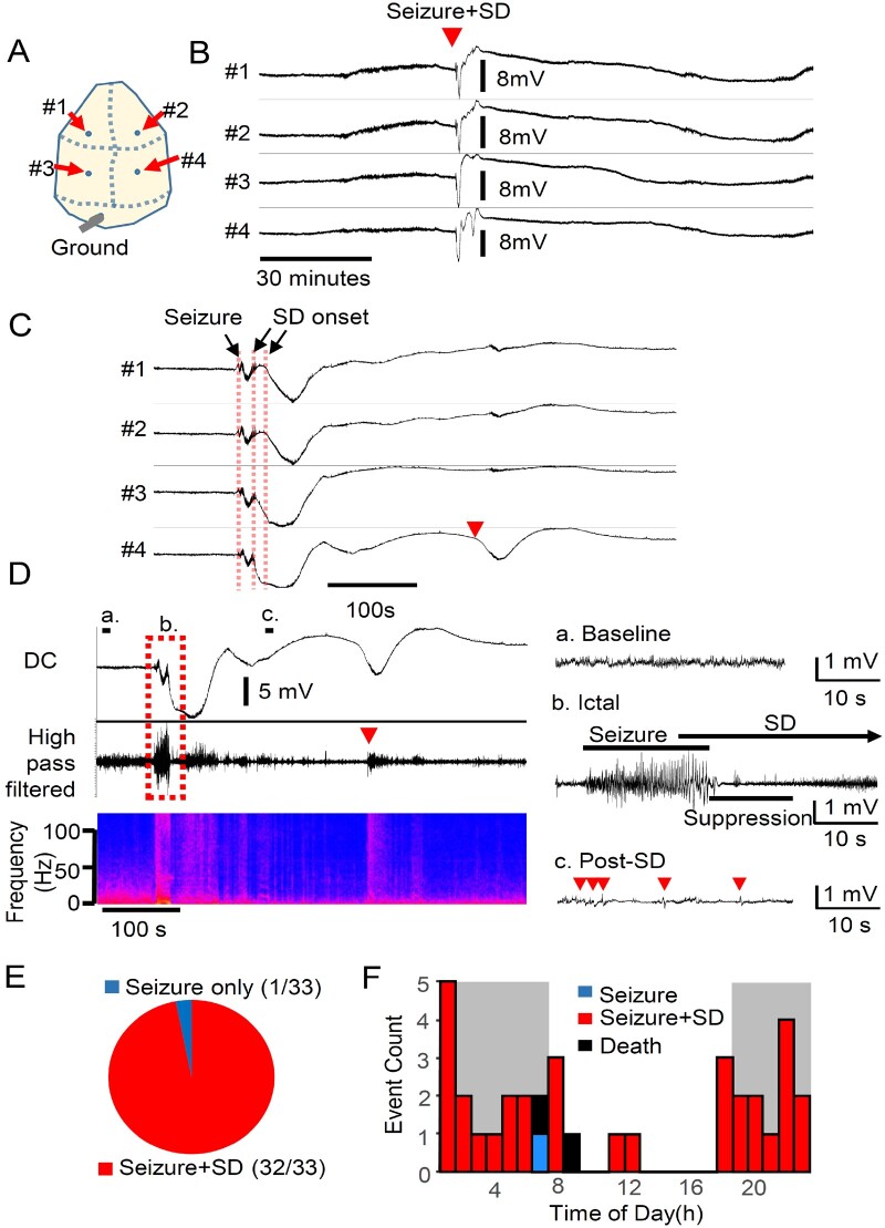
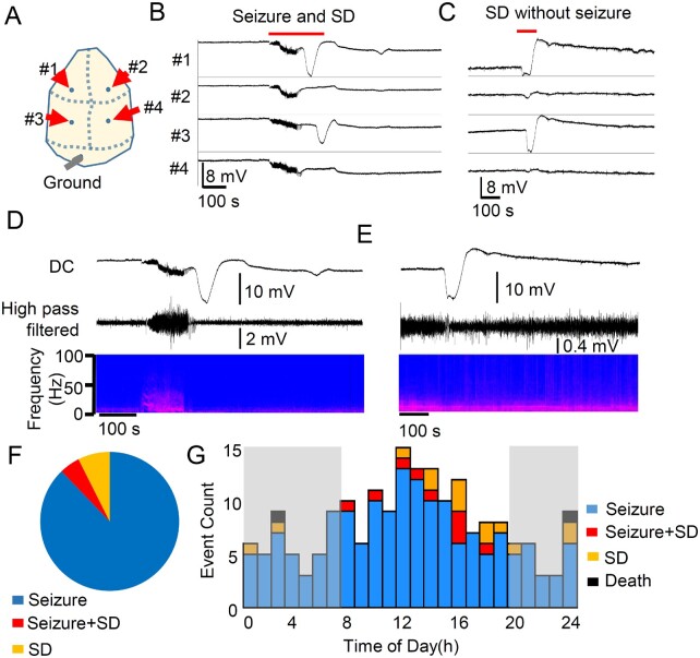
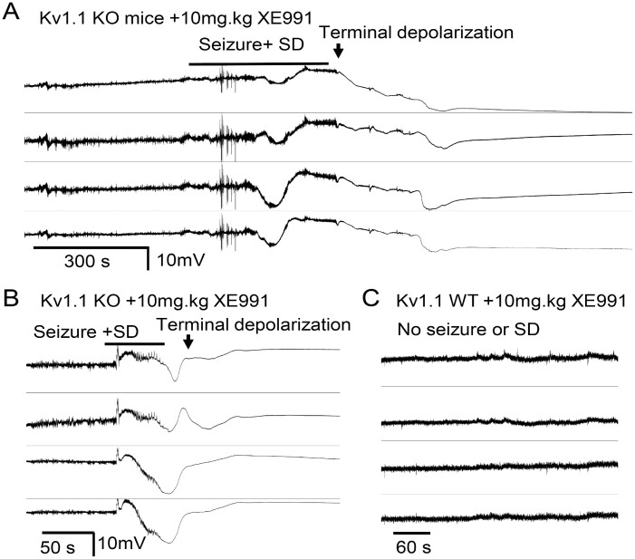
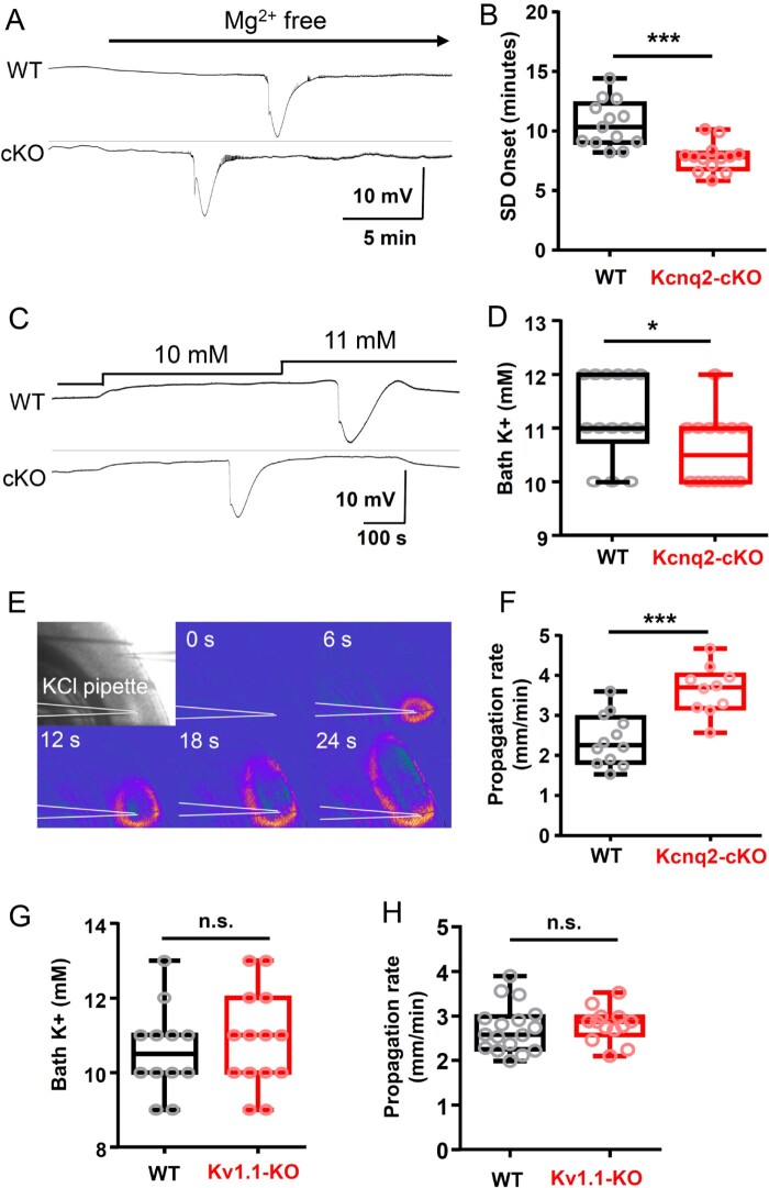
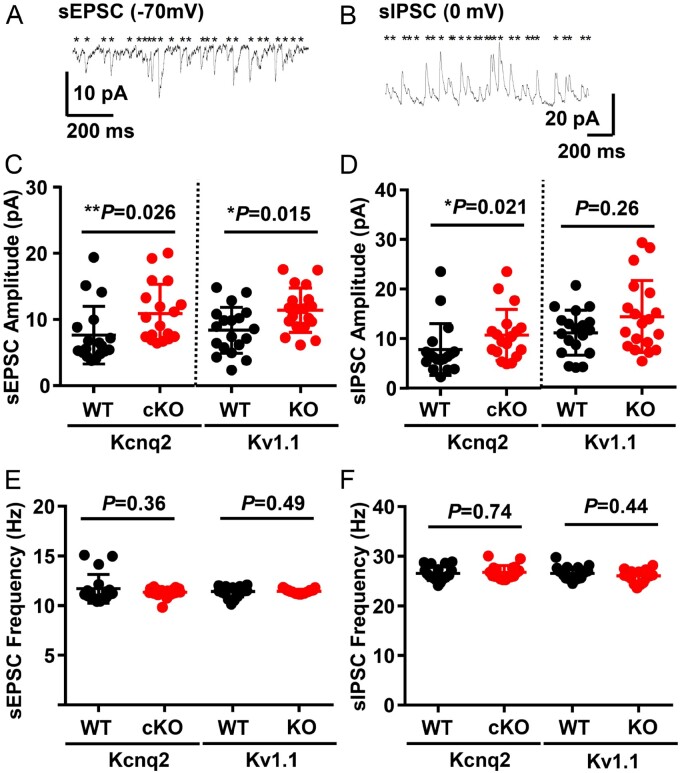
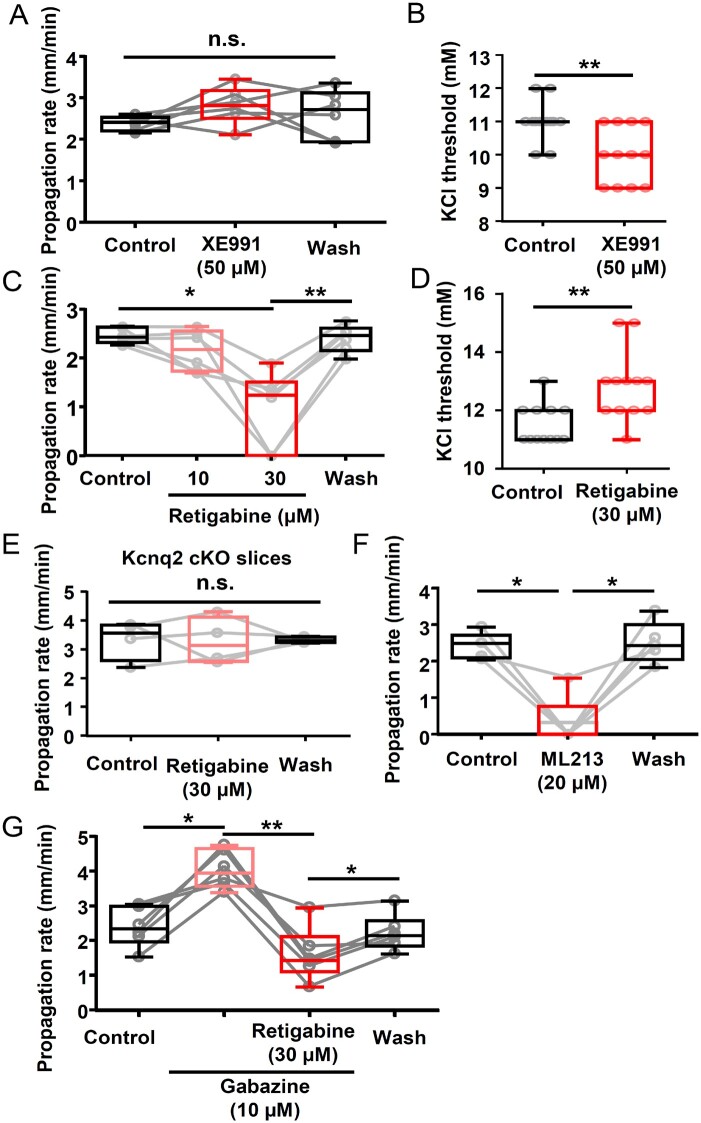
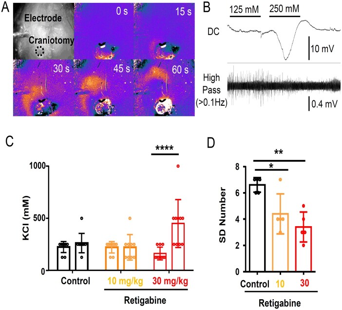
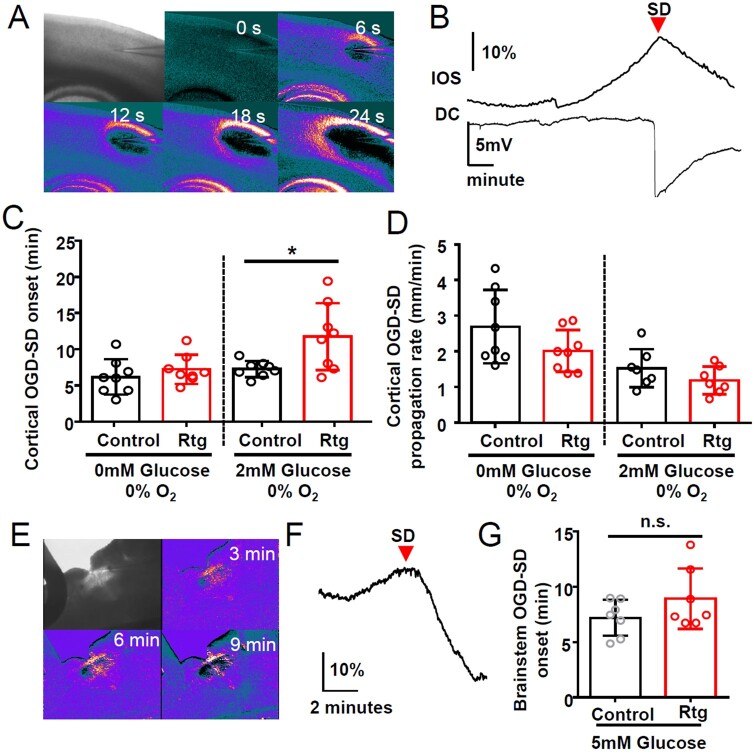
Spreading depolarization is a slowly propagating wave of massive cellular depolarization associated with acute brain injury and migraine aura. Genetic studies link depolarizing molecular defects in Ca2+ flux, Na+ current in interneurons, and glial Na+-K+ ATPase with spreading depolarization susceptibility, emphasizing the important roles of synaptic activity and extracellular ionic homeostasis in determining spreading depolarization threshold. In contrast, although gene mutations in voltage-gated potassium ion channels that shape intrinsic membrane excitability are frequently associated with epilepsy susceptibility, it is not known whether epileptogenic mutations that regulate membrane repolarization also modify spreading depolarization threshold and propagation. Here we report that the Kcnq2/Kv7.2 potassium channel subunit, frequently mutated in developmental epilepsy, is a spreading depolarization modulatory gene with significant control over the seizure-spreading depolarization transition threshold, bi-hemispheric cortical expression, and diurnal temporal susceptibility. Chronic DC-band cortical EEG recording from behaving conditional Kcnq2 deletion mice (Emx1cre/+::Kcnq2flox/flox) revealed spontaneous cortical seizures and spreading depolarization. In contrast to the related potassium channel deficient model, Kv1.1-KO mice, spontaneous cortical spreading depolarizations in Kcnq2 cKO mice are tightly coupled to the terminal phase of seizures, arise bilaterally, and are observed predominantly during the dark phase. Administration of the non-selective Kv7.2 inhibitor XE991 to Kv1.1-KO mice partly reproduced the Kcnq2 cKO-like spreading depolarization phenotype (tight seizure coupling and bilateral symmetry) in these mice, indicating that Kv7.2 currents can directly and actively modulate spreading depolarization properties. In vitro brain slice studies confirmed that Kcnq2/Kv7.2 depletion or pharmacological inhibition intrinsically lowers the cortical spreading depolarization threshold, whereas pharmacological Kv7.2 activators elevate the threshold to multiple depolarizing and hypometabolic spreading depolarization triggers. Together these results identify Kcnq2/Kv7.2 as a distinctive spreading depolarization regulatory gene, and point to spreading depolarization as a potentially significant pathophysiological component of KCNQ2-linked epileptic encephalopathy syndromes. Our results also implicate KCNQ2/Kv7.2 channel activation as a potential adjunctive therapeutic target to inhibit spreading depolarization incidence.

Keywords: Kv1.1; XE991; epilepsy; retigabine; seizure.


Figures

Figure 1
Figure 1
Spontaneous seizure/spreading depolarization complexes predominate in Emx1-Kcnq2cKO mice. Chronic cortical DC recording from juvenile Kcnq2-cKO mouse. (A) Recording configuration. (B and C) Spontaneous seizure/spreading depolarization complex in awake Kcnq2-cKO mouse shown at slow (B) and fast (C) time scales. Spreading depolarization was detected as a sharp negative DC potential shift following a brief generalized seizure detected simultaneously in both hemispheres. A solitary ectopic DC shift was occasionally detected posteriorly (C, arrowhead). (D) Characterization of fast EEG activity during seizure and spreading depolarization. The negative DC shifts of spreading depolarization were detected immediately after or even before the end of seizure activity. Left: Slow time scale. Right: Faster scale. High frequency EEG activity was enhanced during the nadir of the DC shift (b), followed by suppression with occasional spikelets (c). (E) Almost all (97%, n = 34/35) spontaneous seizures were followed by spreading depolarization. No spreading depolarizations were detected without a preceding seizure. (F) Circadian cycle of seizure/spreading depolarization complexes in seven Kcnq2-cKO mice. Histogram of seizure and spreading depolarization incidence during 24 h period (bin size = 1 h, dark grey = dark phase). Spontaneous seizure/spreading depolarization complexes were detected primarily during the dark phase (P < 0.0001, Fisher’s exact test). SD = spreading depolarization.
Figure 2
Figure 2
Spontaneous bilateral seizure with unilateral spreading depolarization and seizure independent spreading depolarization in Kcna1 KO mice. Chronic DC recording in juvenile Kv1.1/Kcna1 KO mice. (A) Representative trace of spreading depolarization following a seizure (postictal spreading depolarization, B) and isolated unilateral spreading depolarization (C). (D and E) EEG characteristics of spreading depolarizations. No clear EEG suppression was detected in these recordings. (F) Unlike Kcnq2-cKO mice, seizure (n = 166) and spreading depolarization (n = 14) were often detected independently, and a seizure + spreading depolarization (SD) complex accounted for 4.8% (9/189) of total events. (G) Histogram of seizure, spreading depolarization, and seizure + spreading depolarization detected from eight Kv1.1 KO mice. There was no clear circadian dependency.
Figure 3
Figure 3
Kv7 inhibitor XE991 triggers bilateral seizure/spreading depolarization complex and death in Kv1.1 KO mice. After baseline recordings, Kv1.1 KO (n = 5) and wild-type (WT, n = 4) mice were administered a single dose of XE991 (5–10 mg/kg i.p.), triggering bilateral seizure-spreading depolarization complexes in all Kv1.1 KO mice. (A) 60% (3/5) of KO mice acutely survived the seizure-spreading depolarization complex but subsequently died. (B) Remaining mice (2/5) died immediately following the bilateral seizure-spreading depolarization. No seizure or spreading depolarization (SD) were detected in four Kv1.1 wild-type control mice and all survived.
Figure 4
Figure 4
Spreading depolarization threshold is lowered in Kcnq2-cKO, but not in Kv1.1 KO cortical slices in vitro. (A and B) Spreading depolarization (SD) onset following exposure to nominally Mg2+-free solution is faster in cKO mouse cortical slices. Representative traces (A) and quantification (B) of wild-type (WT: 10.6 ± 2.0 min, n = 13), and mutant (cKO: 7.7 ± 1.2 min, n = 12) slices. (C and D) Kcnq2-cKO cortical slices had a lowered K+ spreading depolarization threshold. Pairs of slices were incubated and bath K+ concentration was incrementally elevated until spreading depolarization is detected. Spreading depolarization was evoked at lower bath K+ concentration in Kcnq2-cKO (10.6 ± 0.2 mM, n = 14) than wild-type slices (11.2 ± 0.2 mM, n = 14), P < 0.05. (E and F) Spreading depolarization propagation rate was faster in Kcnq2-cKO slices. Spreading depolarization was evoked by focal KCl microinjection and detected by IOS (see ‘Materials and methods’ section). The velocity of spreading depolarization propagation was faster in the Kcnq2-cKO slices (WT: 2.4 ± 0.2 mm/min, n = 12; cKO: 3.6 ± 0.2 mm/min, n = 10, P < 0.005). (G) Spreading depolarization threshold determined by incrementally increasing bath K+ concentrations. (WT: 10.6 ± 0.3 mM, n = 12; KO: 10.9 ± 0.5 mM, n = 14). (H) Spreading depolarization propagation rate was determined by focal KCl microinjection (WT: 2.7 ± 0.1 mm/min, n = 17; KO: 2.8 ± 0.1 mm/min, n = 13). *P < 0.05, ***P < 0.005.
Figure 5
Figure 5
sEPSC and sIPSC balances in the cortical layer 2/3 pyramidal neurons of Kcnq2-cKO and Kv1.1-KO mice. (A and B) Representative traces of sEPSC and sIPSC activity recorded from layer 2/3 cortical pyramidal neurons. (C and D) SEPSC amplitudes were enhanced in both Kcnq2-cKO [wild-type (WT): 7.6 ± 1.0 pA; cKO: 10.9 ± 1.0pA) and Kv1.1-KO (WT: 8.4 ± 0.8 pA; KO: 11.40 ± 0.8pA), while sIPSC amplitudes were increased only in Kcnq2-cKO (WT: 7.8 ± 1.2 pA; cKO: 10.7 ± 1.2pA), not in Kv1.1-KO cortical tissue (WT: 11.0 ± 1.0 pA; KO: 14.2 ± 1.7 pA). (E and F) No differences were detected in the frequencies of sEPSC (Kcnq2-WT: 23.2 ± 0.7 Hz, -cKO: 22.7 ± 0.2 Hz; Kv1.1-WT: 22.7 ± 0.3 Hz, -KO: 22.9 ± 0.1 Hz) and sIPSC (Kcnq2-WT: 26.8 ± 0.3 Hz, -cKO: 27.0 ± 0.4 Hz, Kv1.1-WT: 26.6 ± 0.3 Hz, -KO: 26.1 ± 0.3 Hz). n = 18 for Kcnq2-WT and cKO, n = 19 for Kv1.1 WT and KO neurons. Mann-Whitney U-test was used for the statistical comparisons of these data.
Figure 6
Figure 6
Acute enhancement/inhibition of spreading depolarization in wild-type cortex by Kv7 inhibitor/activator in vitro. (A and B) Effect of XE991 on spreading depolarization propagation rate and K+ threshold. (A) Spreading depolarization propagation rate measured from spreading depolarizations repetitively generated in single slices while incubated in the drug was unchanged. Control: 2.4 ± 0.2 mm/min, XE991: 2.8 ± 0.5 mm/min, Wash: 2.6 ± 0.6 mm/min, n = 6, P = 0.37. n.s. = not significant. (B) Spreading depolarization threshold was decreased by Kcnq inhibitor in K+ bath application model. Control: 11.0 ± 0.2 mM, XE991: 10.0 ± 0.2 mM, n = 12 each, *P < 0.005. (C and D) Retigabine dose-dependently reduced spreading depolarization propagation rate (C,n = 6) and elevated the K+ threshold (vehicle: 11.5 ± 0.2 mM, retigabine: 12.8 ± 0.3 mM, n = 12, P = 0.016) (D, n = 12). (E) Retigabine (30 µM) did not alter spreading depolarization propagation in Kcnq2-cKO slices, n = 4. (F) ML216 (20 µM), another Kv7 activator, also inhibited spreading depolarization propagation in wild-type slices (Control: 2.4 ± 0.4 mm/min, ML216: 0.3 ± 0.7 mm/min, wash: 2.5 ± 0.6 mm/min, n = 5). (G) Retigabine effect on spreading depolarization was analysed in the presence of the GABAAR antagonist gabazine. Gabazine increased the spreading depolarization propagation rate; however, retigabine still reduced the spreading depolarization propagation rate (Control: 2.4 ± 0.6 mm/min, gabazine: 4.1 ± 0.5 mm/min, gabazine + retigabine: 1.6 ± 0.8 mm/min, wash: 2.2 ± 0.5 mm/min, n = 6, *P < 0.05, **P < 0.01).
Figure 7
Figure 7
Spreading depolarization inhibition by retigabine in in vivo anaesthetized wild-type mouse cortex. Spreading depolarization was triggered by repetitively applying a KCl solution (100, 300, 500, 1000 mM for 2 min) to the cortical surface. (A) Spreading depolarization wave was detected with IOS signal shown in a pseudocoloured image, and (B) electrophysiologically with Ag/AgCl electrode. (C) Summary of spreading depolarization threshold measurement. In each animal, spreading depolarization threshold was measured before and after drug injection. Vehicle (0.1% DMSO) and 10 mg/kg retigabine had no effect, while 30 mg/kg retigabine significantly increased K+ evoked spreading depolarization threshold. ****P < 0.001. (D) Number of recurrent spreading depolarization events during continuous 0.5 M KCl application for 30 min. Retigabine 30 mg/kg significantly decreased the regenerative spreading depolarization number. *P < 0.05, **P < 0.01.
Figure 8
Figure 8
Kv7.2 activator retigabine inhibits mild, but not severe OGD-induced spreading depolarization. (A) Image of OGD-spreading depolarization wave in cortical slice triggered by continuous exposure to OGD solution (0% O2, 0 mM glucose), and detected as IOS travelling across cortical tissue. (B) Arrival of IOS signal (top) near electrode coincided with negative DC potential shift (bottom trace). (C and D) Retigabine delayed OGD-spreading depolarization when metabolic stress was mild (2 mM glucose), but had no effect during severe compromise (0 mM glucose). Retigabine did not inhibit spreading depolarization propagation rate (D) n = 8 each, *P < 0.05. (EG) Retigabine had no effect on OGD-spreading depolarization generated in slices of the medulla at the level of the nucleus tractus solitarius (nTS). (E) Spreading depolarization was triggered by 0% O2/5 mM glucose solution with or without 30 µM retigabine. (F and G) Spreading depolarization onset was determined by the IOS signal peak at the lateral margin of the nTS. (n = 7 in all experiments). SD = spreading depolarization.

References

    1. Somjen GG. Mechanisms of spreading depression and hypoxic spreading depression-like depolarization. Physiol Rev. 2001;81(3):1065–1096. - PubMed
    1. Dreier JP. The role of spreading depression, spreading depolarization and spreading ischemia in neurological disease. Nat Med. 2011;17(4):439–447. - PubMed
    1. Dreier JP, Major S, Foreman B, et al.Terminal spreading depolarization and electrical silence in death of human cerebral cortex. Ann Neurol. 2018;83(2):295–310. - PMC - PubMed
    1. Carlson AP, Shuttleworth CW, Major S, Lemale CL, Dreier JP, Hartings JA.. Terminal spreading depolarizations causing electrocortical silencing prior to clinical brain death: Case report. J Neurosurg. 2018;131(6):1773–1779. - PubMed
    1. Dreier JP, Major S, Lemale CL, et al.Correlates of spreading depolarization, spreading depression, and negative ultraslow potential in epidural versus subdural electrocorticography. Front Neurosci. 2019;13:373. - PMC - PubMed

Publication types

MeSH terms

LinkOut - more resources