

A pleural ultrasound image of a collapsed lung surrounded by pleural fluid ("jellyfish sign") may correspond to an intrapericardial mass
- PMID: 33768495
- PMCID: PMC9148363
- DOI: 10.1007/s40477-021-00577-9
A pleural ultrasound image of a collapsed lung surrounded by pleural fluid ("jellyfish sign") may correspond to an intrapericardial mass
Abstract
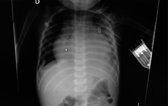
Lung ultrasound has been shown to be a valuable diagnostic tool. It has become the main way to get to the diagnosis of pleural effusion with much more specificity and sensibility than the x-ray. The diagnosis of pleural effusion with ultrasound is easily obtained after the visualization of hypoechoic fluid surrounding the lung. Sometimes it appears as an image of a collapsed lung moving with the surrounded pleural fluid ("jellyfish sign"). Until now this sign was almost pathognomonic of pleural effusion, but we explore a case in which this sign could have led to a misleading diagnosis. We present the case of a child admitted to intensive care with respiratory distress. In the point of care lung ultrasound we believed to see a pleural effusion with a collapsed lung moving into the effusion. Due to the enlargement of the pericardial sac, we did not realize that what we thought to be the pleural space was in fact the pericardial space. Unfortunately, there was a more echogenic area inside the pericardial effusion which led to a misleading fake lung atelectasis with pleural effusion ("jellyfish sign"). The correct diagnosis was properly obtained after assessing a cardiac point of care ultrasound using a four chambers view. The left side of the thorax is more difficult to be sonographed than the right due to the presence of the heart fossa that occupies a significant part of that side. Obtaining the diagnosis of pleural effusion on that side is more difficult for this reason and can sometimes be misleading with a pericardial effusion. The presence of the "jellyfish sign" is not pathognomonic and may lead to an error if we are guided only by the presence of that sign. To avoid such a misleading diagnosis, we highly recommend performing a point of care cardiac ultrasound if a pleural effusion is primarily seen in the lung ultrasound.
Keywords: Flapping lung; Jellyfish sign; Pericardial effusion; Pleural effusion; Ultrasound.
© 2021. Società Italiana di Ultrasonologia in Medicina e Biologia (SIUMB).
Conflict of interest statement
The authors declare that they have no competing interests.
Figures


References
-
- Dénier A. Les ultrasons, leur application au diagnostic. PresseMéd. 1946;22:307–308.
Publication types
MeSH terms
LinkOut - more resources
Full Text Sources
Other Literature Sources
Medical
