. 2021 Sep;26(5):e13037.
doi: 10.1111/adb.13037. Epub 2021 Mar 25.

Cell-type specific expression and behavioral impact of galanin and GalR1 in the locus coeruleus during opioid withdrawal

Affiliations

Cell-type specific expression and behavioral impact of galanin and GalR1 in the locus coeruleus during opioid withdrawal

Stephanie L Foster et al. Addict Biol. 2021 Sep.

Abstract

The neuropeptide galanin is reported to attenuate opioid withdrawal symptoms, potentially by reducing neuronal hyperactivity in the noradrenergic locus coeruleus (LC) via galanin receptor 1 (GalR1). We evaluated this mechanism by using RNAscope in situ hybridization to characterize GalR1 mRNA distribution in the dorsal pons and to compare galanin and GalR1 mRNA expression in tyrosine hydroxylase-positive (TH+) LC cells at baseline and following chronic morphine or precipitated withdrawal. We then used genetically altered mouse lines and pharmacology to test whether noradrenergic galanin (NE-Gal) modulates withdrawal symptoms. RNAscope revealed that, while GalR1 signal was evident in the dorsal pons, 80.7% of the signal was attributable to TH- neurons outside the LC. Galanin and TH mRNA were abundant in LC cells at baseline and were further increased by withdrawal, whereas low basal GalR1 mRNA expression was unaltered by chronic morphine or withdrawal. Naloxone-precipitated withdrawal symptoms in mice lacking NE-Gal (GalcKO-Dbh ) were largely similar to WT littermates, indicating that loss of NE-Gal does not exacerbate withdrawal. Complementary experiments using NE-Gal overexpressor mice (NE-Gal OX) and systemic administration of the galanin receptor agonist galnon revealed that increasing galanin signaling also failed to alter behavioral withdrawal, while suppressing noradrenergic transmission with the alpha-2 adrenergic receptor agonist clonidine attenuated multiple symptoms. These results indicate that galanin does not acutely attenuate precipitated opioid withdrawal via an LC-specific mechanism, which has important implications for the general role of galanin in regulation of somatic and affective opioid responses and LC activity.

Keywords: GalR1; galanin; locus coeruleus; noradrenergic; opioid; withdrawal.


Conflict of interest statement

Conflict of interest: The authors declare no conflict of interest.

Figures

Figure 1.
Figure 1.. GalR1 expression is low in noradrenergic neurons of the LC.
RNAscope was performed to identify GalR1 mRNA (GalR1, green) and noradrenergic neurons of the LC defined by tyrosine hydroxylase mRNA (TH, magenta) along with DAPI nuclear stain (blue). A representative image of a coronal mouse brain section shows strong GalR1 expression around, and little within, the LC (A). The few TH+ cells expressing GalR1 are observed in the LC periphery (white arrows) in comparison to the LC core (B). GalR1 mRNA was also readily observed in control sections of hypothalamus, a region previously shown to strongly express GalR1 mRNA (C). RNAscope for GalR1 mRNA (green) in the rat LC (TH, white) shows a similar pattern with GalR1 primarily outside the LC border (D). IHC for mCherry in a GalR1-mCherry mouse line reveals a pattern of GalR1 protein consistent with mRNA findings (E), and robust signal as expected in positive control sections containing the paraventricular nucleus of the thalamus (PVT, F). All scale bars are 50 μm. Abbreviations: V4 (fourth ventricle); LC (locus coeruleus); D (dorsal); L (lateral); V3 (third ventricle).
Figure 2.
Figure 2.. Pontine GalR1 mRNA expression is higher in LC-adjacent regions than the LC itself.
RNAscope was performed for GalR1 (green) and TH (magenta) in addition to the neuronal marker SNAP25 (yellow) to evaluate GalR1 mRNA expression in TH+ and TH− populations. SNAP25 labels both TH+ LC neurons and TH− neurons in the surrounding field of view (A,C). Enlarged images show low GalR1 signal in LC neurons that are both TH+ (B, arrowheads) and SNAP25+ (D, arrowheads). Higher GalR1 signal can be seen in neurons that are TH− (B, arrows) but SNAP25+ (D, arrows). Merged images with DAPI nuclear stain (blue) highlight robust GalR1 signal in SNAP25+ neurons outside the LC (E,F). The majority of GalR1 signal in each LC image is contained within TH−, rather than TH+ cells (G). Analysis of GalR1 puncta density by cell type shows that within the LC field of view, TH− cells express significantly more GalR1 than TH+ cells (H). n = 6 mice, 3 LC images per mouse. All scale bars are 50 μm. Bar graphs display mean ± SEM. **** p < 0.0001.
Figure 3.
Figure 3.. Morphine withdrawal increases LC expression of TH and galanin, but not GalR1 mRNA.
Representative 40x LC images from mice that received saline injections (Sal / Nlx), chronic morphine injections (Mor / Sal), or underwent naloxone-precipitated withdrawal following induction of morphine dependence (Mor / Nlx). RNAscope was performed with probes for TH (magenta), galanin (Gal, yellow), and GalR1 (green). Compared to saline treatment, chronic morphine and withdrawal increased TH expression, indicated by elevated fluorescent signal intensity (A-C). Withdrawal also increased galanin expression compared to saline treatment (D-F). Baseline GalR1 expression was markedly lower than either TH or Gal, and expression was unaffected by chronic morphine or withdrawal (G-I, LC outlined in gray). Merged images display all probes with DAPI nuclear stain (blue) (J-L). All scale bars are 50 μm. Abbreviations: D (dorsal); M (medial).
Figure 4.
Figure 4.. Low GalR1 mRNA expression in the LC is unaltered by chronic morphine or withdrawal.
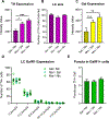
Quantification of TH, Gal, and GalR1 mRNA signal in 40x LC images from mice that received saline injections (Sal / Nlx), chronic morphine injections (Mor / Sal), or underwent withdrawal (Mor / Nlx). Withdrawal and chronic morphine increased TH mRNA expression as measured by fluorescence intensity (A). There were no differences in the number of TH+ LC cells detected per treatment group, indicating that changes in TH intensity did not affect LC quantification for GalR1 analysis (B). Withdrawal increased galanin mRNA expression (C). GalR1 is not expressed in the majority of TH+ LC neurons, and relative proportions of GalR1 expression are not altered by chronic morphine or withdrawal (D). Among cells that expressed any GalR1 puncta, treatment group did not affect the average number of GalR1 puncta per cell (E). n = 6 mice per group, 3 LC images per mouse. All graphs display mean ± SEM. * p < 0.05, ** p < 0.01, *** p < 0.001.
Figure 5.
Figure 5.. Noradrenergic galanin does not modulate precipitated withdrawal symptoms in mice.
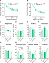
Noradrenergic-specific galanin knockout mice (GalcKO-Dbh), noradrenergic galanin overexpressor mice (NE-Gal OX), and wild-type littermates (WT) received escalating doses of morphine twice daily for 5 days (20, 40, 60, 80, 100 mg/kg, i.p.) to induce dependence. On day 6, mice received a final dose of 100 mg/kg morphine and 2 h later underwent naloxone-precipitated withdrawal (1 mg/kg, s.c.). Neither GalcKO-Dbh nor NE-Gal OX differed from WT littermates in weight lost during the morphine dosing period (A, B). GalcKO-Dbh, NE-Gal OX, and WT littermates exhibited similar occurrences of most withdrawal symptoms (C-G, I, J). Only backwards steps were significantly different between groups, in which GalcKO-Dbh exhibited fewer occurrences than WT mice (H). n = 10–11 for GalcKO-Dbh and NE-Gal OX; n = 20 for WT. All graphs display mean ± SEM. * p < 0 .05, n.s. = not significant.
Figure 6.
Figure 6.. Activation of central galanin receptors does not alter withdrawal symptoms.
Food-deprived mice were pre-treated with either vehicle or galnon (2 mg/kg, i.p.) prior to a feeding test. Galnon-treated mice consumed less food than vehicle treated mice (A), and latency to eat was greater than vehicle but not significantly different (B), indicating that systemic galnon was sufficient to exert central effects on feeding. n = 7 per group. A separate cohort was pre-treated with galnon, clonidine (0.3 mg/kg, i.p.), or respective vehicle before naloxone-precipitated withdrawal. Galnon did not affect any symptoms compared to vehicle, while clonidine significantly reduced multiple symptoms compared to either vehicle or galnon treatment (E-H, J). Clonidine significantly increased weight loss compared to galnon, but not vehicle (C). Jumps and wet dog shakes did not differ from vehicle for either treatment (D, I). n = 10–11 for galnon and clonidine groups; n = 19 for vehicle. All graphs display mean ± SEM. * p < 0.05, ** p < 0.01, *** p < 0.001, **** p < 0.0001, n.s. = not significant.

References

    1. Rasmussen K, Beitner-Johnson DB, Krystal JH, Aghajanian GK, Nestler EJ. Opiate withdrawal and the rat locus coeruleus: behavioral, electrophysiological, and biochemical correlates. J Neurosci 1990;10(7):2308–2317. - PMC - PubMed
    1. Kosten TR, Baxter LE. Review article: Effective management of opioid withdrawal symptoms: A gateway to opioid dependence treatment. Am J Addict 2019;28(2):55–62. - PMC - PubMed
    1. Lang R, Gundlach AL, Holmes FE, et al.Physiology, signaling, and pharmacology of galanin peptides and receptors: three decades of emerging diversity. Pharmacol Rev 2015;67(1):118–175. - PubMed
    1. Zachariou V, Brunzell DH, Hawes J, et al.The neuropeptide galanin modulates behavioral and neurochemical signs of opiate withdrawal. Proc Natl Acad Sci U S A. 2003;100(15):9028–9033. - PMC - PubMed
    1. Holmes FE, Armenaki A, Iismaa TP, et al.Galanin negatively modulates opiate withdrawal via galanin receptor 1. Psychopharmacology (Berl). 2012;220(3):619–625. - PMC - PubMed

Publication types

MeSH terms