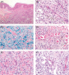
Hemosiderotic dermatofibroma mimicking melanoma: A case report and review of the literature
- PMID: 33768851
- PMCID: PMC7981605
- DOI: 10.1002/ccr3.3780
Hemosiderotic dermatofibroma mimicking melanoma: A case report and review of the literature
Abstract
Hemosiderotic dermatofibroma (HDF) often mimics melanoma clinically. A definite diagnosis relies on histopathological evaluation and immunohistochemistry. As it can progress to aneurysmal dermatofibroma (ADF), complete excision is recommended.
Keywords: aneurysmal dermatofibroma; hemosiderotic dermatofibroma; melanoma; skin.
© 2021 The Authors. Clinical Case Reports published by John Wiley & Sons Ltd.
Conflict of interest statement
The authors declare no conflict of interests.
Figures


Similar articles
-
168 Cases of aneurysmal dermatofibroma and 29 cases of hemosiderotic dermatofibroma: A clinicopathologic study.Am J Clin Pathol. 2024 Mar 1;161(3):232-244. doi: 10.1093/ajcp/aqad136. Am J Clin Pathol. 2024. PMID: 37897209
-
Giant thigh hemosiderotic/aneurysmal dermatofibroma: Case report with radiologic-pathologic correlation.Rev Esp Patol. 2024 Jul-Sep;57(3):217-224. doi: 10.1016/j.patol.2024.04.001. Epub 2024 May 20. Rev Esp Patol. 2024. PMID: 38971622
-
Hemosiderotic dermatofibroma: clinical and dermoscopic presentation mimicking melanoma.J Dermatol Case Rep. 2015 Jun 30;9(2):39-41. doi: 10.3315/jdcr.2015.1198. eCollection 2015 Jun 30. J Dermatol Case Rep. 2015. PMID: 26236411 Free PMC article.
-
Giant Hemosiderotic Adenodermatofibroma: A Case Report and Review of the Literature.Am J Dermatopathol. 2023 Mar 1;45(3):192-195. doi: 10.1097/DAD.0000000000002373. Epub 2023 Jan 10. Am J Dermatopathol. 2023. PMID: 36728283 Review.
-
Giant dermatofibroma. A little-known clinical variant of dermatofibroma.J Am Acad Dermatol. 1994 May;30(5 Pt 1):714-8. J Am Acad Dermatol. 1994. PMID: 8176009 Review.
Cited by
-
Dermatofibroma: Reappraisal and Updated Review.Clin Cosmet Investig Dermatol. 2025 Aug 4;18:1873-1887. doi: 10.2147/CCID.S526191. eCollection 2025. Clin Cosmet Investig Dermatol. 2025. PMID: 40785832 Free PMC article. Review.
References
-
- Senel E, Yuyucu Karabulut Y, Dogruer Senel S. Clinical, histopathological, dermatoscopic and digital microscopic features of dermatofibroma: a retrospective analysis of 200 lesions. J Eur Acad Dermatol Venereol. 2015;29:1958‐1966. - PubMed
-
- Santa Cruz DJ, Kyriakos M. Aneurysmal ("angiomatoid") fibrous histiocytoma of the skin. Cancer. 1981;47:2053‐2061. - PubMed
-
- Calonje E, Mentzel T, Fletcher CD. Cellular benign fibrous histiocytoma. Clinicopathologic analysis of 74 cases of a distinctive variant of cutaneous fibrous histiocytoma with frequent recurrence. Am J Surg Pathol. 1994;18:668‐676. - PubMed
-
- Singh Gomez C, Calonje E, Fletcher CD. Epithelioid benign fibrous histiocytoma of skin: clinico‐pathological analysis of 20 cases of a poorly known variant. Histopathology. 1994;24:123‐129. - PubMed
Publication types
LinkOut - more resources
Full Text Sources
Other Literature Sources
Research Materials

