Transcatheter closure of the aortopulmonary window in a three-month-old infant with a symmetric membranous ventricular septal defect occluder device
- PMID: 33768987
- PMCID: PMC7970074
- DOI: 10.5606/tgkdc.dergisi.2021.20988
Transcatheter closure of the aortopulmonary window in a three-month-old infant with a symmetric membranous ventricular septal defect occluder device
Abstract
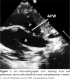
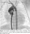
Although most of aortopulmonary window cases are closed surgically, percutaneous closure can be also used in suitable patients. Defects which are far from the pulmonary and aortic valves, coronary artery, and pulmonary artery bifurcation, with adequate septal rims are considered suitable for percutaneous closure. A three-month-old male infant weighing 4 kg was referred to our pediatric cardiology department with the complaints of fatigue while breastfeeding, difficulty in weight gain, heart murmur, and respiratory distress. A large aortopulmonary window (5.3 mm) and left heart chamber dilatation were detected on echocardiography. The large aortopulmonary window was closed using a symmetric membranous ventricular septal defect occluder device. The closure procedure was performed via the antegrade route without forming an arteriovenous loop. In conclusion, the use of a symmetric membranous ventricular septal defect device for closure of large aortopulmonary window seems to be a safe and effective alternative to surgery in selected infants.
Keywords: Aortopulmonary window; heart failure; infant; transcatheter closure.
Copyright © 2021, Turkish Society of Cardiovascular Surgery.
Conflict of interest statement
Conflict of Interest: The authors declared no conflicts of interest with respect to the authorship and/or publication of this article.
Figures


References
-
- Jacobs JP, Quintessenza JA, Gaynor JW, Burke RP, Mavroudis C. Congenital Heart Surgery Nomenclature and Database Project: aortopulmonary window. S44-9Ann Thorac Surg. 2000;69(4 Suppl) - PubMed
-
- Stamato T, Benson LN, Smallhorn JF, Freedom RM. Transcatheter closure of an aortopulmonary window with a modified double umbrella occluder system. Cathet Cardiovasc Diagn. 1995;35:165–167. - PubMed
-
- Campos-Quintero A, García-Montes JA, Zabal-Cerdeira C, Cervantes-Salazar JL, Calderón-Colmenero J, Sandoval JP. Transcatheter device closure of aortopulmonary window. Is there a need for an alternative strategy to surgery. Rev Esp Cardiol (Engl Ed) 2019;72:349–351. - PubMed
-
- Trehan V, Nigam A, Tyagi S. Percutaneous closure of nonrestrictive aortopulmonary window in three infants. Catheter Cardiovasc Interv. 2008;71:405–411. - PubMed
Publication types
LinkOut - more resources
Full Text Sources
Other Literature Sources
