Review
. 2021 Oct 1;101(4):1487-1559.
doi: 10.1152/physrev.00022.2020. Epub 2021 Mar 26.

Regulation of cerebral blood flow in humans: physiology and clinical implications of autoregulation

Affiliations
Review

Regulation of cerebral blood flow in humans: physiology and clinical implications of autoregulation

Jurgen A H R Claassen et al. Physiol Rev. .

Abstract

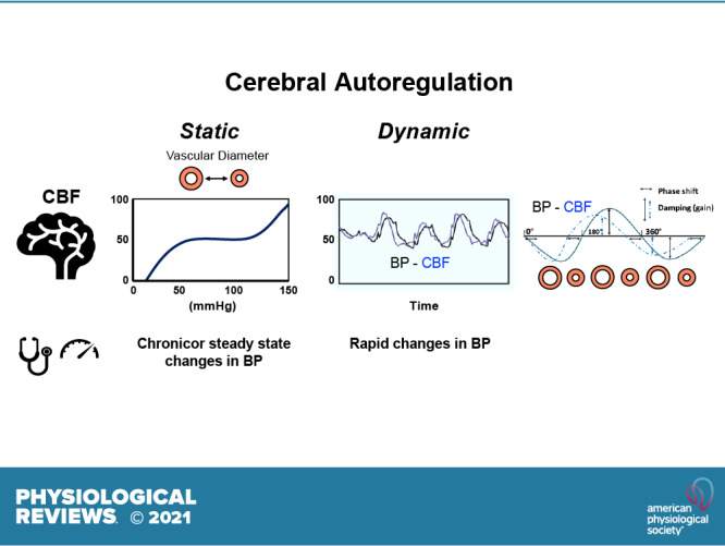
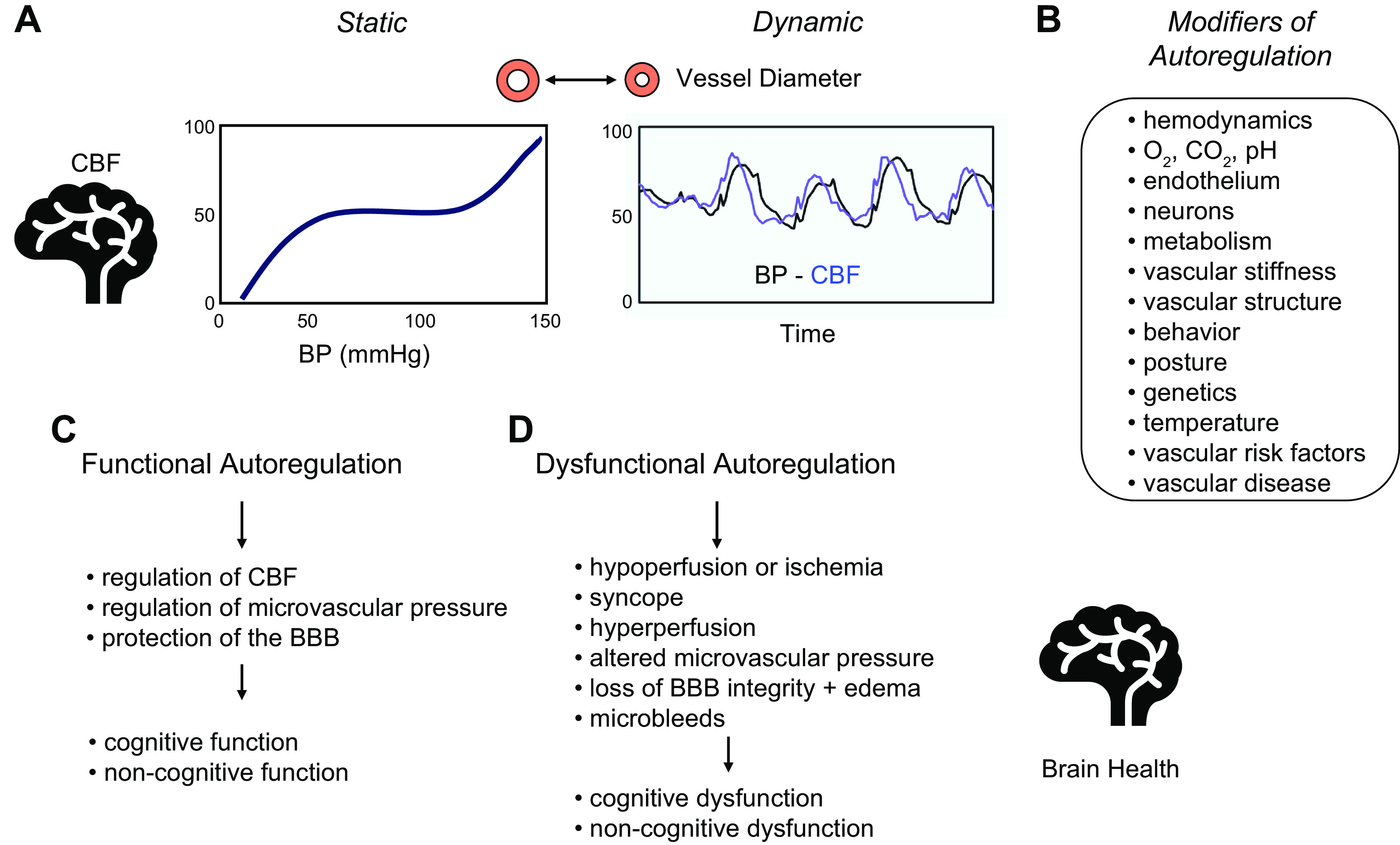
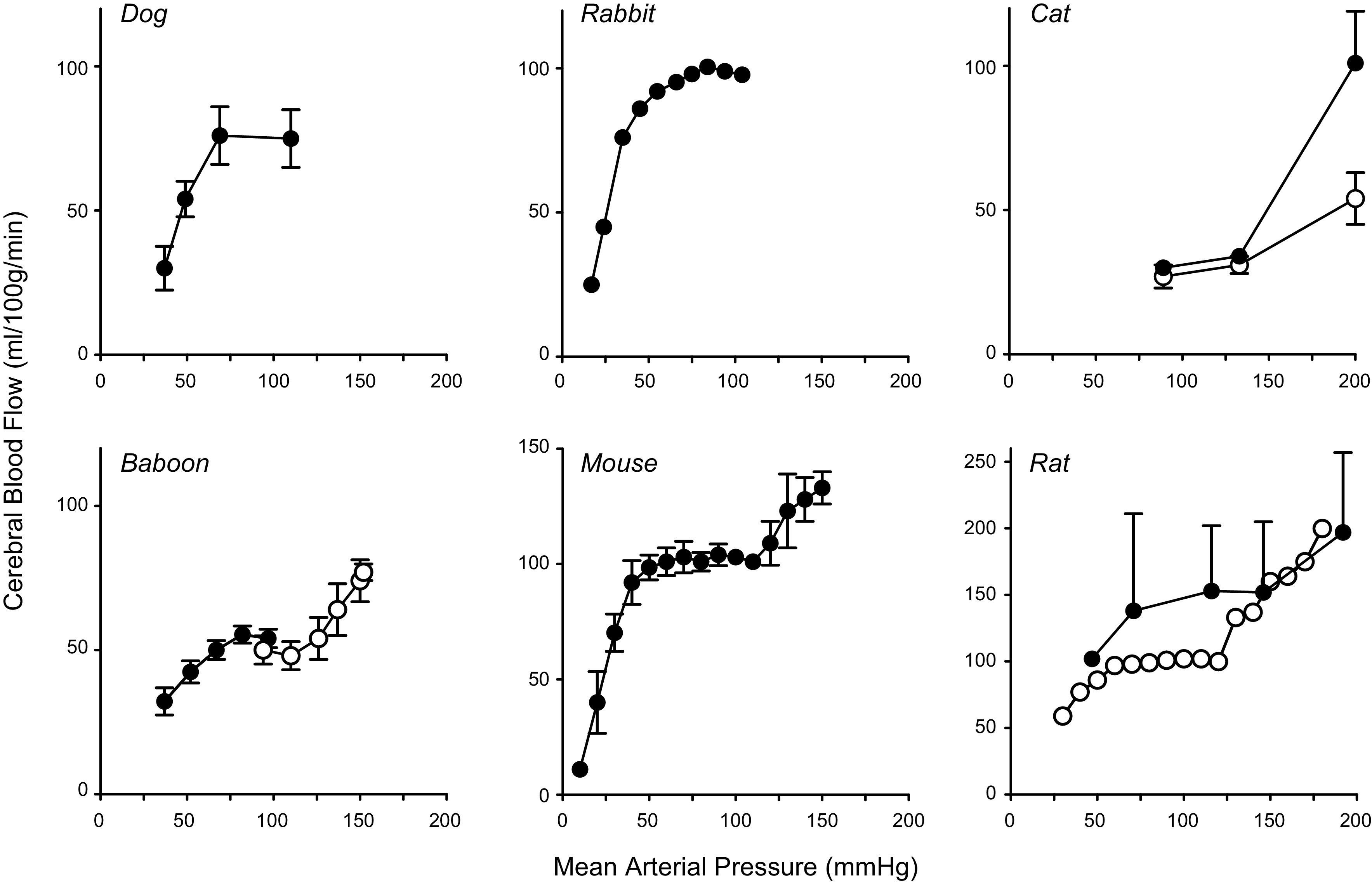
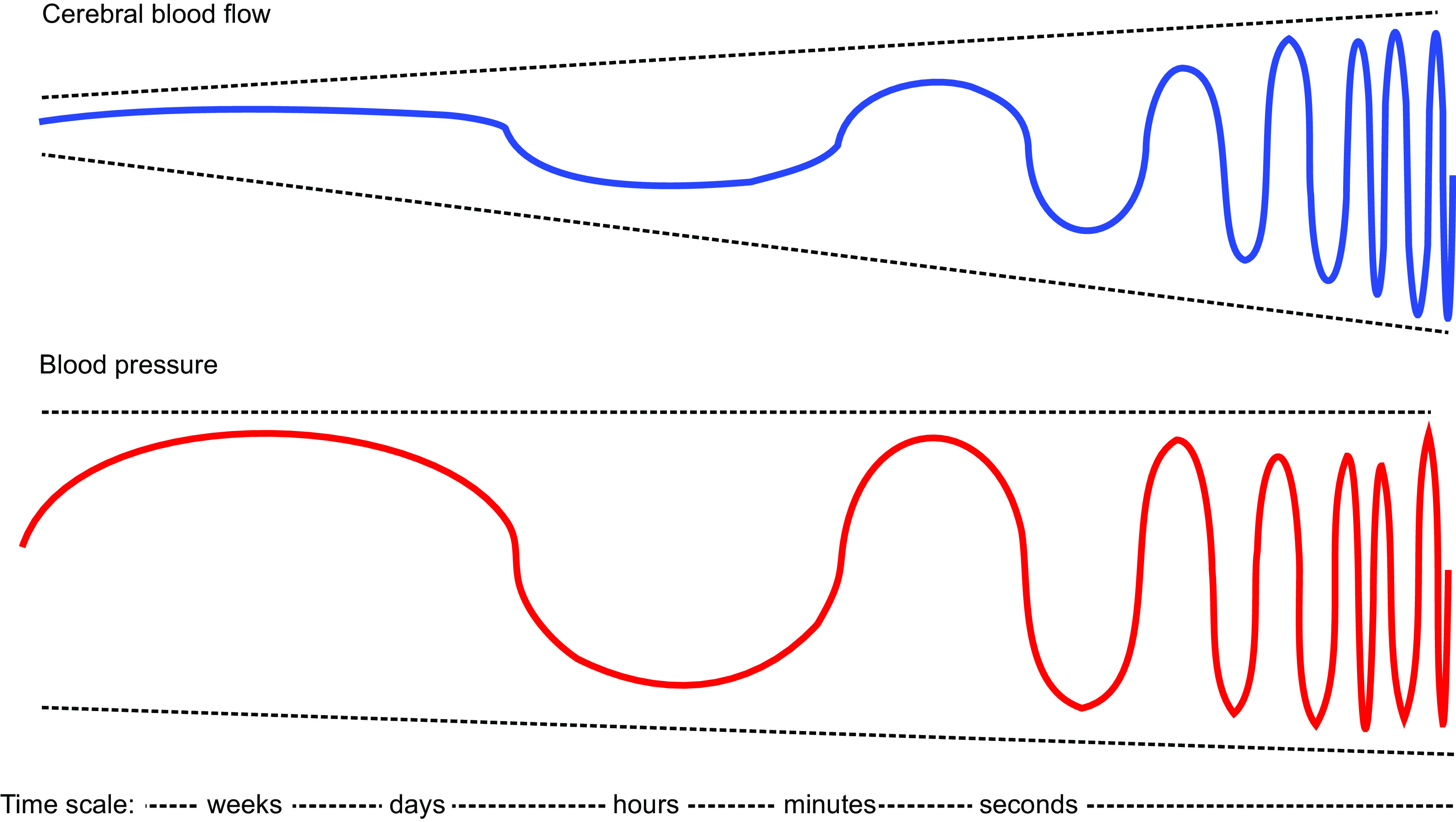
Brain function critically depends on a close matching between metabolic demands, appropriate delivery of oxygen and nutrients, and removal of cellular waste. This matching requires continuous regulation of cerebral blood flow (CBF), which can be categorized into four broad topics: 1) autoregulation, which describes the response of the cerebrovasculature to changes in perfusion pressure; 2) vascular reactivity to vasoactive stimuli [including carbon dioxide (CO2)]; 3) neurovascular coupling (NVC), i.e., the CBF response to local changes in neural activity (often standardized cognitive stimuli in humans); and 4) endothelium-dependent responses. This review focuses primarily on autoregulation and its clinical implications. To place autoregulation in a more precise context, and to better understand integrated approaches in the cerebral circulation, we also briefly address reactivity to CO2 and NVC. In addition to our focus on effects of perfusion pressure (or blood pressure), we describe the impact of select stimuli on regulation of CBF (i.e., arterial blood gases, cerebral metabolism, neural mechanisms, and specific vascular cells), the interrelationships between these stimuli, and implications for regulation of CBF at the level of large arteries and the microcirculation. We review clinical implications of autoregulation in aging, hypertension, stroke, mild cognitive impairment, anesthesia, and dementias. Finally, we discuss autoregulation in the context of common daily physiological challenges, including changes in posture (e.g., orthostatic hypotension, syncope) and physical activity.

Keywords: Alzheimer’s disease; hypertension; microcirculation; neurovascular coupling; stroke.


Conflict of interest statement

No conflicts of interest, financial or otherwise, are declared by the authors.

Figures

None
Graphical abstract
FIGURE 1.
FIGURE 1.
Overview of cerebral autoregulation. General overview of autoregulation and key summary points for this review. A: 2 graphs illustrating the concepts of static and dynamic autoregulation. B: summary of modifiers or factors that have a direct influence on cerebral blood flow (CBF) or on the relationship between blood pressure (BP) and CBF (i.e., autoregulation). Examples include hemodynamics (including the rate of rise or temporal profile of changes in BP) and behavior (sedentary versus exercise). C: summary of the impact of normally functioning autoregulation. D: summary of consequences of impairment in autoregulation in relation to brain health. See text for further details. BBB, blood brain barrier.
FIGURE 2.
FIGURE 2.
Static autoregulation: summary of animal models. Data for a range of experimental models, replotted from the original publications, illustrate the relationship between cerebral blood flow (CBF) and mean arterial pressure (MAP). Many studies either examined the low end or the high end of the autoregulatory curve, but a few quantified both ends of the curve within a single publication. For all species, there was a substantial range of MAP where CBF was relatively stable (,, , , , –84). For clarity, we have not plotted all studies of this type, but for reference cite some additional examples (66, 78, 80). For the panels with data from dogs, cats, baboons, and rats (filled circles), CBF is expressed in ml/min/100 g. For panels with data from rabbits, mice, and rats (open circles), CBF is expressed as percent change with the control value set at 100.
FIGURE 3.
FIGURE 3.
Effect of the time scale of blood pressure (BP) changes on autoregulation of cerebral blood flow (CBF). Schematic representation explaining how the time period in which changes in BP occur affects autoregulation of CBF. Slower changes (e.g., weeks, such as with chronic hypertension or treatment of hypertension) have minimal effects on CBF, whereas with more rapid changes the effects on CBF can increase such that there is essentially no effective autoregulation with very fast BP changes occurring within seconds. This presentation represents a hypothetical model of the transition from static autoregulation (slow changes in BP) to dynamic autoregulation (faster changes in BP).
FIGURE 4.
FIGURE 4.
Oscillations in blood pressure (BP) and cerebral blood flow (CBF) induced by repeated sit to stand maneuvers. An example of repeated sit-to-stand maneuvers to induce strong oscillations in BP and CBF from a patient in a study on autoregulation in Alzheimer’s disease (99). Starting with a baseline recording while seated, the patient was asked to stand up for 10 s, then to sit down for 10 s, followed by standing up for 10 s, and so on [using methods proposed previously (100)]. With a cycle duration of 20 s, large oscillations in BP and CBF (blood velocity in the left and right middle cerebral artery) are induced at 0.05 Hz, a frequency where dynamic autoregulation is active (see text for details). Changes in systolic BP, which had a seated baseline value of ≈150 mmHg, oscillated between ≈110 and ≈190 mmHg and CBF velocity, which had a seated baseline value of ≈50 cm/s, oscillated between 40 and 70 cm/s. SBP, systolic blood pressure recorded using Finapres; CBFV, blood velocity in the left (L) and right (R) middle cerebral arteries, measured using transcranial Doppler.
FIGURE 5.
FIGURE 5.
With the use of a zoomed-in section of the data presented in FIGURE 4, this graph illustrates the relationship between changes in blood pressure (BP; black line) and cerebral blood flow (CBF) (blue line) induced by the repeated sit-to-stand maneuvers at 0.05 Hz, see details in the legend for FIGURE 4. Bottom: the timing of these sit-stand maneuvers is schematically illustrated in the graph. Note that the BP and CBF graphs are not fully synchronous; this is an effect of autoregulation. The leftward shift of CBF [here, represented by the left middle cerebral artery blood velocity (CBFVL) signal recorded with transcranial Doppler] compared with BP [systolic BP (SBP) measured using Finapres] is referred to as phase shift. Phase shift is one of the parameters that follows from transfer function analysis. Top: inserted graph is a schematic explanation of phase shift. If we consider the repeated changes in BP and CBF as an oscillatory signal, resembling a sinusoid with a period of 360°, the leftward shift of CBF can be quantified in degrees (or converted to radians), in this example ∼40–50°, which in this frequency (0.05 Hz, the very-low-frequency range) indicates normal dynamic autoregulation. The parameter gain is also illustrated, representing damping, where effective dynamic autoregulation will result in relatively smaller changes in CBF compared with BP. Because BP and CBF have different physical units, gain is not necessarily below 1.
FIGURE 6.
FIGURE 6.
Comparison of hemodynamic traces from one of Bayliss’s 1895 animal experiments with those from a human experiment performed in 2009. A: original figure from Bayliss et al. (5). The transient increase in carotid arterial blood pressure (BP) appears to be passively followed by the trace labeled cerebral venous pressure (CVP), which was used in this experiment as a proxy for cerebral blood fluid (CBF). Systemic venous pressure (SVP) was recorded simultaneously and remained stable (to indicate that the cerebrovascular changes were not secondary to systemic changes). B: tracings from a human experiment from Claassen et al. (118). The transient increase in BP (evoked by a squat-stand maneuver) also appears to be passively followed by middle cerebral artery blood velocity (CBFV; see also the legends for FIGURES 4 and 5), a proxy for CBF. Closer inspection and analysis, as described in FIGURES 4 and 5, are required to appreciate effects of autoregulation.
FIGURE 7.
FIGURE 7.
Mechanisms that contribute to myogenic responses and the impact of these mechanisms on regulation of vascular tone, the distribution of intravascular pressure in brain, and autoregulation of cerebral blood flow (CBF). Several mechanisms have been implicated in regulation of myogenic responses. Vascular diameter is highly sensitive to changes in the cellular membrane potential (A). Membrane potential is determined by the integrated effects of mechanisms that produce depolarization or hyperpolarization of the cell membrane (A). Two key regulators in the context of pressure-induced changes in vascular tone are voltage-dependent calcium channels (e.g., CaV2.1) and large conductance potassium channels (BKCa) (A). Several molecular mechanotransducers have been proposed to function as sensors for changes in pressure and initiators of depolarization of vascular muscle, resulting in increased intracellular Ca2+ (B). Increases in intracellular Ca2+ can also occur due to Ca2+ release from the sarcoplasmic reticulum (SR) (local Ca2+ signals) resulting in activation of contractile proteins [myosin light chain kinase (MLCK)], phosphorylation of myosin (MLCP), contraction of vascular muscle, and a reduction in diameter of resistance vessels (B). Local release of Ca2+ sparks from the SR can activate BKCa, producing local hyperpolarization and feedback that limits the degree of vasoconstriction. In addition to activating CaV2.1, mechanotransduction activates guanine nucleotide exchange factors that activate RhoA (RhoGEF) and a RhoA target, Rho kinase (ROCK). ROCK exerts inhibitory effects on MLC phosphatase. As discussed in the main text, there are several candidate sensors (or mechanotransducers) for pressure changes (C) as well as modulators of autoregulation (D). Integrated changes in myogenic responses and thus vascular tone influence the distribution of intravascular pressure that normally occurs along the vascular tree in brain (E) as well as the efficacy of autoregulation of CBF (F). See text for further details. GPCR, G protein-coupled receptor; TRP, transient receptor potential.
FIGURE 8.
FIGURE 8.
Effects of antihypertensive treatment on cerebral blood flow (CBF) in hypertensive patients. Summary of studies that measured effects of antihypertensive treatment on blood pressure (BP) and CBF, using different techniques to measure CBF: transcranial Doppler (TCD; A), MRI arterial spin labeling (B), and Xenon-133 CT (C). A: data from a study investigating antihypertensive treatment in patients with mild or moderate hypertension, after 2–3 wk and after 3 mo of treatment (390). B: summary from 3 studies. Two of these studies (label 1: Ref. ; label 3: Ref. 392) investigated effects of standard versus intensive BP-lowering treatment. One study (label 2: Ref. 393) investigated effects of stopping antihypertensive treatment (causing BP to increase). C: the study that compared different β-blocking agents to lower BP (394). See text for details on all these studies. CBFV, middle cerebral artery blood velocity.

References

    1. Willis T. Cerebri Anatome: Cui Accessit Nervorum Descriptio Et Usus. London: James Flesher, Joseph Martyn and James Allestry, 1664.
    1. Monro A. Observations on the Structure and Functions of the Nervous System. Edinburgh, Scotland: William Creech, 1783.
    1. Friedland RP, Iadecola C. Roy and Sherrington (1890): a centennial reexamination of “On the regulation of the blood-supply of the brain”. Neurology 41: 10–14, 1991. doi:10.1212/WNL.41.1.10. - DOI - PubMed
    1. Hill L. The Physiology and Pathology of the Cerebral Circulation. An Experimental Research. London: J&A Churchill, 1896.
    1. Bayliss WM, Hill L, Gulland GL. On intra-cranial pressure and the cerebral circulation: Part I. Physiological; Part II. Histological. J Physiol 18: 334–362, 1895. doi:10.1113/jphysiol.1895.sp000572. - DOI - PMC - PubMed

Publication types

MeSH terms

LinkOut - more resources