. 2021 Jan-Dec;13(1):1-20.
doi: 10.1080/19490976.2021.1902718.

Extracellular vesicles of Fusobacterium nucleatum compromise intestinal barrier through targeting RIPK1-mediated cell death pathway

Affiliations

Extracellular vesicles of Fusobacterium nucleatum compromise intestinal barrier through targeting RIPK1-mediated cell death pathway

Le Liu et al. Gut Microbes. 2021 Jan-Dec.

Abstract

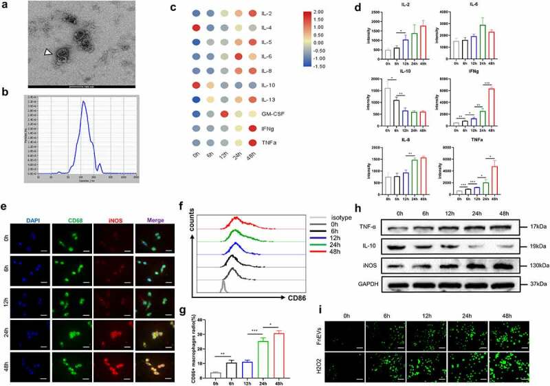
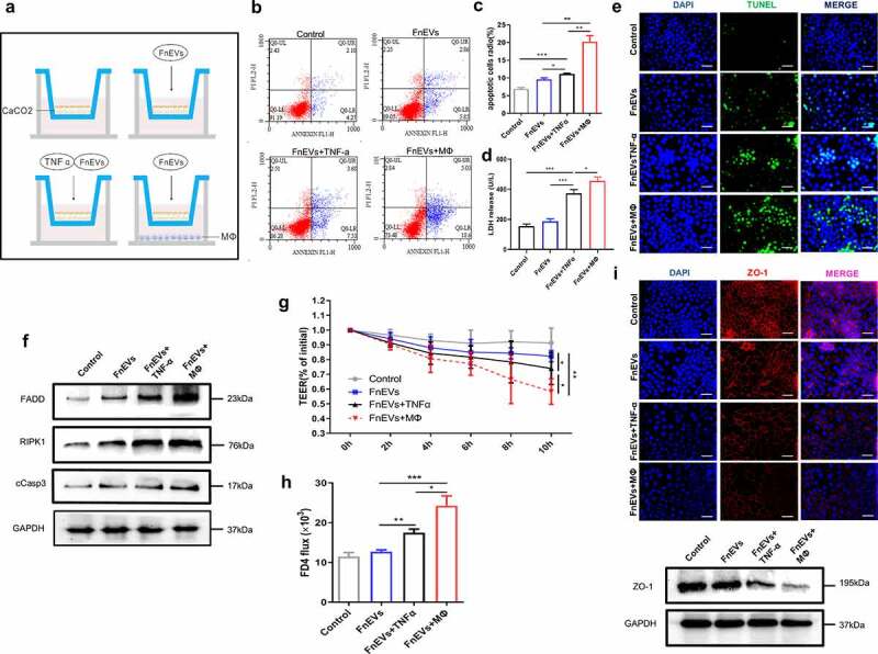
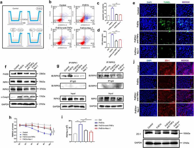
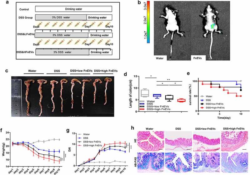
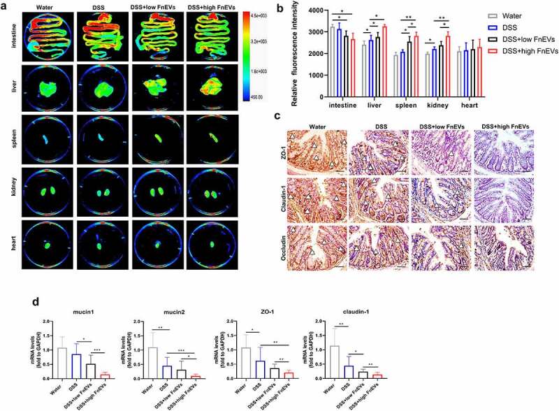
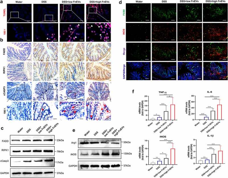
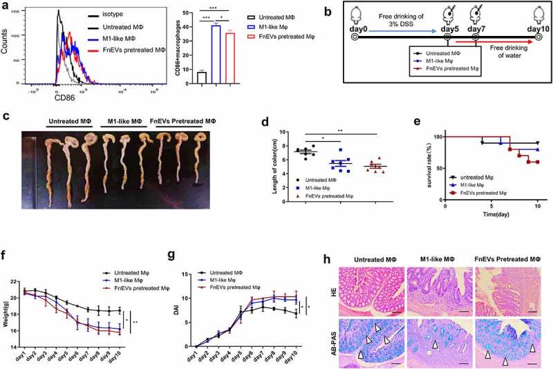
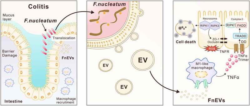
Microbial factors that mediate microbes-host interaction in ulcerative colitis (UC), a chronic disease seriously affecting human health, are not fully known. The emerging oncobacterium Fusobacterium nucleatum (Fn) secretes extracellular vesicles carrying several types of harmful molecules in the intestine which can alter microbes-host interaction, especially the epithelial homeostasis in UC. However, the mechanism is not yet clear. Previously, we isolated EVs by the ultracentrifugation of Fn culture media and characterized them as the potent inducer of pro-inflammatory cytokines. Here, we examined the mechanism in detail. We found that in macrophage/Caco-2 co-cultures, FnEVs significantly promoted epithelial barrier loss and oxidative stress damage, which are related to epithelial necroptosis caused by the activation of receptor-interacting protein kinase 1 (RIPK1) and receptor-interacting protein kinase 3 (RIPK3). Furthermore, FnEVs promoted the migration of RIPK1 and RIPK3 into necrosome in Caco2 cells. Notably, these effects were reversed by TNF-α neutralizing antibody or Necrostatin-1 (Nec-1), a RIPK1 inhibitor. This suggested that FADD-RIPK1-caspase-3 signaling is involved in the process. Moreover, the observed effects were verified in the murine colitis model treated with FnEVs or by adoptive transfer of FnEVs-trained macrophages. In conclusion, we propose that RIPK1-mediated epithelial cell death promotes FnEVs-induced gut barrier disruption in UC and the findings can be used as the basis to further investigate this disease.

Keywords: Fusobacterium nucleatum; extracellular vesicles; macrophages; necroptosis; oncobacterium; ulcerative colitis.


Figures

Figure 1.
Figure 1.
FnEVs induce a significant pro-inflammatory profiles and ROS in macrophages. (a)TEM of isolated FnEVs.(b) Size distribution of FnEVs analyzed by NTA. (c) Heatmap of Luminex analysis of culture supernatants of PBMC-derived macrophages stimulated with FnEVs at 0, 6, 12, 24 and 48 h. (d) Statistically significantly upregulated or downregulated cytokines measured by antibody-protein chip assay. (e) Double staining of CD68 (green) and iNOS (red). Nuclear counterstaining is provided with DAPI (blue). Scale bar = 50 um. (f) Flow cytometry analysis of M1 marker CD86 expression. (g) Bar graphs show the relative mean ratio of CD86+ cells in CD68+ population. (h) Immunoblot analysis of protein extracts from PBMC-derived macrophages with the indicated antibodies. (i) ROS was detected with a DCFH-DA staining. Scale bar = 100 um. *p < .05, **p < .01, ***p < .001. All the results were repeated three times. All data were presented as means ± SD (n = 3). PBMC, peripheral blood mononuclear cell
Figure 2.
Figure 2.
FnEVs promote TNF-α induced Caco2 cells death and barrier loss. (a) Diagram of groups. (b) Apoptosis was analyzed by using the annexin V FITC/PI assay. (c) Bar graphs show the relative mean of Caco2 cells apoptosis rate in different view fields. (d) The releasing levels of LDH were detected. (e) Representative images of TUNEL stainings of Caco-2 cells (green, TUNEL positive; blue, DAPI). Scale bar = 50 um. (f) Immunoblot analysis of protein extracts from Caco2 cells with the indicated antibodies. (g) Caco-2 cells were plated on a permeable membrane for TEER at different time points. (h) FITC-dextran flux was measured using Caco-2 cells grown to maximum TEER. (i) Immunofluorescence results of immunofluorescence localization of the TJ protein ZO-1(red, ZO-1 positive; blue, DAPI) and immunoblot analysis of ZO-1 protein. Scale bar = 50 um. *p < .05, **p < .01, ***p < .001. All the results were repeated three times. All data were presented as means ± SD (n = 3). TEER, transepithelial electrical resistance measurements
Figure 3.
Figure 3.
FnEVs potentiate caco-2 cells barrier loss through RIPK1-mediated cell death pathway. (a) Diagram of groups. (b) Apoptosis was analyzed by using the annexin V FITC/PI assay. (c) Bar graphs show the relative mean of Caco-2 cells apoptosis rate in different view fields. (d) The releasing levels of LDH were detected. (e) Representative images of TUNEL stainings of Caco-2 cells (green, TUNEL positive; blue, DAPI). Scale bar = 50 um. (f) Immunoblot analysis of protein extracts from Caco-2 cells with the indicated antibodies. (g) Immunoprecipitation of RIPK3 with its antibody caused coimmunoprecipitation of RIPK1 in Caco-2 cells. (h) Caco-2 cells were plated on a permeable membrane for TEER at different time points. (i) FITC-dextran flux was measured using Caco-2 cells grown to maximum TEER. (j) Representative images of localization of the TJ protein ZO-1(red, ZO-1 positive; blue, DAPI) stainings of Caco-2 cells and immunoblot analysis of ZO-1 protein. Scale bar = 50 um. *p < .05, **p < .01, ***p < .001. All the results were repeated three times. All data were presented as means ± SD (n = 3). TEER, transepithelial electrical resistance measurements
Figure 4.
Figure 4.
FnEVs aggravate clinical signs of DSS-induced acute colitis. (a) Experimental design outlining the DSS-induced colitis mice model and FnEVs treatment protocol. (b)Localization of FnEVs labeled with Cy7 at 12 h after intragastric administration. (c) Photograph of the representative colon on day 10 after colitis induction. (d) Colon length was measured on day 10 after colitis induction. (e) Effect of FnEVs on the survival rate of DSS-treated mice. (f) Effect of FnEVs on the bodyweight of 3% DSS-treated mice. (g) clinical DAI was assessed following DSS exposure. (h) H&E staining, AB-PAS staining of colon sections on day 10 after colitis induction. Scale bar = 100 um. *p < .05, **p < .01, ***p < .001. All data were presented as the means ± SD (n = 6 mice per group). DSS, dextran sulfate sodium; DAI, disease activity index. H&E, hematoxylin and eosin; AB-PAS, Alcian blue-periodic acid-Schiff
Figure 5.
Figure 5.
FnEVs increase gut barrier leakage in experimental colitis models. (a) Representative images and ex vivo imaging with the intestine, liver, heart, spleen and kidney of mice. (b) Relative fluorescence intensity of translocated EGFP-labeled E.coli in every tissues. (c) Representative images of immunohistochemical stainings of ZO-1, claudin-1 and occludin in the colon on day 3 after colitis induction. Scale bar = 50 um. (d) The relative mRNA level of mucin1, mucin2, ZO-1 and claudin-1 was detected in colon samples on day 3 after colitis induction. *p < .05, **p < .01, ***p < .001. All data were presented as the means ± SD (n = 6 mice per group)
Figure 6.
Figure 6.
RIPK1 mediated epithelial cell death drives exacerbated barrier loss in FnEVs-treated colitis mice. (a) Representative images of TUNEL stainings of colon sections on day 3 after colitis induction (red, TUNEL positive; blue, DAPI). Scale bar = 50 um (up) or 20 um (down). (b) Representative images of immunohistochemical stainings of FADD, RIPK1 and cCASP3 in the colon sections on day 3 after colitis induction. Scale bar = 50 um or 20 um (downmost). (c) Immunoblot analysis of protein extracts from colon samples with the indicated antibodies. (d) Double staining of F4/80 (green) and iNOS (red) on day 3 after colitis induction. Nuclear counterstaining is provided with DAPI (blue). Scale bar = 100 um. (e) Immunoblot analysis of protein extracts from colon samples with the indicated antibodies. (f) The relative mRNA level of TNF-α, IL-6, iNOS and IL-1β was detected in colon samples on day 3 after colitis induction. *p < .05, **p < .01, ***p < .001. All data were presented as the means ± SD (n = 6 mice per group)
Figure 7.
Figure 7.
Adoptive transfer of FnEVs-pretreated macrophages promoted intestinal epithelial death. (a) Flow cytometry analysis of M1 marker CD86 expression and bar graphs show the relative mean ratio of CD86+ cells in F4/80+ population. (b) Experimental design outlining DSS-induced colitis mice model and adoptive transfer assay protocol. (c) Photograph of the representative colon on day 10 after colitis induction. (d) Colon length was measured on day 10 after administration of 3% DSS. (e) Effect of FnEVs-pretreated macrophages on the survival rate of DSS-treated mice. (f) Effect of FnEVs-pretreated macrophages on the bodyweight of 3% DSS-treated mice. (g) clinical DAI was assessed following DSS exposure. (h) H&E staining and AB-PAS staining of colon sections on day 10. Scale bar = 100 um. *p < .05, **p < .01, ***p < .001. All data were presented as the means ± SD (n = 6 mice per group)
Figure 8.
Figure 8.
Proposed mechanism of FnEVs-driven intestinal mucosal barrier dysfunction in UC. Modulation of intestinal mucosal responses by Fn is thought to potentiate both Fn persistence and the development of chronic intestinal inflammation. Release of extracellular vesicles (EVs) by Fn promote macrophages to secrete proinflammatory factors that activate RIPK1 mediated cell death signals in intestinal epithelial cells, leading to the disruption of intercellular tight junctions. Thus, increased bacterial translocation induces an amplification loop of inflammation that inhibited intestinal epithelial renewal, and accelerate the progression of colitis

References

    1. Ungaro R, Mehandru S, Allen PB, Peyrin-Biroulet L, Colombel JF.. Ulcerative colitis. Lancet. 2017;389(10080):1756–20. doi:10.1016/S0140-6736(16)32126-2. - DOI - PMC - PubMed
    1. Colquhoun C, Duncan M, Grant G. Inflammatory bowel diseases: host-microbial-environmentalinteractions in dysbiosis. Diseases. 2020;8(2):13. doi:10.3390/diseases8020013. - DOI - PMC - PubMed
    1. Ducarmon QR, Zwittink RD, Hornung B, Van Schaik W, Young VB, Kuijper EJ. Gut microbiota and colonization resistance against bacterial enteric infection. Microbiol Mol Biol Rev. 2019;83:3. - PMC - PubMed
    1. Gunther C, Neumann H, Neurath MF, Becker C. Apoptosis, necrosis and necroptosis: cell death regulation in the intestinal epithelium. Gut. 2013;62(7):1062–1071. doi:10.1136/gutjnl-2011-301364. - DOI - PubMed
    1. Siggers RH, Hackam DJ. The role of innate immune-stimulated epithelial apoptosis during gastrointestinal inflammatory diseases. Cell Mol Life Sci. 2011;68(22):3623–3634. doi:10.1007/s00018-011-0821-4. - DOI - PMC - PubMed

Publication types

MeSH terms

Substances

LinkOut - more resources