. 2021 Jun 1;320(6):H2211-H2221.
doi: 10.1152/ajpheart.00056.2021. Epub 2021 Mar 26.

Regenerative cross talk between cardiac cells and macrophages

Affiliations

Regenerative cross talk between cardiac cells and macrophages

Alexander J Whitehead et al. Am J Physiol Heart Circ Physiol. .

Abstract

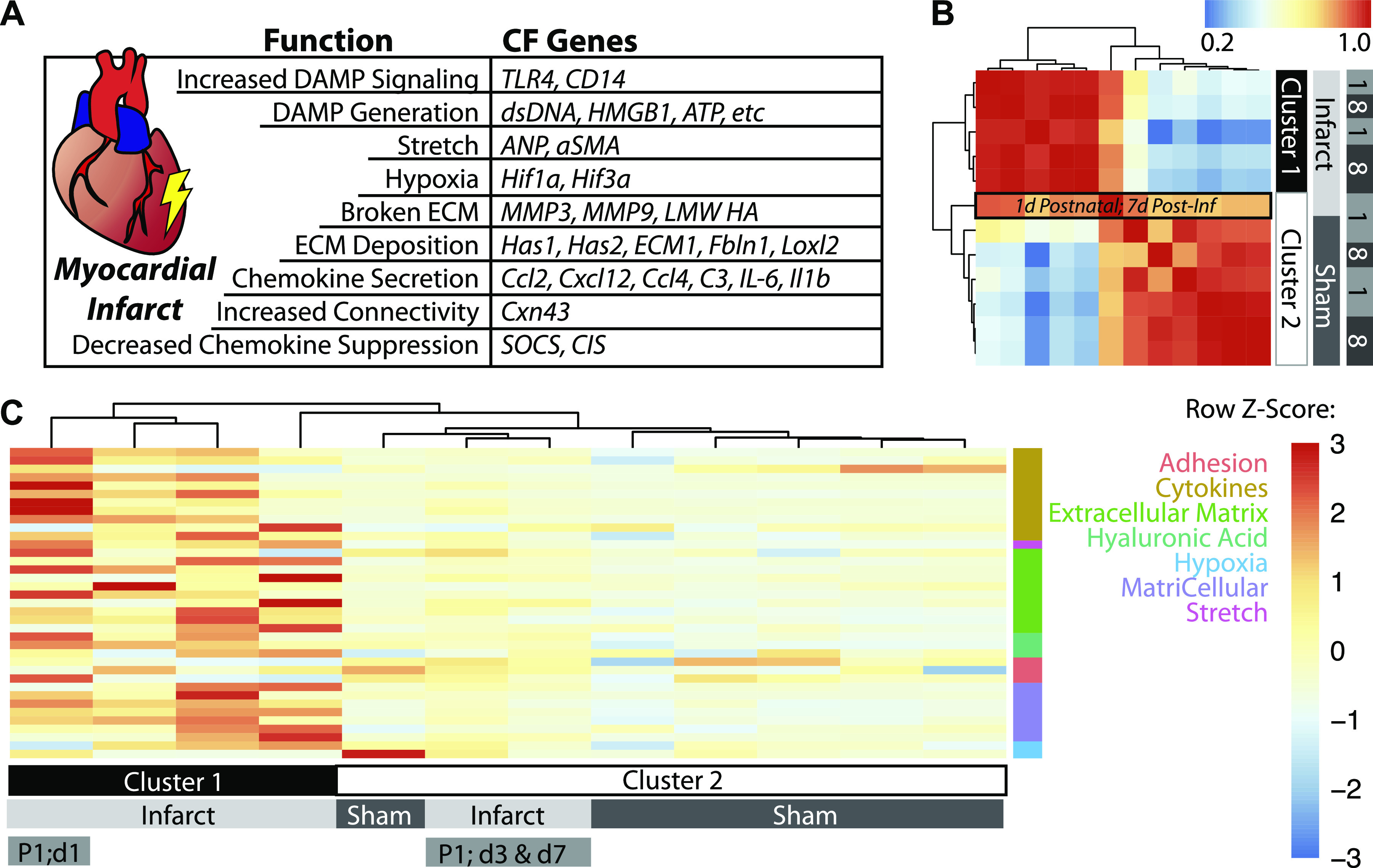
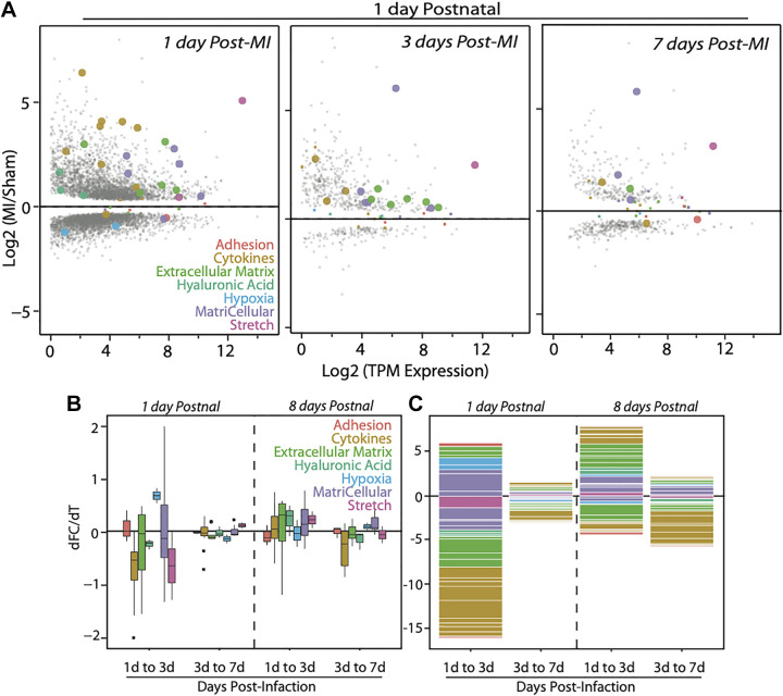
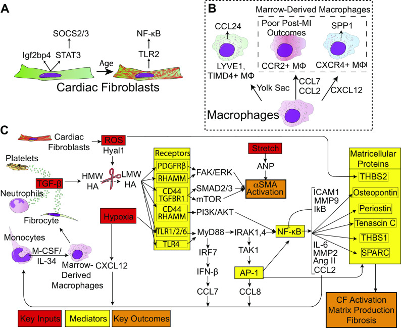
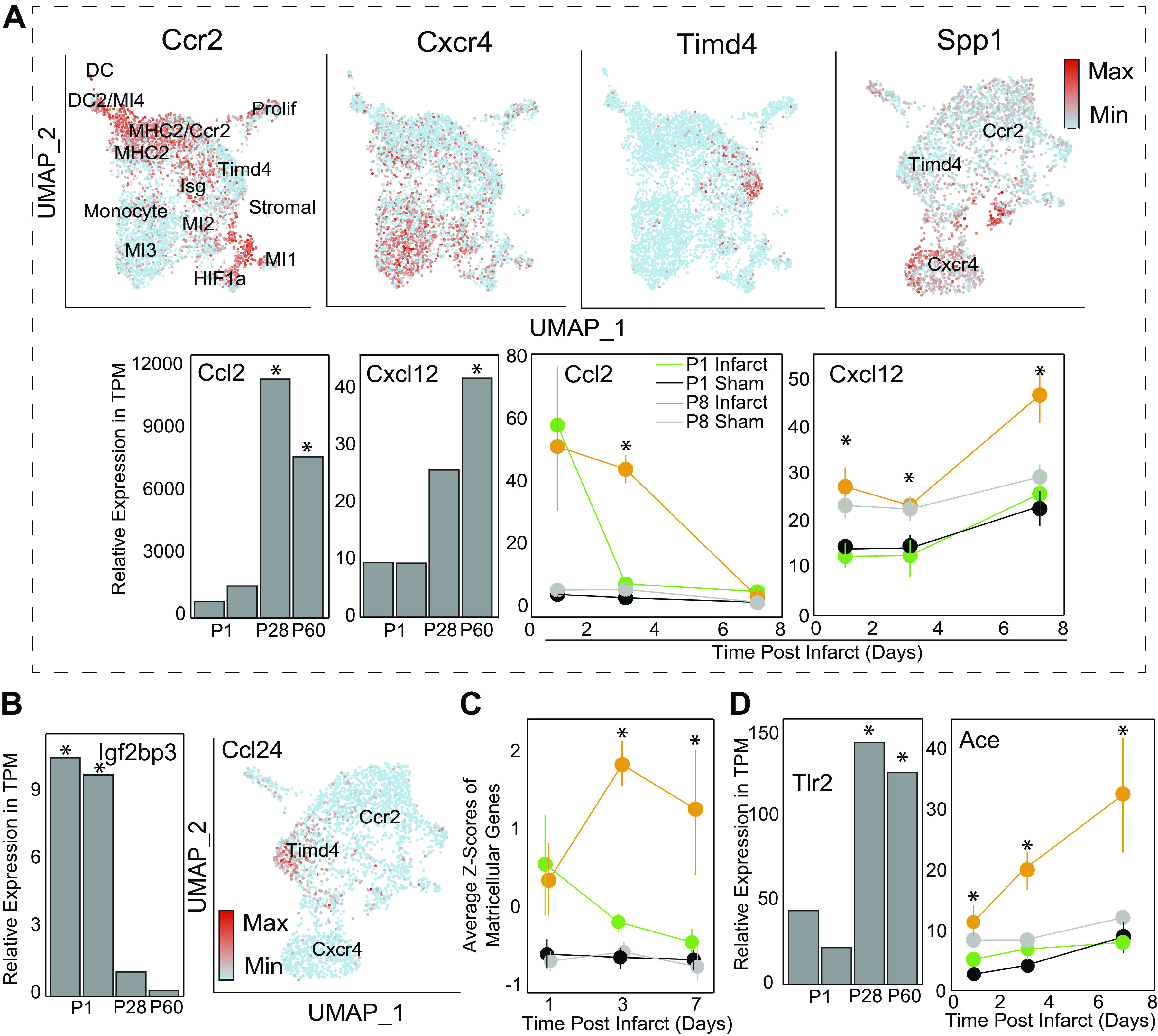
Aside from the first week postnatal, murine heart regeneration is restricted and responses to damage follow classic fibrotic remodeling. Recent transcriptomic analyses have suggested that significant cross talk with the sterile immune response could maintain a more embryonic-like signaling network that promotes acute, transient responses. However, with age, this response-likely mediated by neonatal yolk sac macrophages-then transitions to classical macrophage-mediated, cardiac fibroblast (CF)-based remodeling of the extracellular matrix (ECM) after myocardial infarction (MI). The molecular mechanisms that govern the change with age and drive fibrosis via inflammation are poorly understood. Using multiple ribonucleic acid sequencing (RNA-Seq) datasets, we attempt to resolve the relative contributions of CFs and macrophages in the bulk-healing response of regenerative (postnatal day 1) and nonregenerative hearts (postnatal day 8+). We performed an analysis of bulk RNA-Seq datasets from myocardium and cardiac fibroblasts as well as a single-cell RNA-Seq dataset from cardiac macrophages. MI-specific pathway differences revealed that nonregenerative hearts generated more ECM and had larger matricellular responses correlating with inflammation, produced greater chemotactic gradients to recruit macrophages, and expressed receptors for danger-associated molecular patterns at higher levels than neonates. These changes could result in elevated stress-response pathways compared with neonates, converging at NF-κB and activator protein-1 (AP-1) signaling. Profibrotic gene programs, which greatly diverge on day 3 post MI, lay the foundation for chronic fibrosis, and thus postnatal hearts older than 7 days typically exhibit significantly less regeneration. Our analyses suggest that the macrophage ontogenetic shift in the heart postnatally could result in detrimental stress signaling that suppresses regeneration.NEW & NOTEWORTHY Immediately postnatal mammalian hearts are able to regenerate after infarction, but the cells, pathways, and molecules that regulate this behavior are unclear. By comparing RNA-Seq datasets from regenerative mouse hearts and older, nonregenerative hearts, we are able to identify biological processes that are hallmarks of regeneration. We find that sterile inflammatory processes are upregulated in nonregenerative hearts, initiating profibrotic gene programs 3 days after myocardial infarction that can cause myocardial disease.

Keywords: fibrosis; gene expression and regulation; inflammation; myocardial infarction; myocardial regeneration.


Conflict of interest statement

No conflicts of interest, financial or otherwise, are declared by the authors.

Figures

Figure 1.
Figure 1.
Transcriptomic analyses of infarct and sham bulk highlight changes in specific remodeling pathways. A: table of gene groupings and corresponding genes that literature suggest are differentially expressed with myocardial infarction. B: heatmap of bulk RNA-Seq data (averaged across three mice per group) showing hierarchical clustering of myocardia based on infarction. The only MI group that clustered with sham controls is indicated in the black box and is the 1 day postnatal MI group after 7 days of healing. C: heatmap of the TPM Z-scores of the 37 genes, with rows grouped by functional process and columns clustered using K-means. Cluster 1 denotes all the infarct groups from day 8 postnatal mice (independent of days postinfarct) and the first time point after infarction for day 1 mice. Days 3 and 7 postinfarct groups of postnatal day 1 infarcted hearts clustered with sham samples, indicating their return to baseline in as little as 3 days. MI, myocardial infarction; RNA-Seq, ribonucleic acid sequencing; TPM, transcripts per kilobase million.
Figure 2.
Figure 2.
MI induces the largest transcriptomic changes initially in younger mice but older mice maintain significant transcriptional differences. A: MA plots show the relationship between the MI/sham gene ratio (i.e., fold change) and the transcript per million reads for murine myocardia infarcted 1 day postnatal and chased for up to 7 days post-MI as indicated in each panel. Gene functional groupings listed are annotated in the figure by color and with large data points for visualization (when individual gene is statistically significant by Wald test with Benjamini–Hochberg correction; q < 0.05). Gray data points are coded by size for significance but are not the 37 literature-identified genes used in the rest of the analysis. B: box-and-whisker plot indicate the changes, broken down by category, in gene ratio (MI/sham) between transcriptome sampled over time as indicated at bottom. Data for mice infarcted 1 day and 8 days postnatal are separated by a dashed line. C: stacked bar plot annotates the log2 of the fold change for genes of the indicated ontologies/functions (genes with ratios less than one result in a negative number). MI, myocardial infarction.
Figure 3.
Figure 3.
Model for cellular and molecular changes with age and infarction. A: age-related transcriptional differences in fibroblasts. As fibroblasts progress through development, they upregulate TLR2 expression and downregulate STAT3 and Igf2bp3. B: macrophage composition of the heart shifts from CCL24-producing YS lineage cells to two ontogenies of BMDMs: CXCR4+ and CCR2+. C: proposed molecular mechanism for nonregenerative cardiac fibroblasts. MI in postnatal day 8 hearts generate inflammatory ligands (red) to a greater extent than postnatal day 1 hearts, which are sensed to a greater extent by TLR2, and ultimately result in NF-κB and AP-1 activation (yellow). Lastly, the signal propagation results in excess matricellular protein and ECM deposition (orange). Signaling from receptors and continuing to the right is believed to occur in fibroblasts. AP-1, activator protein-1; BMDM, bone marrow-derived macrophages; ECM, extracellular matrix; MI, myocardial infarction; NF, necrosis factor; TLR, Toll-like receptors.
Figure 4.
Figure 4.
P8 cytokines recruit BMDMs deficient in growth proteins to an increasingly sensitive inflammatory microenvironment. A: post-MI Macrophages were clustered according to original (4) study’s markers. Macrophages either express high levels of CCR2 (classical monocytes), CXCR4 (late-phase monocytes) or Timd4 (YS macrophages). Spp1 graph was generated by examining a subset of cells based on expression of previous three genes and then re-clustering and demonstrates high Spp1 expression by Cxcr4+ macrophages. Bar graphs are from fibroblast dataset, line graphs are from bulk tissue, and UMAPs are from single-cell macrophages. B: growth factors identified from original bulk analysis identified in neonatal CF and Mac populations, respectively. Ccl24 was from same Seurat object that generated Spp1 plot. C: average Z-scores of matricellular genes demonstrating similar trends between genes by group and timepoint. Individual genes and functional groupings are listed in Supplemental Table 1. D: TLR2 expression in fibroblasts on postnatal days 1, 28, and 60 (34) is plotted here. Significance is indicated as *P < 0.05 as determined by exact test with Benjamini–Hochberg correction. Conversely, ACE expression is plotted from the bulk heart dataset (33), with significance determined by Wald test with Benjamini–Hochberg correction, P < 0.05. ACE, angiotensin converting enzyme; BMDM, bone marrow-derived macrophages; CF, cardiac fibroblasts; ECM, extracellular matrix; MI, myocardial infarction; NF, necrosis factor; P8, postnatal day 8; TLR, Toll-like receptors; YS, yolk sac.

Comment in

  • Find the stimulus, save the heart: a heroes' story.
    Westbury B, Bolus D, DeLeon-Pennell KY. Westbury B, et al. Am J Physiol Heart Circ Physiol. 2021 Jun 1;320(6):H2185-H2187. doi: 10.1152/ajpheart.00194.2021. Epub 2021 Apr 23. Am J Physiol Heart Circ Physiol. 2021. PMID: 33891514 Free PMC article. No abstract available.

Similar articles

Cited by

References

    1. Perbellini F, Watson SA, Bardi I, Terracciano CM. Heterocellularity and cellular cross-talk in the cardiovascular system. Front Cardiovasc Med 5: 143, 2018. doi:10.3389/fcvm.2018.00143. - DOI - PMC - PubMed
    1. King KR, Aguirre AD, Ye YX, Sun Y, Roh JD, Ng RP, Kohler RH, Arlauckas SP, Yoshiko V, Savo A, Sadreyev RI, Kelly M, Fitzgibbons TP, Fitzgerald KA, Mitchison T, Libby P, Nahrendorf M, Weissleder R. IRF3 and type i interferons fuel a fatal response to myocardial infarction. Nat Med 23: 1481–1487, 2017. doi:10.1038/nm.4428. - DOI - PMC - PubMed
    1. Prabhu SD, Frangogiannis NG. The biological basis for cardiac repair after myocardial infarction. Circ Res 119: 91–112, 2016. doi:10.1161/CIRCRESAHA.116.303577. - DOI - PMC - PubMed
    1. Dick SA, Macklin JA, Nejat S, Momen A, Clemente-Casares X, Althagafi MG, Chen J, Kantores C, Hosseinzadeh S, Aronoff L, Wong A, Zaman R, Barbu I, Besla R, Lavine KJ, Razani B, Ginhoux F, Husain M, Cybulsky MI, Robbins CS, Epalman S. Self-renewing resident cardiac macrophages limit adverse remodeling following myocardial infarction. Nat Immunol 20: 29–39, 2019. doi:10.1038/s41590-018-0272-2. - DOI - PMC - PubMed
    1. Ma Y, Mouton AJ, Lindsey ML. Cardiac macrophage biology in the steady-state heart, the aging heart, and following myocardial infarction. Transl Res 191: 15–28, 2018. doi:10.1016/j.trsl.2017.10.001. - DOI - PMC - PubMed

Publication types

MeSH terms