. 2021 Mar 26;16(3):e0247335.
doi: 10.1371/journal.pone.0247335. eCollection 2021.

Structure of the EphB6 receptor ectodomain

Affiliations

Structure of the EphB6 receptor ectodomain

Emilia O Mason et al. PLoS One. .

Abstract

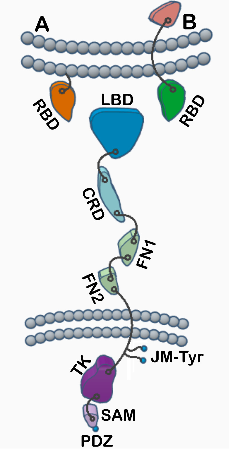
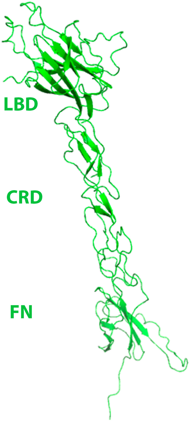
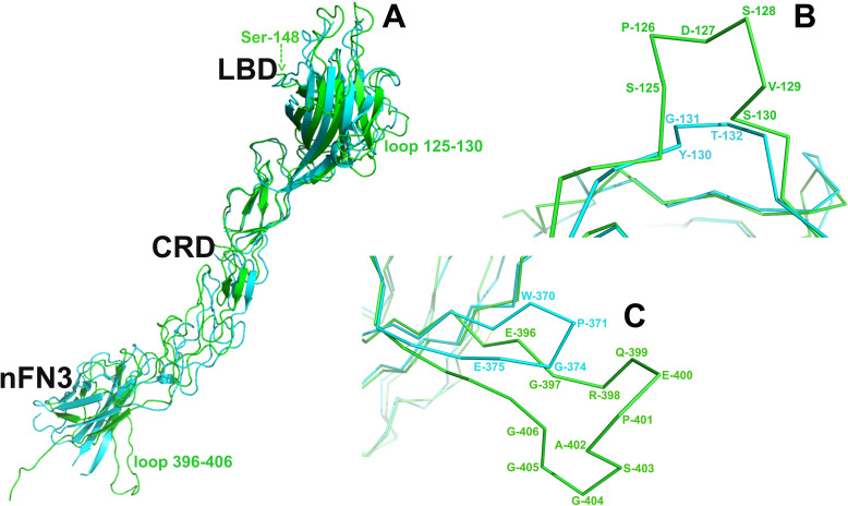
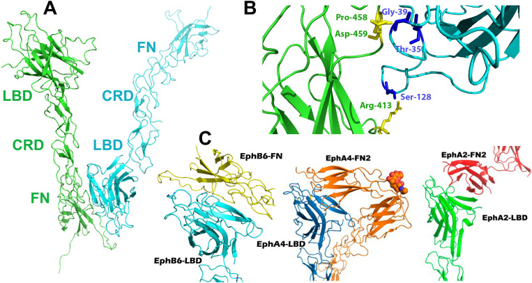
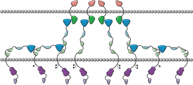
Eph receptors are the largest group amongst the receptor tyrosine kinases and are divided into two subgroups, A and B, based on ligand binding specificities and sequence conservation. Through ligand-induced and ligand-independent activities, Ephs play central roles in diverse biological processes, including embryo development, regulation of neuronal signaling, immune responses, vasculogenesis, as well as tumor initiation, progression, and metastasis. The Eph extracellular regions (ECDs) are constituted of multiple domains, and previous structural studies of the A class receptors revealed how they interact with ephrin ligands and simultaneously mediate Eph-Eph clustering necessary for biological activity. Specifically, EphA structures highlighted a model, where clustering of ligand-bound receptors relies on two distinct receptor/receptor interfaces. Interestingly, most unliganded A class receptors also form an additional, third interface, between the ligand binding domain (LBD) and the fibronectin III domain (FN3) of neighboring molecules. Structures of B-class Eph ECDs, on the other hand, have never been reported. To further our understanding of Eph receptor function, we crystallized the EphB6-ECD and determined its three-dimensional structure using X-ray crystallography. EphB6 has important functions in both normal physiology and human malignancies and is especially interesting because this atypical receptor innately lacks kinase activity and our understanding of the mechanism of action is still incomplete. Our structural data reveals the overall EphB6-ECD architecture and shows EphB6-LBD/FN3 interactions similar to those observed for the unliganded A class receptors, suggesting that these unusual interactions are of general importance to the Eph group. We also observe unique structural features, which likely reflect the atypical signaling properties of EphB6, namely the need of co-receptor(s) for this kinase-inactive Eph. These findings provide new valuable information on the structural organization and mechanism of action of the B-class Ephs, and specifically EphB6, which in the future will assist in identifying clinically relevant targets for cancer therapy.


Conflict of interest statement

The authors have declared that no competing interests exist.

Figures

Fig 1
Fig 1. Domain organization of Eph receptors and ephrin ligands.
Class A and class B receptor-binding domains (RBDs), ligand-binding domain (LBD), cysteine-rich domain (CRD), fibronectin-type-3 region (FN1 and FN2), juxtamembrane tyrosine residues (JM-Tyr), tyrosine kinase (TK), sterile alpha motif (SAM), PDZ-binding motif (PDZ).
Fig 2
Fig 2. Purification and crystallization of EphB6-ECD.
(A) EphB6-ECD-Fc after initial Protein A Sepharose purification; (B) EphB6-ECD upon addition of thrombin and Protein A Sepharose beads; (C) The finally purified EphB6-ECD, showing the expected molecular weight of ~65 kDa; (D) EphB6-ECD protein crystals.
Fig 3
Fig 3. Binding of the EphB6-ECD to the ephrin-B2-Fc ligand.
(A) EphB6-ECD binds to ephrin-B2-Fc with a Kd value of 8.85 E-8 M as measured on a Blitz instrument; (B) Size-exclusion FPLC on a Superdex200 column reveals a 1:1 stoichiometry for the EphB6-ECD/ephrin-B2 complex.
Fig 4
Fig 4. EphB6-ECD structure showing the ligand binding (LBD), cysteine rich (CRD), and fibronectin (FN) domains.
The ectodomain has a rigid, rod-like conformation with limited flexibility between the subdomains. The LBD has a jelly-roll folding topology, while the CRD includes β-strands arranged as a β-sandwich, with several disulfide bonds stabilizing the structure. The N-terminal FN adopts a typical immunoglobulin-like fold.
Fig 5
Fig 5
(A) Superimposition of the EphB6 and EphA2 ectodomains. The rmsd value between the C-alpha atoms is 3.803Å. The EphB6-ECD is colored in green and the EphA2-ECD—in cyan. Arrow shows the location of Ser-148 preceding the ‘Super Serine’ loop (residues 151–161) that is not visible in our electron density map. LBD, ligand-binding domain; CRD, cysteine-rich domain; nFN3, N-terminal fibronectin 3 domain. (B) Close-up of the superimposed LBD H-I loop of the two molecules (EphB6 residues 125–130). (C) Close-up of the superimposed FN domain loop (EphB6 residues 396–406) of the two molecules.
Fig 6
Fig 6
(A) Head-to-tail interactions in EphB6-ECD. Similar to the previously published A-class ectodomain structures, the LBD (cyan) and FN (green) domains of neighboring EphB6 molecules form an interface in the unliganded receptors. (B) The zoom-in shows the interacting amino acid residues. (C) Comparison of the head-to-tail interactions in EphB6-, A4- and A2-ECDs. (Left) EphB6-LBD (cyan) bound to EphB6-FN of a neighboring molecule (yellow) in the crystals of the unliganded EphB6-ECD; (Center) EphA4-LBD (blue) bound to the EphA4-FN of a neighboring molecule (orange) in the crystals of unliganded EphA4-ECD; (Right) EphA2-LBD (green) bound to the EphA2-FN of a neighboring molecule (red) in the crystals of unliganded EphA2-ECD.
Fig 7
Fig 7. Schematic representation of the head to tail Eph-ECD interactions.
The Eph receptors are in blue/green/purple and are interacting with the ephrins in green/orange. The LBDs of the ephrin-free Ephs (in blue) and the FN3 regions (in light green) of their neighbors are interacting. Intracellular, juxtamembrane Tyrosines are shown as small circles. They get fully phosphorylated only when biologically active heterotetramers and higher-order Eph/ephrin assemblies form after the initial ligand binding events. The cell membrane of the two interacting cells are in grey.

References

    1. Lisabeth EM, Falivelli G, Pasquale EB. Eph Receptor Signaling and Ephrins. Cold Spring Harb Perspect Biol. 2013;5. 10.1101/cshperspect.a009159 - DOI - PMC - PubMed
    1. Himanen JP. Ectodomain structures of Eph receptors. Semin Cell Dev Biol. 2012;23: 35–42. 10.1016/j.semcdb.2011.10.025 - DOI - PubMed
    1. Janes PW, Nievergall E, Lackmann M. Concepts and consequences of Eph receptor clustering. Semin Cell Dev Biol. 2012;23: 43–50. 10.1016/j.semcdb.2012.01.001 - DOI - PubMed
    1. Himanen J-P, Nikolov DB. Eph signaling: a structural view. Trends Neurosci. 2003;26: 46–51. 10.1016/s0166-2236(02)00005-x - DOI - PubMed
    1. Pasquale EB. Eph receptor signalling casts a wide net on cell behaviour. Nat Rev Mol Cell Biol. 2005;6: 462–475. 10.1038/nrm1662 - DOI - PubMed

Publication types

LinkOut - more resources