. 2021 Mar 26;16(3):e0249324.
doi: 10.1371/journal.pone.0249324. eCollection 2021.

Prioritization of candidate genes for a South African family with Parkinson's disease using in-silico tools

Affiliations

Prioritization of candidate genes for a South African family with Parkinson's disease using in-silico tools

Boiketlo Sebate et al. PLoS One. .

Abstract

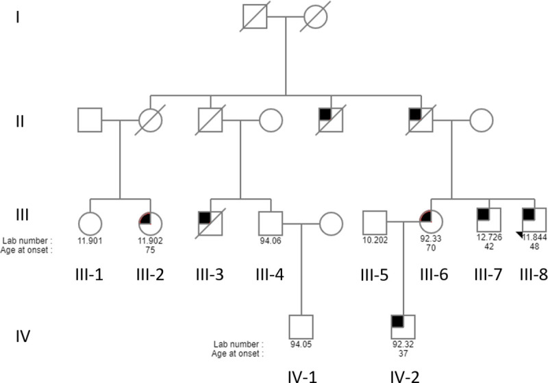
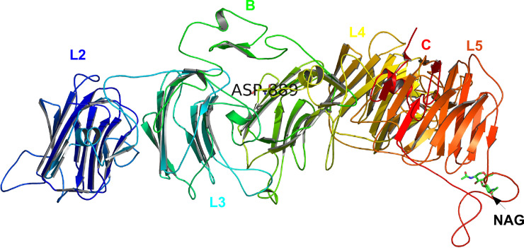
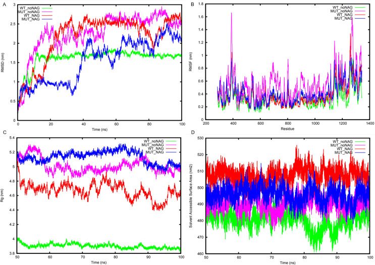
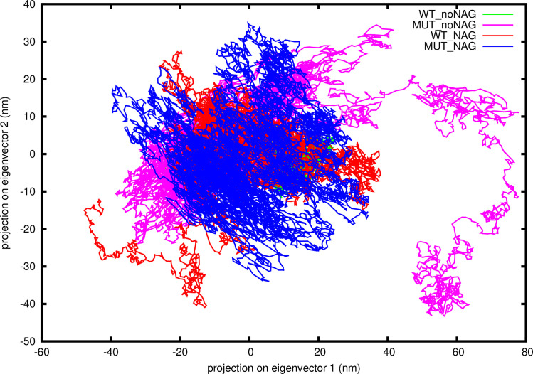
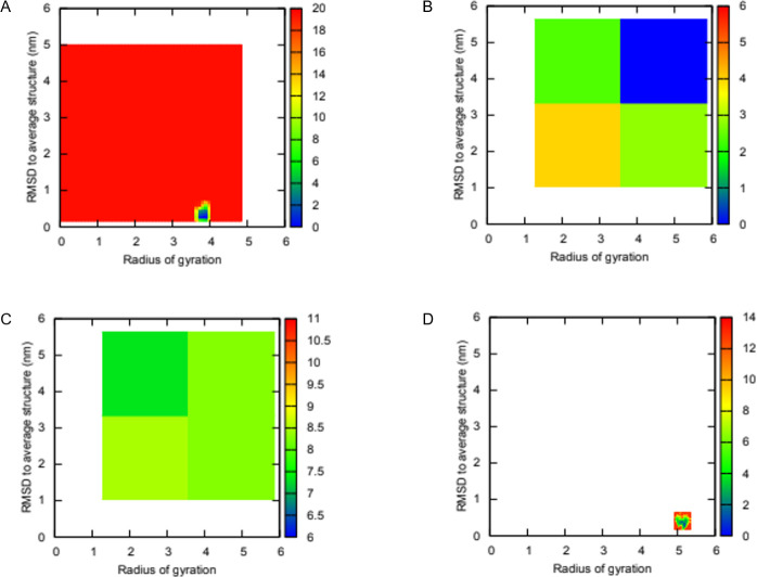
Parkinson's disease (PD) is a neurodegenerative disorder exhibiting Mendelian inheritance in some families. Next-generation sequencing approaches, including whole exome sequencing (WES), have revolutionized the field of Mendelian disorders and have identified a number of PD genes. We recruited a South African family with autosomal dominant PD and used WES to identify a possible pathogenic mutation. After filtration and prioritization, we found five potential causative variants in CFAP65, RTF1, NRXN2, TEP1 and CCNF. The variant in NRXN2 was selected for further analysis based on consistent prediction of deleteriousness across computational tools, not being present in unaffected family members, ethnic-matched controls or public databases, and its expression in the substantia nigra. A protein model for NRNX2 was created which provided a three-dimensional (3D) structure that satisfied qualitative mean and global model quality assessment scores. Trajectory analysis showed destabilizing effects of the variant on protein structure, indicated by high flexibility of the LNS-6 domain adopting an extended conformation. We also found that the known substrate N-acetyl-D-glucosamine (NAG) contributed to restoration of the structural stability of mutant NRXN2. If NRXN2 is indeed found to be the causal gene, this could reveal a new mechanism for the pathobiology of PD.


Conflict of interest statement

The authors have declared that no competing interests exist.

Figures

Fig 1
Fig 1. Pedigree of the South African family ZA253.
For readability and confidentiality, the pedigree is greatly simplified. Circles denote females and squares depict males. The filled in symbols indicate affected individuals. The diagonal line indicates that the person is deceased, and the arrow indicates the proband. The numbers below each individual is the laboratory ID number and age at onset of disease. Branches without clinically confirmed PD or without DNA samples were omitted, but all known living family members with PD diagnoses are included.
Fig 2
Fig 2. Swissmodel predicted 3-dimensional (3D) structure for human NRXN2 with mutation p.G849D (p.G889D) in complex with N-acetyl-D-glucosamine (NAG).
Each domain is colour coded, EGF-like domains are labelled B and C, substrate NAG are labelled as well as the mutation, p.G889D in our protein model.
Fig 3
Fig 3. Trajectory analysis of wild type (WT) NRNX2 and mutant (MUT) NRNX2, both with and without NAG.
(A) root mean square deviation (RMSD) deviation of the backbone atoms. MEAN + STDEV (1.61mm ± 0.26, 2.26mm ± 0.52, 2.16mm ± 0.60 and 1.54mm ± 0.55). (B) The average RMSF fluctuation per-residue. MEAN + STDEV (0.31mm ± 0.14, 0.54mm ± 0.24, 0.37mm ± 0.18 and 0.40mm ± 0.19). (C) Radius of gyration of the backbone atoms. MEAN + STDEV (3.90mm ± 0.03, 5.01mm ± 0.10, 4.67mm ± 0.11 and 5.13mm ± 0.08). (D) Solvent accessible surface area of the protein. MEAN + STDEV (3.90mm ± 0.03, 5.01mm ± 0.10, 4.67mm ± 0.11 and 5.13mm ± 0.08). Line colors: WT_noNAG = green, MUT_noNAG = light magenta, WT_NAG = red and MUT_NAG = blue.
Fig 4
Fig 4. Two-dimensional (2D) projections of the first and second principal components for the WT NRNX2, MUT NRNX2 without NAG and WT NRNX2, MUT NRNX2 with NAG systems.
Covariance matrix after diagonalization values for each system; 237.82 nm, 612.38 nm, 508.19 nm and 554.72 nm. Line colors: WT_noNAG = green, MUT_noNAG = light magenta, WT_NAG = red and MUT_NAG = blue.
Fig 5
Fig 5. Two-dimensional (2D) free energy landscapes plotted for NRXN2 along two order parameters root mean square deviation (RMSD) to average structure and Rg.
(A) WT without NAG (B) MUT without NAG (C) WT with NAG (D) MUT with NAG. Blue regions represent low energy conformational states while red indicate high energy states.

References

    1. Barone P, Antonini A, Colosimo C, Marconi R, Morgante L, Avarello TP, et al.. The PRIAMO study: A multicenter assessment of nonmotor symptoms and their impact on quality of life in Parkinson’s disease. Mov Disord. 2009;24: 1641–1649. 10.1002/mds.22643 - DOI - PubMed
    1. Lill CM. Genetics of Parkinson’s disease. Mol Cell Probes. 2016;3: 386–396. 10.1016/j.mcp.2016.11.001 - DOI - PubMed
    1. Puschmann A. New Genes Causing Hereditary Parkinson’s Disease or Parkinsonism. Curr Neurol Neurosci Rep. 2017;17: 66. 10.1007/s11910-017-0780-8 - DOI - PMC - PubMed
    1. Zimprich A, Benet-Pagès A, Struhal W, Graf E, Eck SH, Offman MN, et al.. A mutation in VPS35, encoding a subunit of the retromer complex, causes late-onset parkinson disease. Am J Hum Genet. 2011;89: 168–175. 10.1016/j.ajhg.2011.06.008 - DOI - PMC - PubMed
    1. Vilariño-Güell C, Wider C, Ross OA, Dachsel JC, Kachergus JM, Lincoln SJ, et al.. VPS35 mutations in parkinson disease. Am J Hum Genet. 2011;89: 162–167. 10.1016/j.ajhg.2011.06.001 - DOI - PMC - PubMed