. 2021 Jul;64(7):1674-1689.
doi: 10.1007/s00125-021-05431-5. Epub 2021 Mar 26.

Fasting and fasting-mimicking treatment activate SIRT1/LXRα and alleviate diabetes-induced systemic and microvascular dysfunction

Affiliations

Fasting and fasting-mimicking treatment activate SIRT1/LXRα and alleviate diabetes-induced systemic and microvascular dysfunction

Sandra S Hammer et al. Diabetologia. 2021 Jul.

Abstract

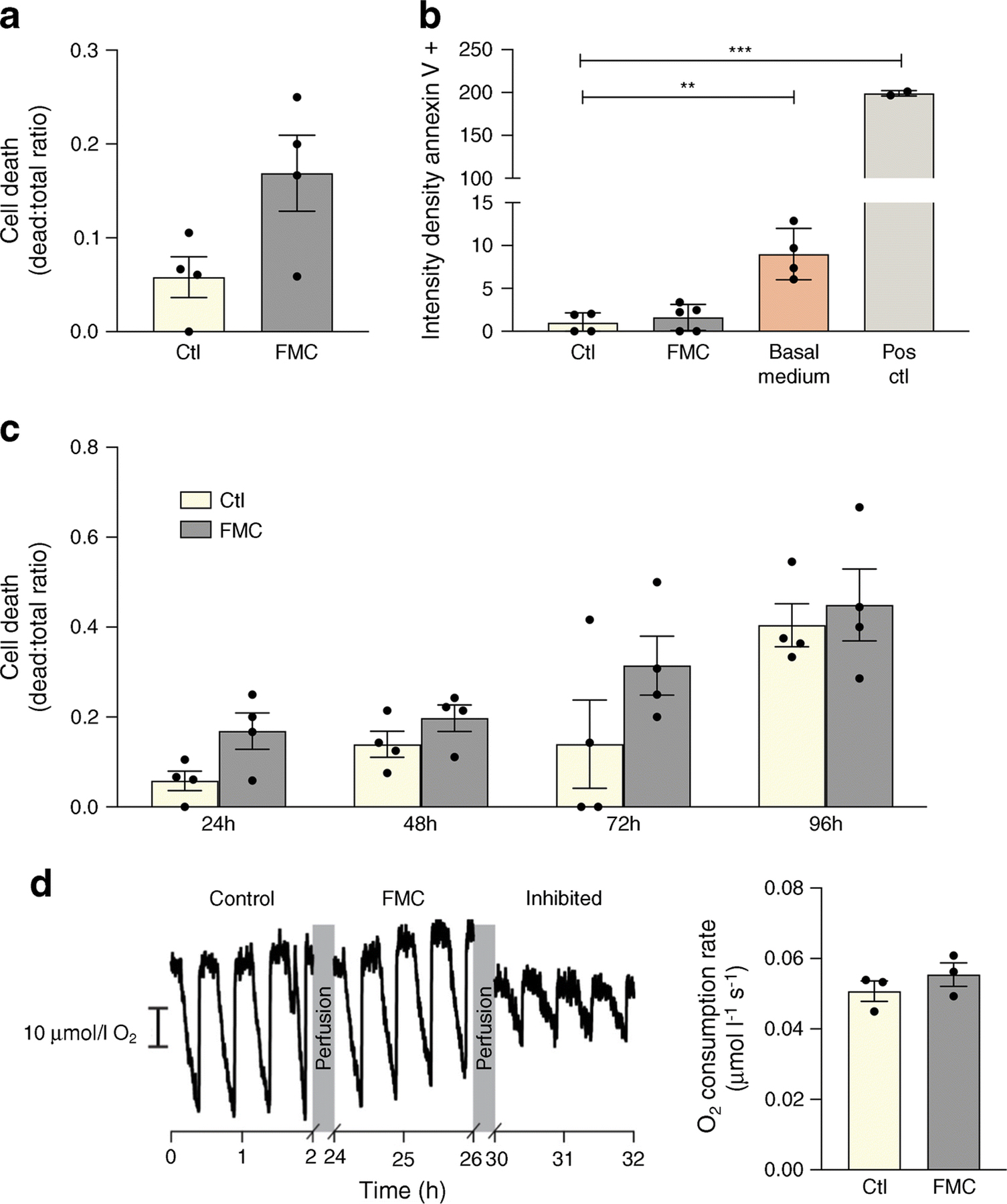
Aims/hypothesis: Homo sapiens evolved under conditions of intermittent food availability and prolonged fasting between meals. Periods of fasting are important for recovery from meal-induced oxidative and metabolic stress, and tissue repair. Constant high energy-density food availability in present-day society contributes to the pathogenesis of chronic diseases, including diabetes and its complications, with intermittent fasting (IF) and energy restriction shown to improve metabolic health. We have previously demonstrated that IF prevents the development of diabetic retinopathy in a mouse model of type 2 diabetes (db/db); however the mechanisms of fasting-induced health benefits and fasting-induced risks for individuals with diabetes remain largely unknown. Sirtuin 1 (SIRT1), a nutrient-sensing deacetylase, is downregulated in diabetes. In this study, the effect of SIRT1 stimulation by IF, fasting-mimicking cell culture conditions (FMC) or pharmacological treatment using SRT1720 was evaluated on systemic and retinal metabolism, systemic and retinal inflammation and vascular and bone marrow damage.

Methods: The effects of IF were modelled in vivo using db/db mice and in vitro using bovine retinal endothelial cells or rat retinal neuroglial/precursor R28 cell line serum starved for 24 h. mRNA expression was analysed by quantitative PCR (qPCR). SIRT1 activity was measured via histone deacetylase activity assay. NR1H3 (also known as liver X receptor alpha [LXRα]) acetylation was measured via western blot analysis.

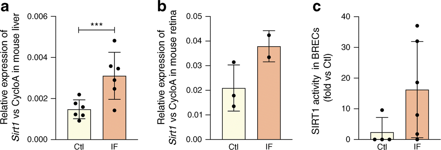
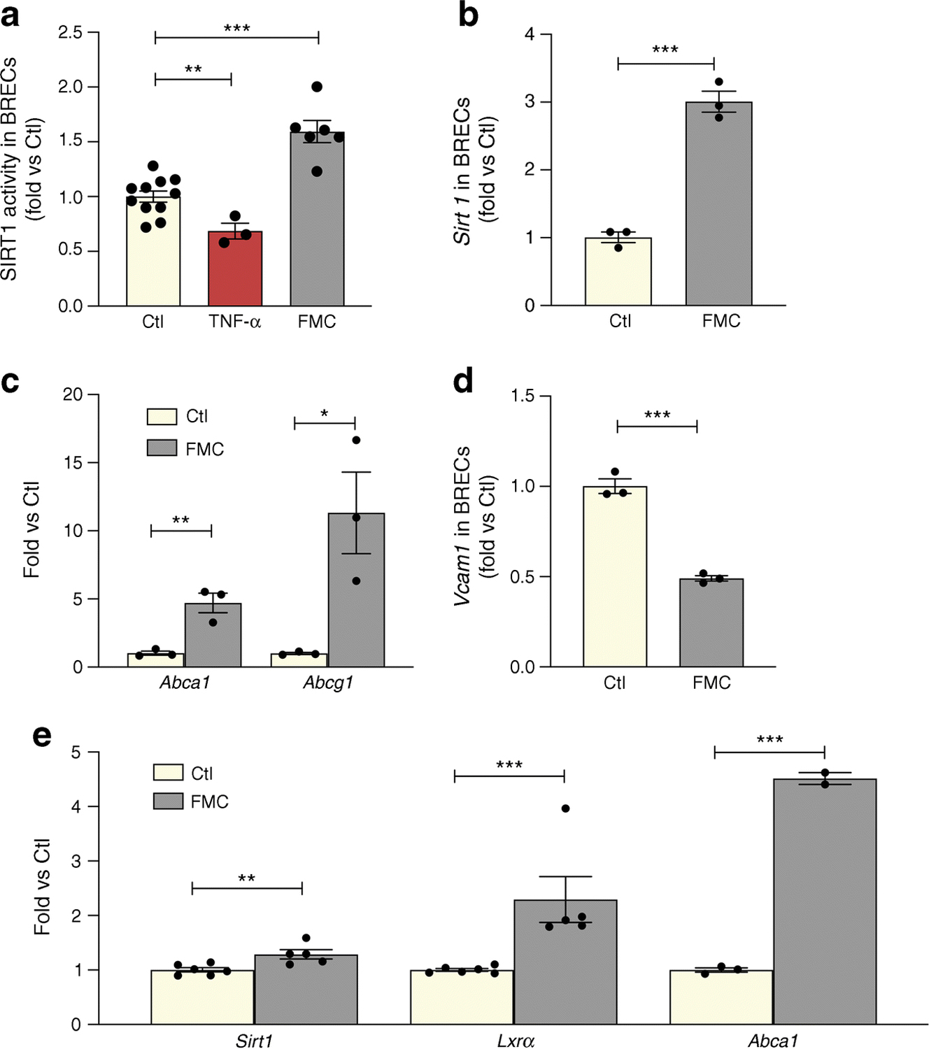
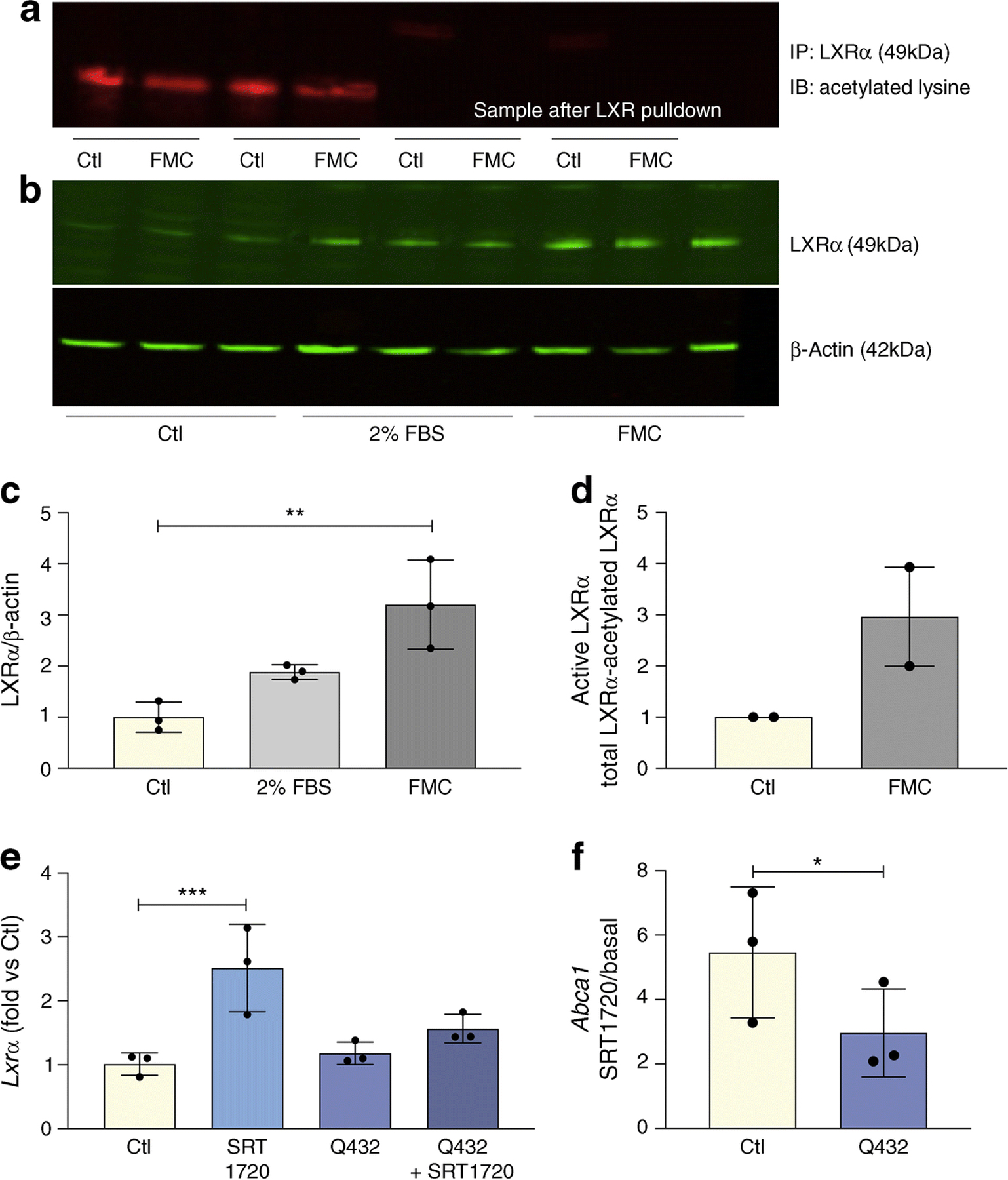
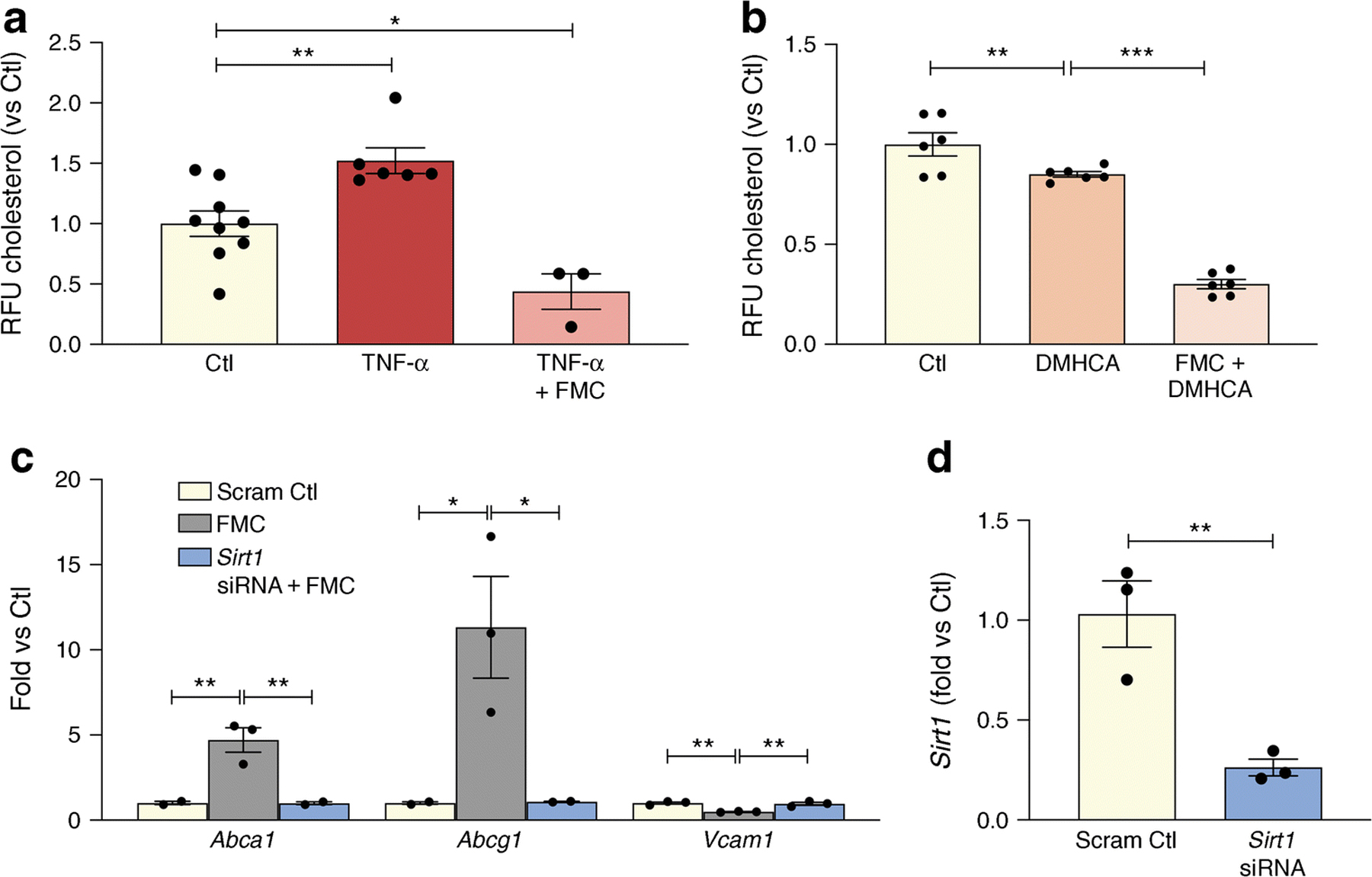
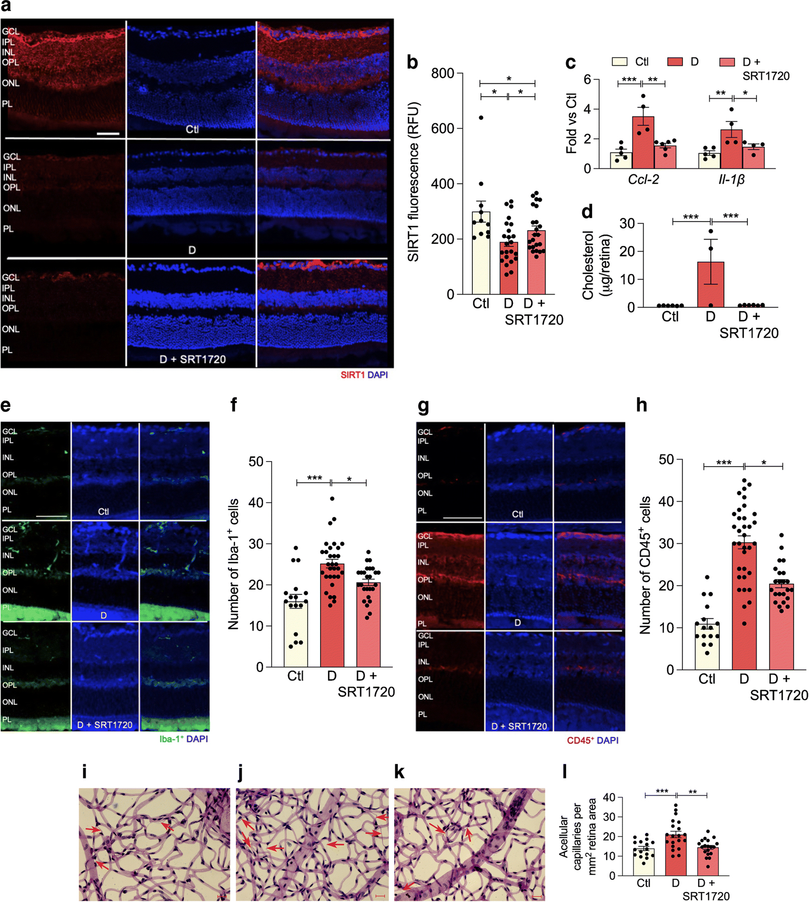
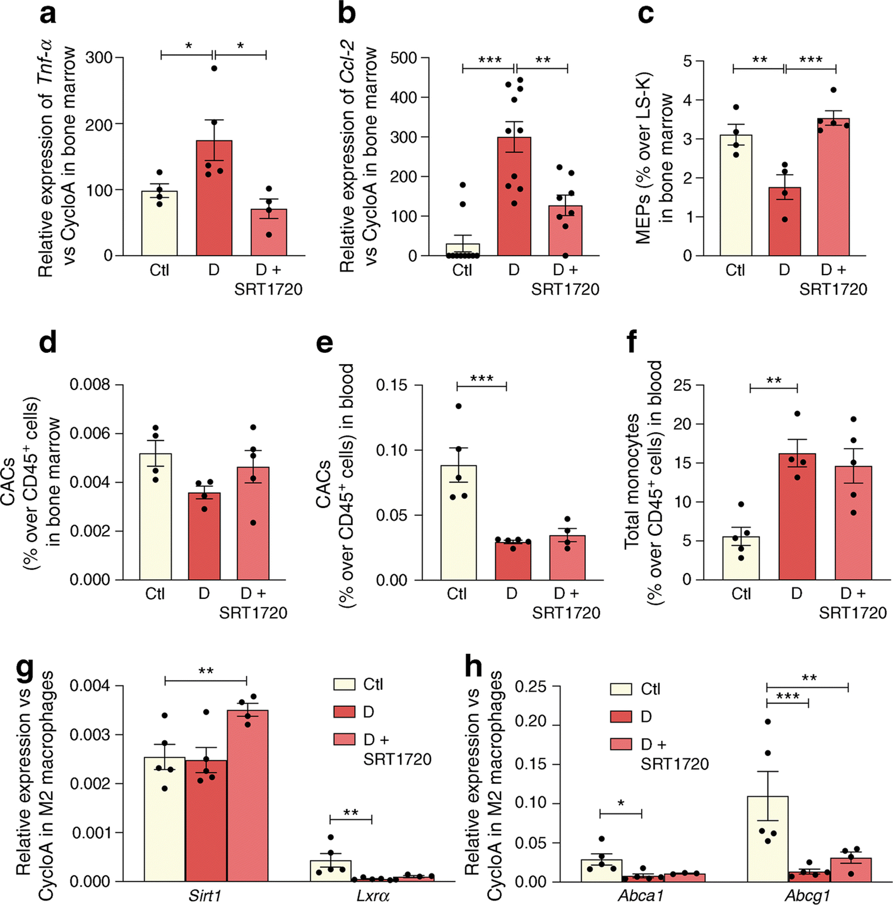
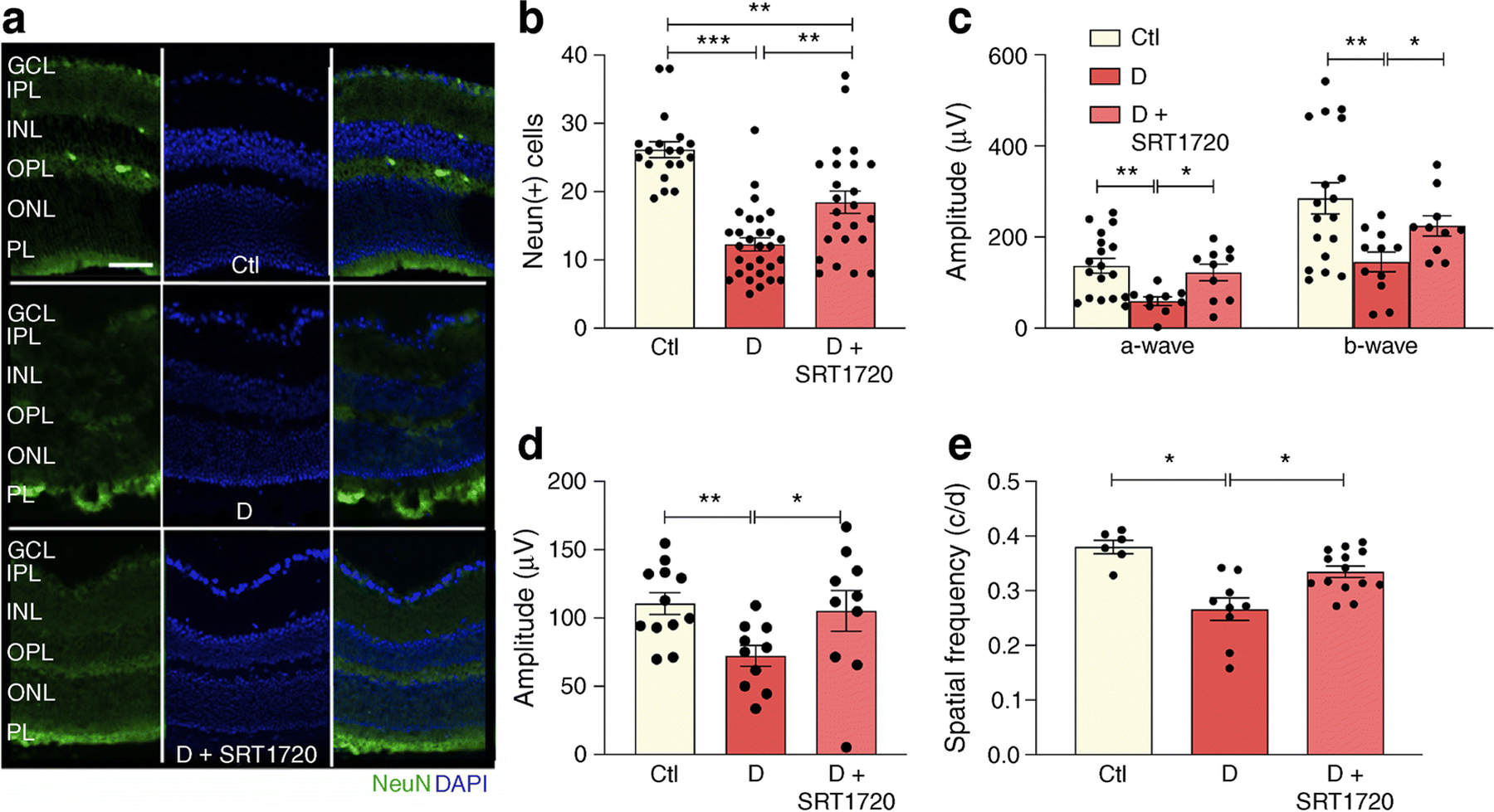
Results: IF increased Sirt1 mRNA expression in mouse liver and retina when compared with non-fasted animals. IF also increased SIRT1 activity eightfold in mouse retina while FMC increased SIRT1 activity and expression in retinal endothelial cells when compared with control. Sirt1 expression was also increased twofold in neuronal retina progenitor cells (R28) after FMC treatment. Moreover, FMC led to SIRT1-mediated LXRα deacetylation and subsequent 2.4-fold increase in activity, as measured by increased mRNA expression of the genes encoding ATP-binding cassette transporter (Abca1 and Abcg1). These changes were reduced when retinal endothelial cells expressing a constitutively acetylated LXRα mutant were tested. Increased SIRT1/LXR/ABC-mediated cholesterol export resulted in decreased retinal endothelial cell cholesterol levels. Direct activation of SIRT1 by SRT1720 in db/db mice led to a twofold reduction of diabetes-induced inflammation in the retina and improved diabetes-induced visual function impairment, as measured by electroretinogram and optokinetic response. In the bone marrow, there was prevention of diabetes-induced myeloidosis and decreased inflammatory cytokine expression.

Conclusions/interpretation: Taken together, activation of SIRT1 signalling by IF or through pharmacological activation represents an effective therapeutic strategy that provides a mechanistic link between the advantageous effects associated with fasting regimens and prevention of microvascular and bone marrow dysfunction in diabetes.

Keywords: ABCA1; ABCG1; Cholesterol; Deacetylation; Diabetic retinopathy; Endothelial cell; Intermittent fasting; LXR; RPE; SIRT1.


Figures

Fig. 1
Fig. 1
Fasting increases Sirt1 expression and activity. (a) Liver and (b) retinal Sirt1 mRNA expression is increased by IF in mice (48 h fast) when compared with non-fasting controls. (c) IF increases SIRT1 HDAC activity in mouse retinal tissue when compared with non-fasting controls, ***p<0.001. Data are represented as mean ± SEM. Ctl, control; CycloA, cyclophilin A; F355/F460, fluorescence at 355/460 nm
Fig. 2
Fig. 2
FMC activates the SIRT1/LXR signalling pathway in BRECs and neuronal (R28) cells. FMC (0% FBS, 24 h) in BRECs activates (a) SIRT1 HDAC activity, with TNF-α used as a positive control, (b) Sirt1 mRNA expression, and (c) increased expression of RCT genes (Abca1 and Abcg1). (d) Vcam1 is decreased after treatment with FMC conditions. (e) Treatment of R28 cells with FMC increases expression of Sirt1 and Lxrα mRNA, as well as LXRα activity, as measured by Abca1 mRNA expression *p<0.05, **p<0.01, ***p<0.001. Data are represented as mean ± SEM. Ctl, control
Fig. 3
Fig. 3
FMC increases LXRα activity via deacetylation. FMC (0% FBS, 24 h) in BRECs (a) decreases levels of non-active, deacetylated LXRα; and (b) increases total LXRα protein levels, quantification shown in (c). The data for active LXR quantified by subtracting acetylated LXRα in (a) from total LXRα in (b) is presented in (d) as mean ± SEM. BRECs were treated with SRT1720 (1 μmol/l) and/or infected with constitutively acetylated LXRα (Q432) for 24 h. Expression of Lxrα (e) or SIRT1-dependent activation of LXRα (f) presented as SRT1720-induced increase in Abca1 expression in BRECs (Ctl) and BRECs infected with Q432. Ratios determined from three independent experiments,*p<0.05, **p<0.01, ***p<0.001. Data are represented as mean ± SEM. Ctl, control; IB, immunoblot; IP, immunoprecipitation
Fig. 4
Fig. 4
SIRT1 plays an important role in fasting mediated decrease of cholesterol levels in BRECs. (a) TNF-α treatment causes a significant increase in cholesterol levels while FMC (0% FBS, 24 h) prevents this TNF-α -induced increase. (b) This decrease is amplified by administration of the LXRα activator, DMHCA. (c) SIRT1 was reduced by exposure of the BRECs to Sirt1 siRNA. Administration of Sirt1 siRNA prevented serum-induced upregulation of Abca1 and Abcg1 and inhibited the downregulation of the proinflammatory gene, Vcam1. (d) Sirt1 knockdown efficiency. *p<0.05, **p<0.01, ***p<0.001. Data are represented as mean ± SEM. Ctl, control; CycloA, cyclophilin A, Scram Ctl; scrambled Sirt1 siRNA sequence used in 4c and 4d
Fig. 5
Fig. 5
FMC does not significantly impact cell death or mitochondrial substrate-supported respiration in BRECs. (a, b) FMC (0% FBS, 24 h) does not significantly increase cell death. Basal media is comprised of only MCB131 media without the addition of growth factors or supplements (m8537). Positive control (Pos ctl): 50°C for 10 min. (c) Time course of cell death assay. (d) Respiratory activity of BRECs cultured in control or FMC media after 24 h, **p<0.01, ***p<0.001, Data are represented as mean ± SEM. Scale bar, 10 µmol/l O2. Ctl, control
Fig. 6
Fig. 6
SRT1720 increases SIRT1 expression in retina of db/db mice with 6 month duration of diabetes. (a) Retinal sections from non-diabetic controls (top), diabetic mice fed normal chow (middle) or chow containing SRT1720 (bottom) were stained with anti-SIRT1 antibody (red) and DAPI was used to stain nuclei (blue). Quantification is shown in (b); several replicate samples were stained from n=4 mice. (c, d) Diabetes significantly increases proinflammatory markers Ccl-2, Il-1β mRNA expression (c) and significantly increases retinal cholesterol levels (mass spectrometry analysis) (d); the SIRT1 agonist SRT1720 restores Ccl-2 and Il-1β (c) and cholesterol (d), to non-diabetic levels; n=4–5 mice. (eh) Diabetes increases, while SRT1720 normalises, the cell number of Iba-1+ (e, f) and CD45+ (g, h) cells in the retina, n=5 from 3 to 5 sections at 100 µm interval for each eye with a minimum of four images for section. (il) Acellular capillary formation (red arrows) was examined in non-diabetic animals (i), and in diabetic animals fed control chow (j) or chow containing SRT1720 (k). Diabetes significantly increases acellular capillary formation (j) while administration of the SIRT1 agonist prevents diabetes-induced acellular capillary formation (k). Quantification is shown in (l); n=5 from 4 to 5 images per mm2 retina area; *p<0.05, **p<0.01,***p<0.001. Data are represented as mean ± SEM. Scale bars, 20 µm. Ctl, control (db/m); D, diabetic (db/db); GCL, ganglion cell layer; INL, inner nuclear layer; IPL, inner plexiform layer; ONL, outer nuclear layer; OPL, outer plexiform layer; PL, photoreceptor layer. RFU, relative fluorescence units
Fig. 7
Fig. 7
SRT1720 normalises inflammatory and reparative cell damage in the bone marrow and blood of db/db mice with 6 month duration of diabetes. (a, b) Tnf-α (n=4–5 mice) (a) and Ccl-2 (n=5 mice carried out in duplicate) (b) expression in the bone marrow of non-diabetic controls, and diabetic mice fed normal chow or chow containing SRT1720. (c, d) Diabetes significantly decreases the number of MEPs (c) and CACs (d) in the bone marrow, while SRT1720 restores MEP to non-diabetic levels, n=4–5. (e, f) In the blood, diabetes significantly decreases the number of reparative CACs (e) and increases the number of inflammatory circulating total monocytes (f); n=4–5. (g, h) In M2 macrophages, diabetes induces decrease in the expression of Lxrα and LXR-controlled target genes (Abca1 and Abcg1). SRT1720 increases Sirt1 expression in M2 macrophages (g), leading to upregulation of LXR activity as shown by an increase in Abcg1 expression (h); n=4–5; *p<0.05, **p<0.01, ***p<0.001. Data are represented as mean ± SEM. Ctl, control (db/m); CycloA, cyclophilin A; D, diabetic (db/db); LS-K, LinScaKit+
Fig. 8
Fig. 8
SRT1720 prevents diabetes-induced NeuN+ retinal decrease and improves visual and OKN response in db/db mice. (a, b) Chronic diabetes is associated with neuronal loss in the retina as demonstrated by reduced NeuN+ expression (green) in db/db mice when compared with non-diabetic db/m controls (a). Pharmacological SIRT1 activation using STR1720 in diabetic mice partly restores NeuN+ expression to control levels (a). Quantification shown in (b); n=4 from 3–5 sections at 100 µm interval for each eye with a minimum of four images per section; NeuN+ cells were quantified only in the GCL. (c) Analysis of the ERG showed diabetes-induced reduction of scotopic a- and b-waves; an increase and improvement in scotopic a- and b-wave was observed in diabetic mice treated with SRT1720 compared with diabetic mice on control chow. (d) An improvement in photopic b-wave was observed in diabetic mice treated with SRT1720 compared with diabetic mice on control chow; n=5–6 carried out from right and left eyes. (e) In diabetic mice, the OKN response is reduced compared with age-matched non-diabetic control mice. Diabetic mice treated with SRT1720 showed an improvement in visual acuity compared with diabetic mice on control chow; n=4–6 carried out from right and left eyes; *p<0.05, **p<0.01 ***p<0.001. Data are represented as mean ± SEM. Scale bar, 50 µm. Ctl, control (db/m); D, diabetic (db/db); GCL, ganglion cell layer; INL, inner nuclear layer; IPL, inner plexiform layer; ONL, outer nuclear layer; OPL, outer plexiform layer; PL, photoreceptor layer

References

    1. Eshaq RS, Aldalati AMZ, Alexander JS, Harris NR (2017) Diabetic retinopathy: Breaking the barrier. Pathophysiology 24(4): 229–241. 10.1016/j.pathophys.2017.07.001 - DOI - PMC - PubMed
    1. Kowluru RA, Santos JM, Zhong Q (2014) Sirt1, a negative regulator of matrix metalloproteinase-9 in diabetic retinopathy. Invest Ophthalmol Vis Sci 55(9): 5653–5660. 10.1167/iovs.14-14383 - DOI - PMC - PubMed
    1. Karbasforooshan H, Karimi G (2018) The role of SIRT1 in diabetic retinopathy. Biomed Pharmacother 97: 190–194. 10.1016/j.biopha.2017.10.075 - DOI - PubMed
    1. Wang RH, Kim HS, Xiao C, Xu X, Gavrilova O, Deng CX (2011) Hepatic Sirt1 deficiency in mice impairs mTorc2/Akt signaling and results in hyperglycemia, oxidative damage, and insulin resistance. The Journal of clinical investigation 121(11): 4477–4490. 10.1172/JCI46243 - DOI - PMC - PubMed
    1. Baur JA, Pearson KJ, Price NL, et al. (2006) Resveratrol improves health and survival of mice on a high-calorie diet. Nature 444(7117): 337–342. 10.1038/nature05354 - DOI - PMC - PubMed

Publication types

MeSH terms

LinkOut - more resources