

Roles of autophagy in orthodontic tooth movement
- PMID: 33771430
- PMCID: PMC10911631
- DOI: 10.1016/j.ajodo.2020.01.027
Roles of autophagy in orthodontic tooth movement
Abstract
Introduction: Orthodontic tooth movement (OTM) relies on efficient remodeling of alveolar bone. While a well-controlled inflammatory response is essential during OTM, the mechanism regulating inflammation is unknown. Autophagy, a conserved catabolic pathway, has been shown to protect cells from excess inflammation in disease states. We hypothesize that autophagy plays a role in regulating inflammation during OTM.
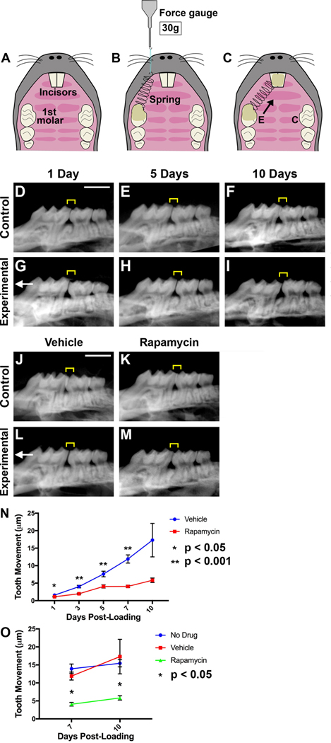
Methods: A split-mouth design was used to force load molars in adult male mice, carrying a GFP-LC3 transgene for in vivo detection of autophagy. Confocal microscopy, Western blot, and quantitative polymerase chain reaction analyses were used to evaluate autophagy activation in tissues of loaded and control molars at time points after force application. Rapamycin, a Food and Drug Administration-approved immunosuppressant, was injected to evaluate induction of autophagy.
Results: Autophagy activity increases shortly after loading, primarily on the compression side of the tooth, and is closely associated with inflammatory cytokine expression and osteoclast recruitment. Daily administration of rapamycin, an autophagy activator, led to reduced tooth movement and osteoclast recruitment, suggesting that autophagy downregulates the inflammatory response and bone turnover during OTM.
Conclusions: This is the first demonstration that shows that autophagy is induced by orthodontic loading and plays a role during OTM, likely via negative regulation of inflammatory response and bone turnover. Exploring roles of autophagy in OTM holds great promise, as aberrant autophagy is associated with periodontal disease and its related systemic inflammatory disorders.
Copyright © 2020. Published by Elsevier Inc.
Conflict of interest statement
Declaration of Interests: The authors have no conflicts of interest to declare.
Figures


Comment in
-
Hypoxia-inducible factor-1α may be the first host response in orthodontic tooth movement.Am J Orthod Dentofacial Orthop. 2021 Aug;160(2):163-164. doi: 10.1016/j.ajodo.2021.04.011. Am J Orthod Dentofacial Orthop. 2021. PMID: 34332685 No abstract available.
-
Author's response.Am J Orthod Dentofacial Orthop. 2021 Aug;160(2):164-165. doi: 10.1016/j.ajodo.2021.04.012. Am J Orthod Dentofacial Orthop. 2021. PMID: 34332686 No abstract available.
References
-
- Koyama Y, Mitsui N, Suzuki N, et al. Effect of compressive force on the expression of inflammatory cytokines and their receptors in osteoblastic Saos-2 cells. Arch. Oral Biol 2008;53(5):488–96. - PubMed
-
- Andrade IJ, Silva TA, Silva GAB, Teixeira AL, Teixeira MM. The role of tumor necrosis factor receptor type 1 in orthodontic tooth movement. J. Dent. Res 2007;86(11):1089–94. - PubMed
-
- Maeda A, Soejima K, Bandow K, et al. Force-induced IL-8 from periodontal ligament cells requires IL-1beta. J. Dent. Res 2007;86(7):629–34. - PubMed
-
- Yamaguchi M, Yoshii M, Kasai K. Relationship between substance P and interleukin-1beta in gingival crevicular fluid during orthodontic tooth movement in adults. Eur. J. Orthod 2006;28(3):241–6. - PubMed
-
- Yamaguchi M, Ozawa Y, Mishima H, Aihara N, Kojima T, Kasai K. Substance P increases production of proinflammatory cytokines and formation of osteoclasts in dental pulp fibroblasts in patients with severe orthodontic root resorption. Am. J. Orthod. Dentofacial Orthop 2008;133(5):690–8. - PubMed
MeSH terms
Grants and funding
LinkOut - more resources
Full Text Sources
Other Literature Sources
