. 2021 May 14;433(10):166953.
doi: 10.1016/j.jmb.2021.166953. Epub 2021 Mar 24.

Prion-like C-Terminal Domain of TDP-43 and α-Synuclein Interact Synergistically to Generate Neurotoxic Hybrid Fibrils

Affiliations

Prion-like C-Terminal Domain of TDP-43 and α-Synuclein Interact Synergistically to Generate Neurotoxic Hybrid Fibrils

Shailendra Dhakal et al. J Mol Biol. .

Abstract

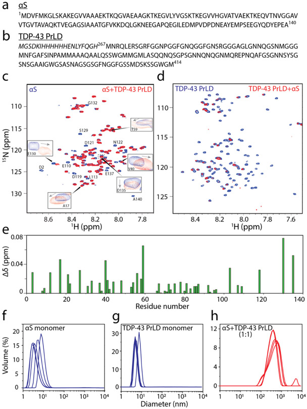
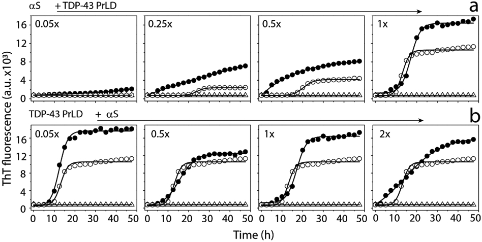
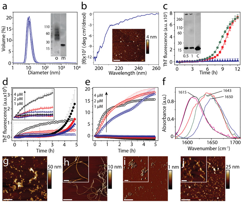
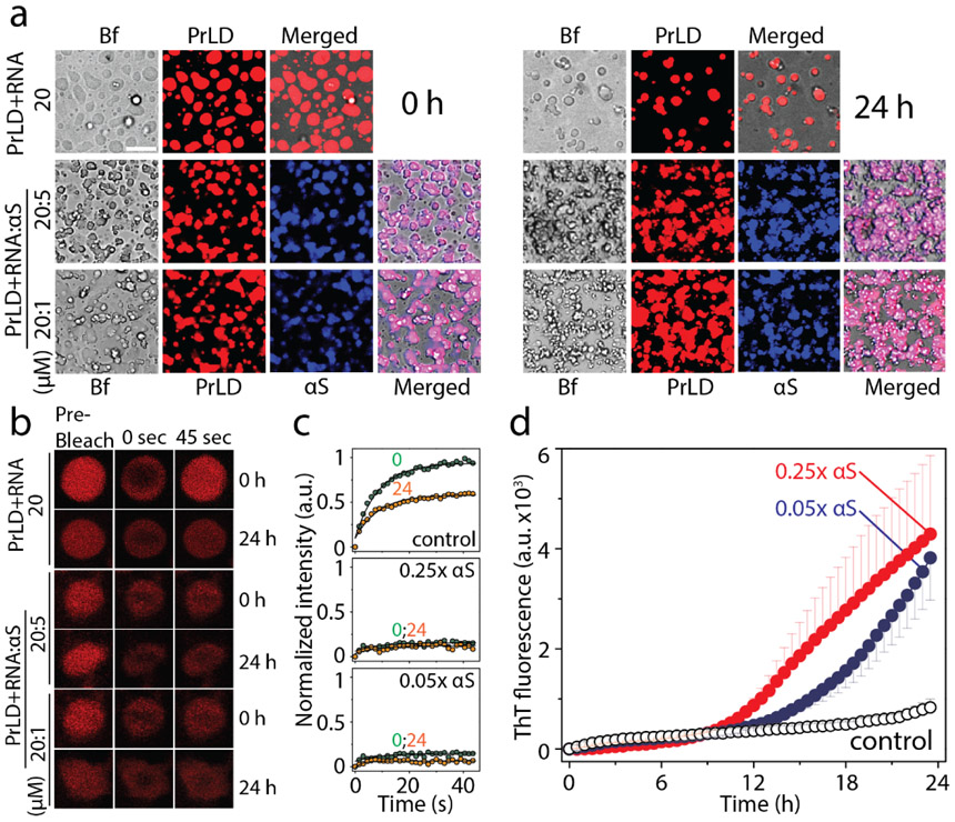
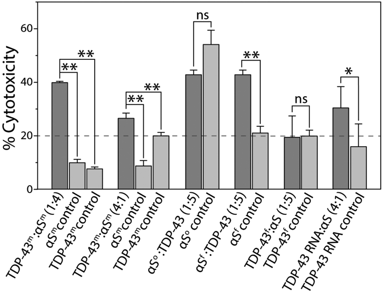
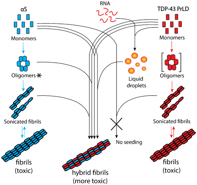
Aberrant aggregation and amyloid formation of tar DNA binding protein (TDP-43) and α-synuclein (αS) underlie frontotemporal dementia (FTD) and Parkinson's disease (PD), respectively. Amyloid inclusions of TDP-43 and αS are also commonly co-observed in amyotrophic lateral sclerosis (ALS), dementia with Lewy bodies (DLB) and Alzheimer disease (AD). Emerging evidence from cellular and animal models show colocalization of the TDP-43 and αS aggregates, raising the possibility of direct interactions and co-aggregation between the two proteins. In this report, we set out to answer this question by investigating the interactions between αS and prion-like pathogenic C-terminal domain of TDP-43 (TDP-43 PrLD). PrLD is an aggregation-prone fragment generated both by alternative splicing as well as aberrant proteolytic cleavage of full length TDP-43. Our results indicate that two proteins interact in a synergistic manner to augment each other's aggregation towards hybrid fibrils. While monomers, oligomers and sonicated fibrils of αS seed TDP-43 PrLD monomers, TDP-43 PrLD fibrils failed to seed αS monomers indicating selectivity in interactions. Furthermore, αS modulates liquid droplets formed by TDP-43 PrLD and RNA to promote insoluble amyloid aggregates. Importantly, the cross-seeded hybrid aggregates show greater cytotoxicity as compared to the individual homotypic aggregates suggesting that the interactions between the two proteins have a discernable impact on cellular functions. Together, these results bring forth insights into TDP-43 PrLD - αS interactions that could help explain clinical and pathological presentations in patients with co-morbidities involving the two proteins.

Keywords: TDP-43; amyloid; fibrillization; liquid–liquid phase separation; α-synuclein.


Conflict of interest statement

Declaration of Competing Interest The authors declare that they have no known competing financial interests or personal relationships that could have appeared to influence the work reported in this paper. Declaration of Competing Interest The authors declare that they have no known competing financial interests or personal relationships that could have appeared to influence the work reported in this paper.

Figures

Figure 1.
Figure 1.
1H-15N HMQC spectroscopy of αS and TDP-43 PrLD along with DLS of respective reactions. a-b) Sequence of full-length 1-140 amino acid residues αS (a) and 267-414 amino acid residues TDP-43 PrLD used in the experiment. c) 1H-15N HMQC of 10 μM 15N-labeled αS monomers alone (blue) or in the presence of unlabeled 10 μM TDP-43 PrLD monomers (red) in 20 mM MES buffer pH 6.0 at 37 °C. Some of the residues undergoing chemical shift perturbations (CSPs) are shown in boxes while those with lost peaks are indicated by letters without boxes. d)1H-15N HMQC of 10 μM 15N-labeled TDP-43 PrLD monomers alone (blue) or in presence of unlabeled αS (red). e) CSPs for equimolar incubations of αS and TDP-43 PrLD that were calculated using the equation, Δδ=(δH)2+0.14(δN)2 from the spectrum in (c). f-g) DLS histograms of 10 μM αS monomer control, TDP-43 PrLD monomer control and co-incubated mixture 1:1 αS and TDP-43 PrLD in the same buffer and temperature conditions taken within 10 minutes of incubation.
Figure 2.
Figure 2.
ThT aggregation kinetics of monomeric αS and TDP-43 PrLD in 20 mM MES buffer at pH 6.0 a) ThT fluorescence of 20 μM αS (△) in presence of 0.05 to 1 molar ratio of TDP-43 PrLD to αS (●) and respective TDP-43 PrLD controls (○). b) ThT fluorescence of 20 μM TDP-43 PrLD (○) in presence of 0.05 to 2 molar ratio of αS to TDP-43 PrLD (●) and respective αS controls (△). The data were fit with Boltzmann’s sigmoidal function (see Materials and Methods) to derive lag time information.
Figure 3.
Figure 3.
Interaction of monomeric TDP-43 PrLD and αS in 20 mM MES buffer pH 6.0. a) ThT fluorescence kinetics of 50 μM αS alone (▼) and in presence of 0.04 (■) and 0.1 (●) molar ratio of TDP-43 PrLD to αS, and respective 0.04 (♦) and 0.1 (▲) TDP-43 PrLD controls. b) Western blot of ThT reactions using αS antibodies. Aliquot of sample from the reaction at 72 hours was subjected to western blot as total sample (T), and supernatant (S) after centrifuging at 18,000 xg c) Representative fluorescence microscopic images of Thioflavin S (ThS) stained αS and TDP-43 PrLD aggregation reactions at 72 hours after centrifuging at 18,000 x g (Scale bar = 20 μm). d-e) Relative quantification of αS and TDP-43 PrLD to the Cytochrome C internal standard in pellet of reactions and control at 72 hours after centrifugation at 18,000 xg; 0.1x and 0.04x indicates molar ratio of TDP-43 PrLD to αS.
Figure 4.
Figure 4.
Cross-seeding of oligomers and sonicated fibrils with monomers. a) DLS analysis of DOPAL-derived αS oligomers isolated from SEC. (inset) SEC fraction containing the oligomer ‘o’ used in the study; ‘m’ refers to control monomer. b) CD spectra and AFM height image (inset) of DOPAL-derived αS oligomers used for cross-seeding reaction (scale bar = 1μm). Cross-seeding reactions with monomers of 15 μM TDP-43 PrLD alone (▲) or in the presence of 0.5 μM (■) or 1 μM (●) DOPAL-derived αS oligomers in the presence of 10 μM ThT. (inset) immunoblot of the reaction after 12 h probed with TDP-43 antibody; ‘c’ refers to TDP-43 PrLD monomer control. d) Seeding of 20 μM αS monomers with 4 μM (○), 2 μM (◇), and 1 μM (△) of αS sonicated fibrils, and 20 μM TDP-43 PrLD monomers seeded with 4 μM (●), 2 μM (♦), 1 μM (▲) of αS sonicated fibrils. e) Seeding of 20 μM TDP-43 PrLD monomers with 4 μM (○), 2 μM (◇), and 1 μM (△) of TDP-43 PrLD sonicated fibrils and seeding of 20 μM αS monomers with 4 μM (●), 2 μM (♦), 1 μM (▲) of TDP-43 PrLD seed. f) FTIR analysis of cross-seeding reactions from (c-e). TDP-43 PrLD monomers cross-seeded with 1 μM DOPAL-derived αS oligomers after 12 h of incubation (—); TDP-43 PrLD monomers seeded with 1 μM αS sonicated fibrils (—); αS monomers seeded with 1 μM TDP-43 PrLD sonicated fibrils (—) along with controls such as αS monomers (—) and TDP-43 PrLD monomers (—) (g-j) AFM height image of αS sonicated fibrils (g), αS sonicated fibrils seeded TDP-43 PrLD monomers (h), TDP-43 PrLD sonicated fibrils (i), and TDP-43 PrLD sonicated fibrils seeded αS monomers (j) (scale bar = 1 μm, inset = 200 nm).
Figure 5.
Figure 5.
Modulation of TDP-43 PrLD LLPS by αS. a) Timestamped confocal images of the co-incubations of Hilyte-647 labelled TDP-43 PrLD (20 μM) and RNA (40 μug/mL) in the absence and presence of Hilyte 405 labelled αS (5 or 1 μM) in 20 mM MES buffer pH 6.0 at 37 °C; Bf represents ‘bright field’, b) FRAP analysis on the selected droplets from the reactions before (pre-bleach), during (0 sec), and after photobleaching (45 sec), immediately after incubation (0 h) and after 24 h. c) Normalized kinetics of fluorescence recovery data obtained from FRAP intensity; TDP-43 PrLD and RNA control reaction along with 5 μM and 1 μM αS at 0h (●) and after 24 h (●). The data was fit to a first order exponential growth equation (solid lines). d) Corresponding ThT fluorescence kinetics of the reactions; TDP-43 PrLD and RNA (○) control reaction along with sub-stoichiometric, 1 μM (●) or 5 μM (●) αS incubations.
Figure 6.
Figure 6.
Cytotoxicity of monomeric αS and TDP-43 PrLD species along with homotypic and heterotypic fibrillar species of αS and TDP-43 in SH-SY5Y cells by XTT assay. ‘m’, ‘o’ and ‘f’ in the superscript represents monomer, oligomer, and sonicated fibril respectively. All the data were obtained in triplicates, * represents p<0.1 and **represents p<0.01 based on one-way ANOVA analysis.
Figure 7.
Figure 7.
Schematic diagram summarizing the collective results from this work. The square parenthesis indicates theoretical transient oligomers and not used in this study. ‘*’ DOPAL-derived de facto intermediate oligomers but shown along the aggregation pathway for simplicity.

References

    1. Soto C & Estrada LD (2008). Protein misfolding and neurodegeneration. Arch. Neurol 65, 184–189. - PubMed
    1. Nakamura T & Lipton SA (2009). Cell death: protein misfolding and neurodegenerative diseases. Apoptosis. 14, 455–468. - PubMed
    1. Sengupta U, Guerrero-Muñoz MJ, Castillo-Carranza DL, Lasagna-Reeves CA, Gerson JE, Paulucci-Holthauzen AA, Krishnamurthy S, Farhed M, Jackson GR & Kayed R (2015). Pathological interface between oligomeric alpha-synuclein and tau in synucleinopathies. Biol. psychiatry 78, 672–683. - PubMed
    1. Galvin JE, Lee VM-Y & Trojanowski JQ (2001). Synucleinopathies: clinical and pathological implications. Arch. Neurol 58, 186–190. - PubMed
    1. Saha P & Sen N (2019). Tauopathy: A common mechanism for neurodegeneration and brain aging. Mech. Ageing Dev 178, 72–79. - PMC - PubMed

Publication types

MeSH terms

LinkOut - more resources