. 2021 May 26;87(12):e0041221.
doi: 10.1128/AEM.00412-21. Epub 2021 May 26.

Mechanistic Insights into Substrate Recognition and Catalysis of a New Ulvan Lyase of Polysaccharide Lyase Family 24

Affiliations

Mechanistic Insights into Substrate Recognition and Catalysis of a New Ulvan Lyase of Polysaccharide Lyase Family 24

Fei Xu et al. Appl Environ Microbiol. .

Abstract

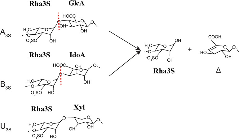
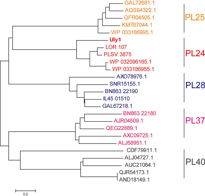
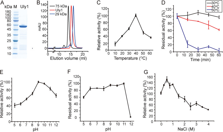
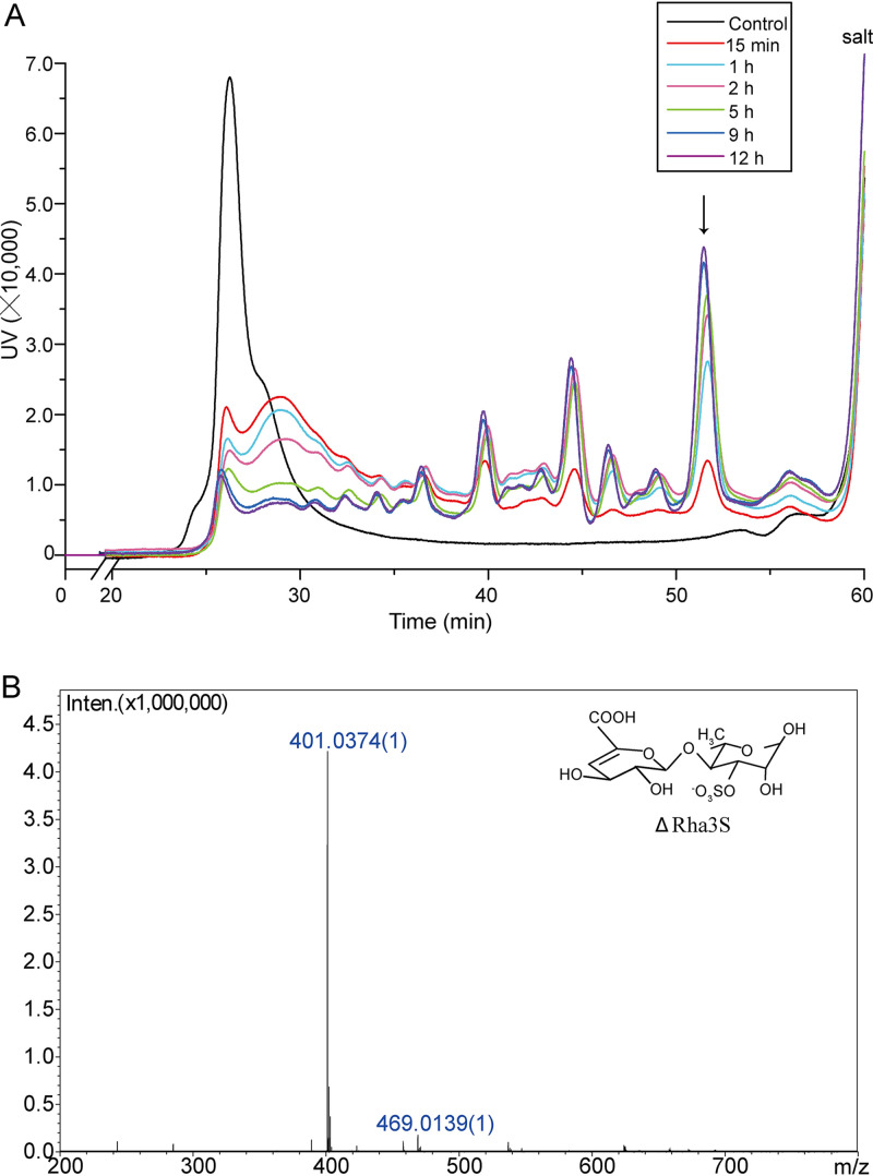
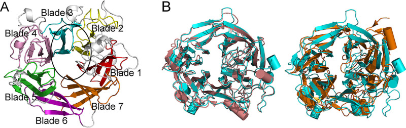
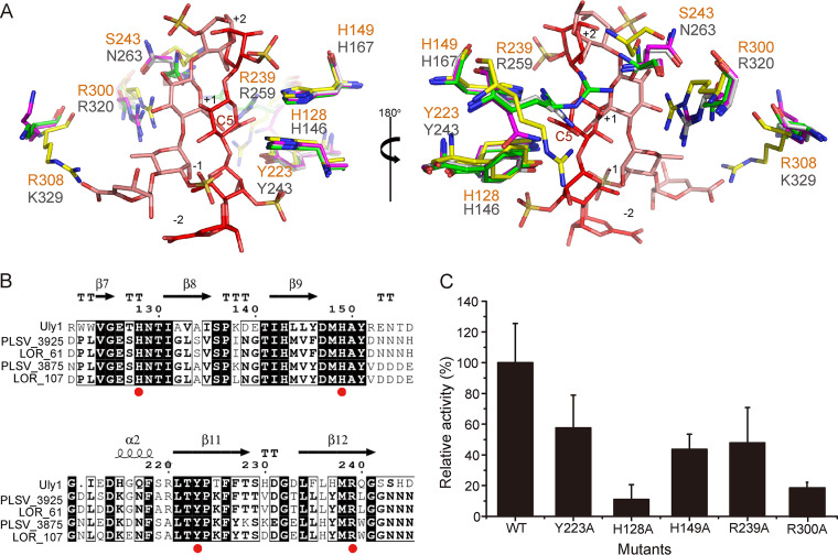
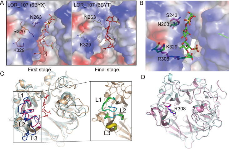
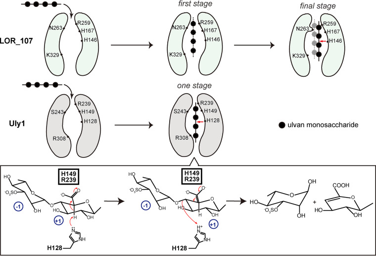
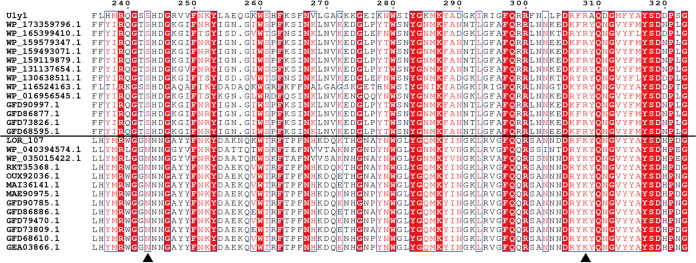
Ulvan is an important marine polysaccharide. Bacterial ulvan lyases play important roles in ulvan degradation and marine carbon cycling. Until now, only a small number of ulvan lyases have been characterized. Here, a new ulvan lyase, Uly1, belonging to polysaccharide lyase family 24 (PL24) from the marine bacterium Catenovulum maritimum, is characterized. The optimal temperature and pH for Uly1 to degrade ulvan are 40°C and pH 9.0, respectively. Uly1 degrades ulvan polysaccharides in the endolytic manner, mainly producing ΔRha3S, consisting of an unsaturated 4-deoxy-l-threo-hex-4-enopyranosiduronic acid and a 3-O-sulfated α-l-rhamnose. The structure of Uly1 was resolved at a 2.10-Å resolution. Uly1 adopts a seven-bladed β-propeller architecture. Structural and site-directed mutagenesis analyses indicate that four highly conserved residues, H128, H149, Y223, and R239, are essential for catalysis. H128 functions as both the catalytic acid and base, H149 and R239 function as the neutralizers, and Y223 plays a supporting role in catalysis. Structural comparison and sequence alignment suggest that Uly1 and many other PL24 enzymes may directly bind the substrate near the catalytic residues for catalysis, different from the PL24 ulvan lyase LOR_107, which adopts a two-stage substrate binding process. This study provides new insights into ulvan lyases and ulvan degradation. IMPORTANCE Ulvan is a major cell wall component of green algae of the genus Ulva. Many marine heterotrophic bacteria can produce extracellular ulvan lyases to degrade ulvan for a carbon nutrient. In addition, ulvan has a range of physiological bioactivities based on its specific chemical structure. Ulvan lyase thus plays an important role in marine carbon cycling and has great potential in biotechnological applications. However, only a small number of ulvan lyases have been characterized over the past 10 years. Here, based on biochemical and structural analyses, a new ulvan lyase of polysaccharide lyase family 24 is characterized, and its substrate recognition and catalytic mechanisms are revealed. Moreover, a new substrate binding process adopted by PL24 ulvan lyases is proposed. This study offers a better understanding of bacterial ulvan lyases and is helpful for studying the application potentials of ulvan lyases.

Keywords: catalytic mechanism; marine bacterium; polysaccharide lyase family 24; substrate recognition; ulvan; ulvan lyase.


Figures

FIG 1
FIG 1
The disaccharide repetition units encountered in ulvan and the degradation product of ulvan lyases on ulvan. Three disaccharide repetition units, A3S, B3S, and U3S, exist in ulvan. Only A3S and B3S can be decomposed by ulvan lyases, leading to a reducing end on one fragment and an unsaturated ring (Δ, 4-deoxy-l-threo-hex-4-enopyranosiduronic acid) on the nonreducing end of the other fragment.
FIG 2
FIG 2
Phylogenetic analysis of the ulvan lyases from the PL24, PL25, PL28, PL37 and PL40 families. The sequences are from the CAZy database, including all the characterized ulvan lyase sequences. The unrooted phylogenetic tree was constructed by neighbor joining with a Poisson model.
FIG 3
FIG 3
Biochemical characterization of Uly1. (A) SDS-PAGE analysis of the purified recombinant Uly1. M, molecular mass marker. (B) Gel filtration analysis of the form of Uly1 in solution. Conalbumin (75 kDa; GE Healthcare) and carbonic anhydrase (29 kDa; GE Healthcare) were used as protein size markers. The predicted molecular mass of Uly1 without the signal peptide is 55.8 kDa (https://web.expasy.org/protparam/). (C) Effect of temperature on Uly1 activity. The highest activity of Uly1 at 40°C was taken as 100%. (D) Effect of temperature on Uly1 stability. Uly1 was incubated at 30, 40, and 50°C for 0 to 60 min. The residual activity was determined at 40°C and pH 9.0. The activity of Uly1 incubated at 4°C was taken as 100%. (E) Effect of pH on Uly1 activity. Experiments were performed at 40°C in Britton-Robinson buffer ranging from pH 5.0 to 12.0. The highest activity at pH 9.0 was taken as 100%. (F) Effect of pH on Uly1 stability. Uly1 was incubated in Britton-Robinson buffer ranging from pH 5.0 to 12.0 for 1 h. The residual activity was determined at 40°C and pH 9.0. The highest activity of Uly1 incubated at pH 11.0 was taken as 100%. (G) Effect of salinity on Uly1 activity. The activity of Uly1 at 0 M NaCl was taken as 100%. All experiments were repeated three times.
FIG 4
FIG 4
The enzymatic products of ulvan generated by Uly1 over a time course of 0 to 12 h. (A) Gel filtration analysis of the enzymatic products over a time course of 0 to 12 h. Gel filtration chromatography analysis was performed using a Superdex peptide 10/300 GL column monitored at a wavelength of 210 nm. Ulvan treated with Uly1 that was preinactivated by boiling was taken as the control. (B) ESI-MS analysis of the major peak indicated by an arrow in panel A. The major peak has an m/z value of 401, consistent with the molecular weight of the disaccharide ΔRha3S.
FIG 5
FIG 5
Overall structure of Uly1. (A) Overall structure of Uly1 monomer. The seven blades of Uly1 structure are shown in different colors. The catalytic canyon is circled. (B) Structural alignment of Uly1 and the ulvan lyases LOR_107 and PLSV_3936. Uly1, LOR_107, and PLSV_3936 are colored in cyan, salmon, and orange, respectively.
FIG 6
FIG 6
Analysis of the important residues in the active site of Uly1. (A) Structural alignment of the important residues of Uly1 and the ulvan lyase LOR_107. The amino acid residues in the structure of Uly1, wild-type LOR_107 structure, and the complex structures of the LOR_107 mutant (R259N) with tetrasaccharide (PDB code 6BYX) and LOR_107 mutant (R320N) with tetrasaccharide (PDB code 6BYT) are colored in yellow, gray, purple, and green, respectively. The bound tetrasaccharides in the complex structures of R259N and R320N occupying the −2 to +2 subsites are shown as salmon and red sticks, respectively. The residues of Uly1 and LOR_107 are labeled in orange and gray letters, respectively. (B) Sequence alignment of Uly1 and characterized PL24 ulvan lyases from the CAZy database. Identical and similar amino acid residues are shaded. Red dots indicate the residues involved in catalysis. The noncatalytic domains of the long ulvan lyases (ulvan lyases LOR_61 and PLSV_3925) are not included. (C) Enzymatic activities of WT Uly1 and its mutants toward ulvan. The activity of WT Uly1 was taken as 100%.
FIG 7
FIG 7
Comparison of the residues involved in the substrate binding in Uly1 with those in LOR_107. (A) The two-stage substrate binding of LOR_107. The figures were generated based on the structures of LOR_107 (PDB codes 6BYX and 6BYT) (18). (B) Structural alignment of Uly1 and the LOR_107 mutant R259N with the tetrasaccharide substrate (PDB code 6BYX). The structure of Uly1 is shown as the electrostatic surface view. The side chains of the amino acid residues of Uly1 are shown as purple sticks. Based on the LOR_107 complex structure, N263, K329, and the bound tetrasaccharide of LOR_107 are modeled into the active center of Uly1 and shown as green sticks. (C) Comparison of the loops on the catalytic canyon of Uly1 with those of LOR_107. The overall structures of Uly1 and LOR_107 are shown in light blue and wheat, respectively. Loops (L1, L2, and L3) of Uly1 and LOR_107 are shown in pink and blue, respectively. In Uly1, L1 includes the residues 300 to 314, L2 includes the residues 361 to 369, and L3 includes the residues 389 to 392. The enlarged view shows the B factor of the three loops of Uly1. (D) Comparison of the side chains of R308 of Uly1 in the WT structure (blue) and the SeMet structure (pink).
FIG 8
FIG 8
Schematic diagram of the proposed substrate binding and catalytic mechanisms of Uly1. The reported substrate binding process of LOR_107 is a two-stage process. The catalytic canyon of LOR_107 is wide. The ulvan substrate is first bound to the left of the canyon at the first stage. Then, N263 moves its side chain, narrowing the canyon and locking the substrate to the active site, which is the final stage. However, the catalytic canyon of Uly1 is narrow, and especially, the long side chain of R308 (corresponds to K329 of LOR_107) further narrows one end of the catalytic canyon, resulting in the inability of Uly1 to bind the saccharide to the left of the canyon. Moreover, the side chain of S243 of Uly1, spatially corresponding to N263 of LOR_107, is too short to push the substrate to the other side of the catalytic canyon. Therefore, in Uly1, the substrate is most likely to be directly bound near the catalytic residues and then catalyzed by the catalytic residues. The red arrows indicate the cleavage site of the tetrasaccharide. During the catalytic process, H128 functions as both the catalytic acid and base, and H149 and R239 neutralize the negative charge of the +1 carboxyl. The glucuronic acid at the +1 subsite in the catalytic mechanism diagram is taken as a saccharide example.
FIG 9
FIG 9
Sequence alignment of Uly1 and the other PL24 lyases. S243 and R308 of Uly1 and the corresponding residues of the other PL24 lyases are indicated with black triangles.

References

    1. Zhu Y, Thomas F, Larocque R, Li N, Duffieux D, Cladiere L, Souchaud F, Michel G, McBride MJ. 2017. Genetic analyses unravel the crucial role of a horizontally acquired alginate lyase for brown algal biomass degradation by Zobellia galactanivorans. Environ Microbiol 19:2164–2181. 10.1111/1462-2920.13699. - DOI - PubMed
    1. Lahaye M, Robic A. 2007. Structure and functional properties of ulvan, a polysaccharide from green seaweeds. Biomacromolecules 8:1765–1774. 10.1021/bm061185q. - DOI - PubMed
    1. Robic A, Gaillard C, Sassi JF, Lerat Y, Lahaye M. 2009. Ultrastructure of ulvan: a polysaccharide from green seaweeds. Biopolymers 91:652–664. 10.1002/bip.21195. - DOI - PubMed
    1. Collen PN, Jeudy A, Sassi JF, Groisillier A, Czjzek M, Coutinho PM, Helbert W. 2014. A novel unsaturated beta-glucuronyl hydrolase involved in ulvan degradation unveils the versatility of stereochemistry requirements in family GH105. J Biol Chem 289:6199–6211. 10.1074/jbc.M113.537480. - DOI - PMC - PubMed
    1. Bikker P, van Krimpen MM, van Wikselaar P, Houweling-Tan B, Scaccia N, van Hal JW, Huijgen WJJ, Cone JW, Lopez-Contreras AM. 2016. Biorefinery of the green seaweed Ulva lactuca to produce animal feed, chemicals and biofuels. J Appl Phycol 28:3511–3525. 10.1007/s10811-016-0842-3. - DOI - PMC - PubMed

Publication types

Supplementary concepts

LinkOut - more resources