. 2021 Mar 26;12(1):1910.
doi: 10.1038/s41467-021-22178-0.

Cardiac macrophages prevent sudden death during heart stress

Affiliations

Cardiac macrophages prevent sudden death during heart stress

Junichi Sugita et al. Nat Commun. .

Abstract

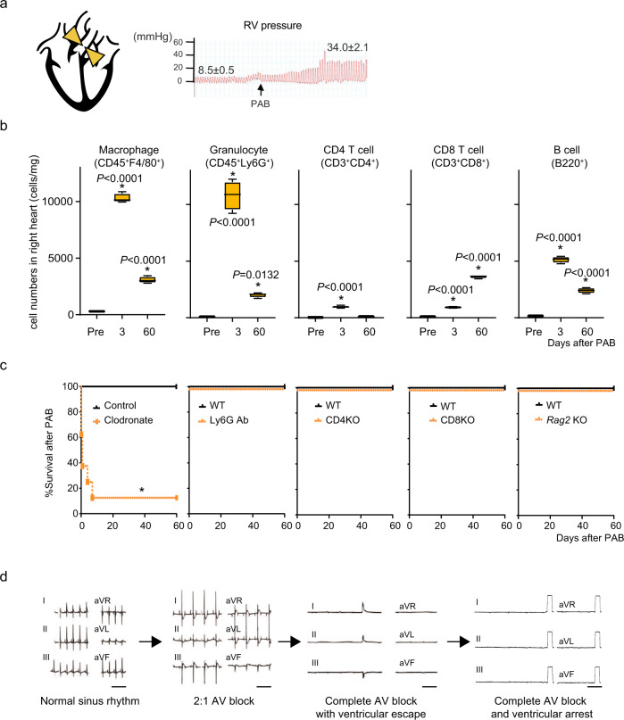
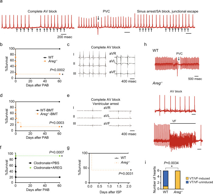
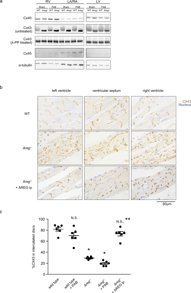
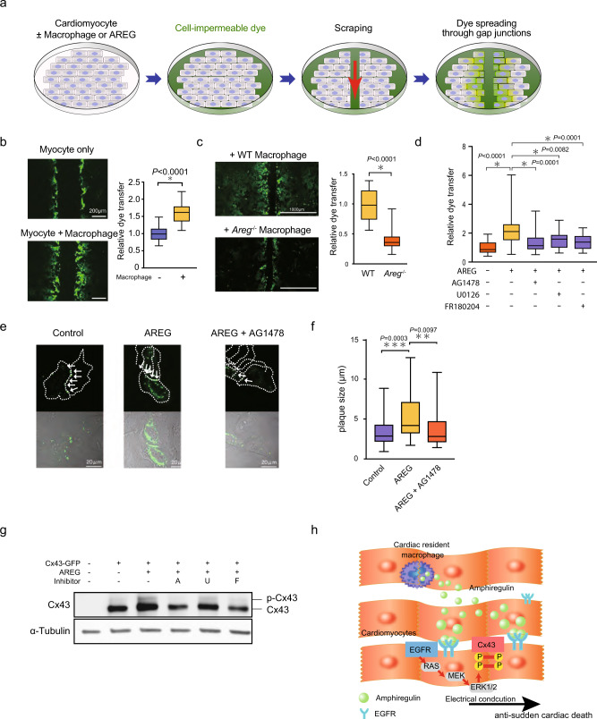
Cardiac arrhythmias are a primary contributor to sudden cardiac death, a major unmet medical need. Because right ventricular (RV) dysfunction increases the risk for sudden cardiac death, we examined responses to RV stress in mice. Among immune cells accumulated in the RV after pressure overload-induced by pulmonary artery banding, interfering with macrophages caused sudden death from severe arrhythmias. We show that cardiac macrophages crucially maintain cardiac impulse conduction by facilitating myocardial intercellular communication through gap junctions. Amphiregulin (AREG) produced by cardiac macrophages is a key mediator that controls connexin 43 phosphorylation and translocation in cardiomyocytes. Deletion of Areg from macrophages led to disorganization of gap junctions and, in turn, lethal arrhythmias during acute stresses, including RV pressure overload and β-adrenergic receptor stimulation. These results suggest that AREG from cardiac resident macrophages is a critical regulator of cardiac impulse conduction and may be a useful therapeutic target for the prevention of sudden death.


Conflict of interest statement

The authors declare no competing interests.

Figures

Fig. 1
Fig. 1. Macrophage depletion causes sudden death after right heart stress.
a Mouse model of right heart pressure overload (PAB). The pulmonary artery was ligated to increase the RV pressure to 30-35 mmHg from 5-10 mmHg. b Flow cytometric analysis of immune cells in the right atrial (RA) and RV free wall in wild-type (WT) mice after PAB. n = 4 in each group. One-way ANOVA followed by Tukey’s post-hoc test. *P < 0.05 vs. pre: steady-state. Box plots show center line as median, box limits as upper and lower quartiles, whiskers as minimum to maximum values. c Effects of depleting macrophages (clodronate), granulocytes (Ly6G Ab), CD4+ (CD4KO) or CD8 T+ (CD8KO) cells, or T and B cells (Rag2KO) on survival after PAB. *P < 0.0001 (log-rank test). d Representative six-channel ECGs from clodronate-treated mice after PAB. Shown is the representative sequence of arrhythmia progression observed in a mouse. Bars, 400 msec.
Fig. 2
Fig. 2. Areg deficiency leads to abnormal cardiac electrical conduction at steady-state and lethal arrhythmias under cardiac stress.
a ECGs from freely moving Areg−/− mice recorded using telemetry. Spontaneous AV block, PVCs, and sinus arrest or SA block were observed in Areg−/− mice. Bars indicate 200 msec. b Survival rates among WT and Areg−/− mice after PAB. *P < 0.001, log-rank test. c Representative ECG from an Areg−/− mouse showing complete AV block and ventricular arrest after PAB. d, e BM chimeric mice that received WT (WT-BMT) or Areg−/− (Areg−/−-BMT) BM were subjected to PAB. Survival rates after PAB are in d. *P < 0.001, log-rank test. In e, a representative ECG from an Areg−/−-BMT mouse after PAB is shown. f Survival curves for clodronate-treated mice with or without continuous administration of AREG (5 μg/day) from 24 h before PAB. n = 10 in each group. *P < 0.01, log-rank test. g, h WT and Areg−/− mice were intraperitoneally administered isoproterenol (bolus, 5 mg/kg). Survival rates are shown in g. In h, representative ECGs from WT and Areg−/− mice are shown. *P < 0.05, log-rank test. i Numbers of Langendorff-perfused hearts that developed VT/VF. *P < 0.01 (χ2-test).
Fig. 3
Fig. 3. AREG promotes gap junctional connection between cardiomyocytes through Cx43.
a Western blot analysis of Cx40, Cx43, and Cx45. Representative immunoblots are shown. α-Tubulin was used as a loading control. Note the mobility shift of Cx43 bands in WT mouse hearts. b Immunohistochemical staining of Cx43 (brown) in the myocardium. Nuclei (blue) were counterstained with methylene blue. WT and Areg−/− mice were intraperitoneally administered either vehicle (PBS) or recombinant AREG (5 μg), and the hearts were harvested 30 min later. WT and Areg−/− mice not receiving AREG were administrated vehicle (PBS) 30 min before sacrifice. The bar indicates 60 µm. The images show representative immunohistochemically stained hearts from WT or Areg−/− mice at steady-state. c Fractions of Cx43 localized to the intercalated discs in cardiomyocytes in RVs. n = 6 mice in each group. One-way ANOVA followed by Tukey’s post-hoc test. *P < 0.0001 vs. WT. **P < 0.0001 vs. Areg−/−. Data are presented as mean values ± SEM.
Fig. 4
Fig. 4. AREG facilitates gap junctional intercellular communication by stabilizing Cx43 gap junctions.
a Schematic of the dye transfer assay. Mouse neonatal cardiomyocytes were cultured with or without cardiac resident macrophages. The cell-impermeable dye was loaded by scraping that caused tearing of the plasma membrane, and the level of dye transfer between cardiomyocytes was evaluated as the distance of dye-stained cells from the scraped edge after 15 min. b, and c Mouse neonatal cardiomyocytes were cultured with or without WT or Areg−/− cardiac macrophages. Shown are representative images of dye-stained cells spreading from the scrape and the relative extent of dye transfer. Effects of macrophage coculture (b) and the lack of AREG in cocultured macrophages (c) on dye transfer were analyzed. n = 24 and 18, n = 22 and 23 (images) *P < 0.0001, two-tailed unpaired Student’s t-test. d Effects of AREG (100 ng/mL) and the indicated signal inhibitors (10 µmol/l, each) on dye transfer. n = 28, 28, 24, 24, and 30; *P < 0.05, post-hoc Dunnett’s test. e, f A Cx43-GFP plasmid was transfected into HeLa cells, and the cells were treated for 5 h with or without AREG (100 ng/mL) or AG1478 (10 µmol/l), as indicated. In e, formation of Cx43-GFP gap junction plaques (white arrows) at the cell-cell borders is shown. Cell borders are marked by dashed lines in upper panels. Scale bars, 20 µm. In f, plaque size is shown. n = 47, 39, and 36; **p < 0.01, ***p < 0.001, post-hoc Tukey’s test comparing cell adhesion between two-cardiomyocytes. g Western blotting of Cx43-GFP transfected HeLa cells treated for 6 h with AREG and 10 µmol/l AG1478 (A), U0126 (U), or FR180204 (F), as indicated. The positions of unphosphorylated and phosphorylated forms of Cx43 are shown. h Schematic model of how amphiregulin derived from cardiac macrophage suppresses arrhythmia. Throughout, box-and-whisker plots represent the median, the first and third quartiles, and the minimum and maximum values.

References

    1. Ziaeian B, Fonarow GC. Epidemiology and aetiology of heart failure. Nat. Rev. Cardiol. 2016;13:368. - PMC - PubMed
    1. Dunlay SM, Roger VL, Redfield MM. Epidemiology of heart failure with preserved ejection fraction. Nat. Rev. Cardiol. 2017;14:591. doi: 10.1038/nrcardio.2017.65. - DOI - PubMed
    1. Naksuk N, et al. Right ventricular dysfunction and long-term risk of sudden cardiac death in patients with and without severe left ventricular dysfunction. Circulation: Arrhythmia Electrophysiol. 2018;11:e006091. - PubMed
    1. Swirski FK, Nahrendorf M. Cardioimmunology: the immune system in cardiac homeostasis and disease. Nat. Rev. Immunol. 2018;18:733–744. doi: 10.1038/s41577-018-0065-8. - DOI - PubMed
    1. Fujiu K, et al. A heart-brain-kidney network controls adaptation to cardiac stress through tissue macrophage activation. Nat. Med. 2017;23:611–622. doi: 10.1038/nm.4326. - DOI - PubMed

Publication types

MeSH terms