. 2021 Mar 26;11(1):6986.
doi: 10.1038/s41598-021-86414-9.

Compound heterozygous mutations in TGFBI cause a severe phenotype of granular corneal dystrophy type 2

Affiliations

Compound heterozygous mutations in TGFBI cause a severe phenotype of granular corneal dystrophy type 2

Ikhyun Jun et al. Sci Rep. .

Abstract

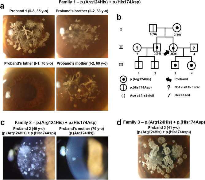
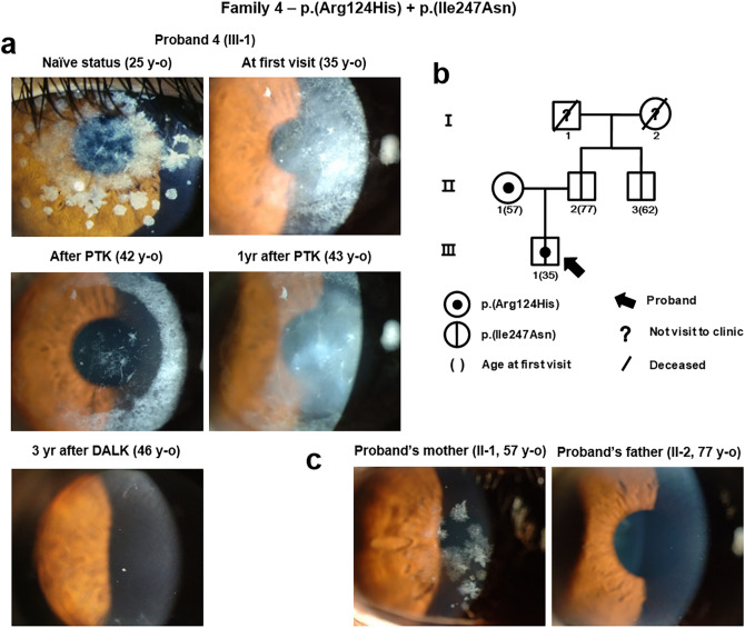
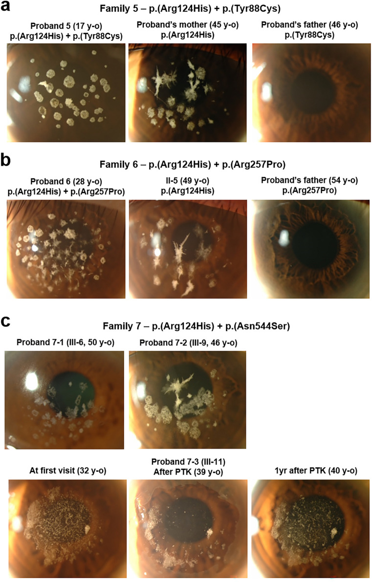
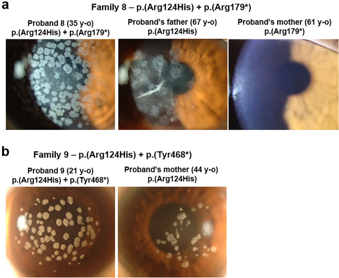
We investigated the clinical and genetic features of patients with severe phenotype of granular corneal dystrophy type 2 (GCD2) associated with compound heterozygosity in the transforming growth factor-β-induced (TGFBI) gene. Patients with severe GCD2 underwent ophthalmic examination (best-corrected visual acuity test, intraocular pressure measurement, slit-lamp examination, and slit-lamp photograph analysis) and direct Sanger sequencing of whole-TGFBI. The patient's family was tested to determine the pedigrees. Five novel mutations (p.(His174Asp), p.(Ile247Asn), p.(Tyr88Cys), p.(Arg257Pro), and p.(Tyr468*)) and two known mutations (p.(Asn544Ser) and p.(Arg179*)) in TGFBI were identified, along with p.(Arg124His), in the patients. Trans-phase of TGFBI second mutations was confirmed by pedigree analysis. Multiple, extensive discoid granular, and increased linear deposits were observed in the probands carrying p.(Arg124His) and other nonsense mutations. Some patients who had undergone phototherapeutic keratectomy experienced rapid recurrence (p.(Ile247Asn) and p.(Asn544Ser)); however, the cornea was well-maintained in a patient who underwent deep anterior lamellar keratoplasty (p.(Ile247Asn)). Thus, compound heterozygosity of TGFBI is associated with the phenotypic variability of TGFBI corneal dystrophies, suggesting that identifying TGFBI second mutations may be vital in patients with extraordinarily severe phenotypes. Our findings indicate the necessity for a more precise observation of genotype-phenotype correlation and additional care when treating TGFBI corneal dystrophies.


Conflict of interest statement

EKK is a medical advisory board member of Avellino Lab, USA. All other authors declare no competing interests.

Figures

Figure 1
Figure 1
TGFBI p.(His174Asp) variant aggravates granular corneal dystrophy 2 caused by p.(Arg124His) mutation in a compound heterozygote. (a) Slit-lamp photographs of the members of Family 1. Severe confluent granular deposits with lattice deposits were observed in Proband 1. Her brother’s eyes also showed severe snowflake-like corneal deposits. (b) Pedigree of Family 1 showed both proband and her brother to have inherited the mutations in trans-phase. (c) Slit-lamp photographs of Family 2. The cornea of Proband 2 showed intensive opacities, whereas that of his mother showed mild phenotype. (d) Slit-lamp photograph of Proband 3 showed coarse granular and lattice deposits.
Figure 2
Figure 2
Compound heterozygous mutations, p.(Ile247Asn) and p.(Arg124His), in TGFBI. (a) Slit-lamp photographs of Proband 4 (Family 4). Intensive snowflake-like corneal deposits were noted at the age of 25 years, and rapid recurrence occurred after phototherapeutic keratectomy (PTK). Following deep anterior lamellar keratoplasty (DALK), the cornea has been well-maintained since the past 3 years. (b) Pedigree of Family 4 confirmed compound heterozygosity. (c) Slit-lamp photographs of the parents of Proband 4. The mother, who only had heterozygous p.(Arg124His) mutation in TGFBI, showed milder phenotypes than the son, and the father, who had heterozygous p.(Ile247Asn) mutation in TGFBI, showed a clear cornea. Although TGFBI p.(Ile247Asn) mutation itself was silent, when accompanying p.(Arg124His) mutation, phenotypes of granular corneal dystrophy 2 became extensive and recurrence occurred rapidly after PTK.
Figure 3
Figure 3
TGFBI p.(Tyr88Cys), p.(Arg257Pro), and p.(Asn544Ser) mutations were accompanied by p.(Arg124His) mutation in patients with severe phenotypes. (a) Slit-lamp photographs of Proband 5 and her parents (Family 5). The proband showed numerous large discoid granular deposits. However, her mother carrying heterozygous p.(Arg124His) variant showed age-appropriate disease pattern, while the father carrying heterozygous p.(Tyr88Cys) variant showed no disease phenotype. (b) Slit-lamp photographs of the members of Family 6. Compound heterozygosity of TGFBI p.(Arg124His) and p.(Arg257Pro) mutations caused severe GCD2 phenotypes compared to the heterozygosity of TGFBI p.(Arg124His) mutation alone, although heterozygosity of TGFBI p.(Arg257Pro) mutation did not cause disease. (c) Slit-lamp photographs of Proband 7 carrying compound heterozygous mutations of TGFBI (p.(Arg124His) and p.(Asn544Ser)). The recurrence was very rapid after phototherapeutic keratectomy (PTK).
Figure 4
Figure 4
Nonsense mutations of TGFBI (p.(Arg179*) and p.(Tyr468*)) manifested severe granular corneal dystrophy 2 (GCD2) phenotypes. (a) Slit-lamp photographs of members of Family 8. Extensive multiple discoid granular deposits, with some annular deposits, were detected, resembling the features of homozygous GCD2 in Proband 8 carrying TGFBI p.(Arg179*) and p.(Arg124His) mutations. Her father carrying heterozygous TGFBI p.(Arg124His) mutation showed milder phenotypes than the proband, and the mother carrying heterozygous TGFBI p.(Arg179*) showed no corneal opacity. (b) Slit-lamp photographs of Proband 9 and his mother. Proband 9 harboured compound heterozygous TGFBI p.(Tyr468*) and p.(Arg124His) mutations and showed more severe discoid granular deposits than his mother who harboured heterozygous TGFBI p.(Arg124His) mutation.

Similar articles

Cited by

References

    1. Runager K, Klintworth GK, Karring H, Enghild JJ. The insoluble TGFBIp fraction of the cornea is covalently linked via a disulfide bond to type XII collagen. Biochemistry. 2013;52:2821–2827. doi: 10.1021/bi400212m. - DOI - PMC - PubMed
    1. Lakshminarayanan R, et al. Biochemical properties and aggregation propensity of transforming growth factor-induced protein (TGFBIp) and the amyloid forming mutants. Ocul Surf. 2015;13:9–25. doi: 10.1016/j.jtos.2014.04.003. - DOI - PubMed
    1. Courtney DG, et al. Protein composition of TGFBI-R124C- and TGFBI-R555W-associated aggregates suggests multiple mechanisms leading to lattice and granular corneal dystrophy. Invest. Ophthalmol. Vis. Sci. 2015;56:4653–4661. doi: 10.1167/iovs.15-16922. - DOI - PubMed
    1. Kim TI, Kim T, Kim SW, Kim EK. Comparison of corneal deposits after LASIK and PRK in eyes with granular corneal dystrophy type II. J. Refract. Surg. 2008;24:392–395. doi: 10.3928/1081597X-20080401-13. - DOI - PubMed
    1. Han KE, et al. Pathogenesis and treatments of TGFBI corneal dystrophies. Prog. Retin Eye Res. 2016;50:67–88. doi: 10.1016/j.preteyeres.2015.11.002. - DOI - PubMed

Substances

Supplementary concepts

LinkOut - more resources