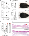
Restoration of the healing microenvironment in diabetic wounds with matrix-binding IL-1 receptor antagonist
- PMID: 33772102
- PMCID: PMC7998035
- DOI: 10.1038/s42003-021-01913-9
Restoration of the healing microenvironment in diabetic wounds with matrix-binding IL-1 receptor antagonist
Abstract
Chronic wounds are a major clinical problem where wound closure is prevented by pathologic factors, including immune dysregulation. To design efficient immunotherapies, an understanding of the key molecular pathways by which immunity impairs wound healing is needed. Interleukin-1 (IL-1) plays a central role in regulating the immune response to tissue injury through IL-1 receptor (IL-1R1). Generating a knockout mouse model, we demonstrate that the IL-1-IL-1R1 axis delays wound closure in diabetic conditions. We used a protein engineering approach to deliver IL-1 receptor antagonist (IL-1Ra) in a localised and sustained manner through binding extracellular matrix components. We demonstrate that matrix-binding IL-1Ra improves wound healing in diabetic mice by re-establishing a pro-healing microenvironment characterised by lower levels of pro-inflammatory cells, cytokines and senescent fibroblasts, and higher levels of anti-inflammatory cytokines and growth factors. Engineered IL-1Ra has translational potential for chronic wounds and other inflammatory conditions where IL-1R1 signalling should be dampened.
Conflict of interest statement
M.M.M. is inventor on U.S. Patent 9,879,062 which covers one of the technologies reported in this article. Monash University has filed for patent protection on the molecular design described herein, and Z.J. and M.M.M. are named as inventors. The other authors declare no competing interests.
Figures


Similar articles
-
Targeting Imbalance between IL-1β and IL-1 Receptor Antagonist Ameliorates Delayed Epithelium Wound Healing in Diabetic Mouse Corneas.Am J Pathol. 2016 Jun;186(6):1466-80. doi: 10.1016/j.ajpath.2016.01.019. Epub 2016 Apr 22. Am J Pathol. 2016. PMID: 27109611 Free PMC article.
-
Local Administration of Interleukin-1 Receptor Antagonist Improves Diabetic Wound Healing.Ann Plast Surg. 2018 May;80(5S Suppl 5):S317-S321. doi: 10.1097/SAP.0000000000001417. Ann Plast Surg. 2018. PMID: 29553981 Free PMC article.
-
Role of interleukin-1/interleukin-1 receptor antagonist family of cytokines in long-term continuous glucose monitoring in vivo.J Diabetes Sci Technol. 2013 Nov 1;7(6):1538-46. doi: 10.1177/193229681300700614. J Diabetes Sci Technol. 2013. PMID: 24351180 Free PMC article.
-
Interleukin-1 receptor antagonist (IL-1Ra) and IL-1Ra producing mesenchymal stem cells as modulators of diabetogenesis.Autoimmunity. 2010 Jun;43(4):255-63. doi: 10.3109/08916930903305641. Autoimmunity. 2010. PMID: 19845478 Review.
-
IL-1 receptor antagonist in metabolic diseases: Dr Jekyll or Mr Hyde?FEBS Lett. 2006 Nov 27;580(27):6289-94. doi: 10.1016/j.febslet.2006.10.061. Epub 2006 Nov 3. FEBS Lett. 2006. PMID: 17097645 Review.
Cited by
-
Inflammasome modulation with P2X7 inhibitor A438079-loaded dressings for diabetic wound healing.Front Immunol. 2024 Feb 15;15:1340405. doi: 10.3389/fimmu.2024.1340405. eCollection 2024. Front Immunol. 2024. PMID: 38426101 Free PMC article.
-
Human 3D nucleus pulposus microtissue model to evaluate the potential of pre-conditioned nasal chondrocytes for the repair of degenerated intervertebral disc.Front Bioeng Biotechnol. 2023 Feb 14;11:1119009. doi: 10.3389/fbioe.2023.1119009. eCollection 2023. Front Bioeng Biotechnol. 2023. PMID: 36865027 Free PMC article.
-
The Immunological Impact of IL-1 Family Cytokines on the Epidermal Barrier.Front Immunol. 2021 Dec 23;12:808012. doi: 10.3389/fimmu.2021.808012. eCollection 2021. Front Immunol. 2021. PMID: 35003136 Free PMC article. Review.
-
Therapeutic Properties of M2 Macrophages in Chronic Wounds: An Innovative Area of Biomaterial-Assisted M2 Macrophage Targeted Therapy.Stem Cell Rev Rep. 2025 Feb;21(2):390-422. doi: 10.1007/s12015-024-10806-3. Epub 2024 Nov 18. Stem Cell Rev Rep. 2025. PMID: 39556244 Review.
-
ER-mitochondria association negatively affects wound healing by regulating NLRP3 activation.Cell Death Dis. 2024 Jun 11;15(6):407. doi: 10.1038/s41419-024-06765-9. Cell Death Dis. 2024. PMID: 38862500 Free PMC article.
References
Publication types
MeSH terms
Substances
LinkOut - more resources
Full Text Sources
Other Literature Sources

