Review
. 2021 Jun;22(6):425-438.
doi: 10.1038/s41580-021-00354-w. Epub 2021 Mar 26.

MicroRNA-mediated regulation of glucose and lipid metabolism

Affiliations
Review

MicroRNA-mediated regulation of glucose and lipid metabolism

Pamela Agbu et al. Nat Rev Mol Cell Biol. 2021 Jun.

Abstract

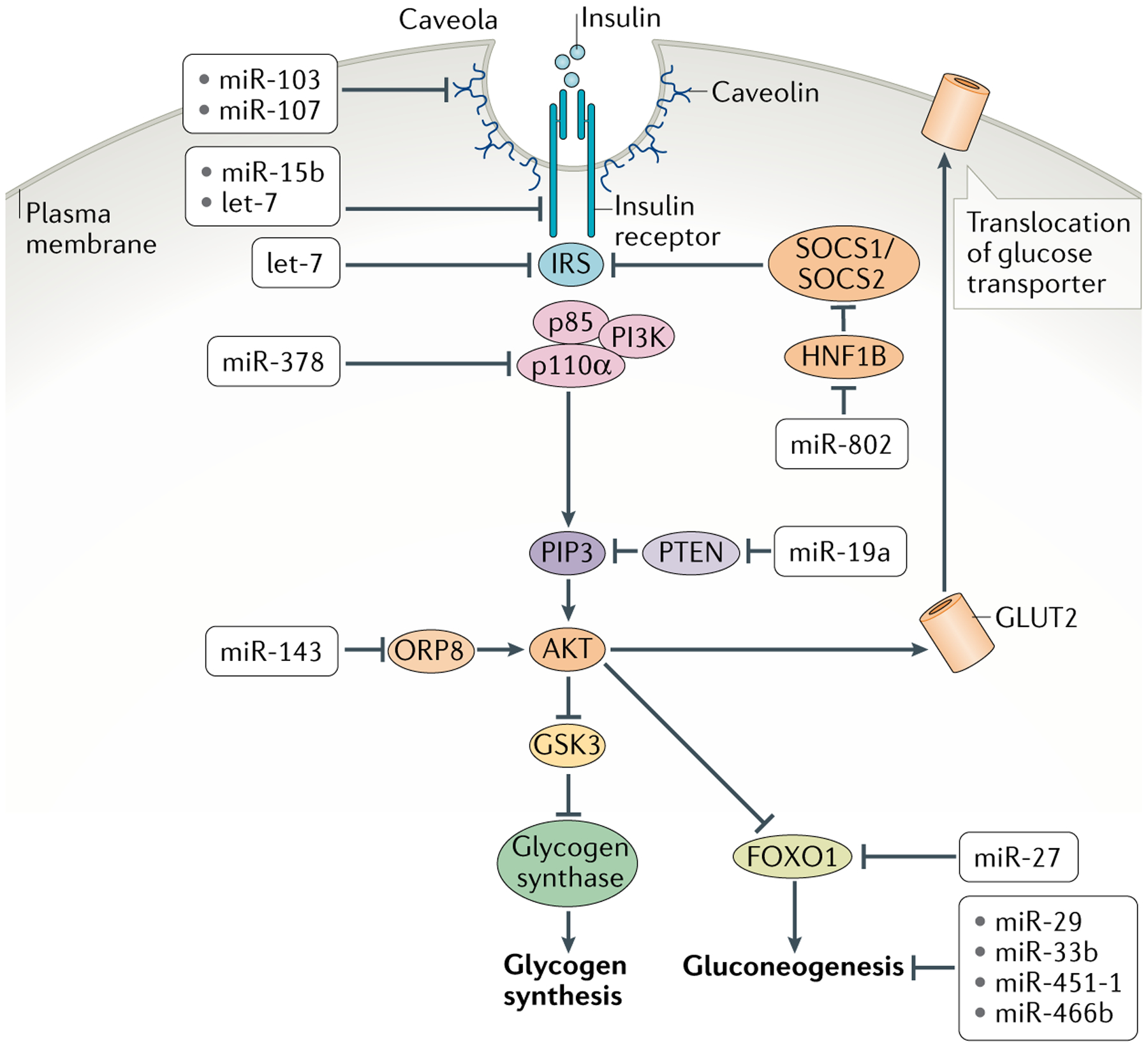
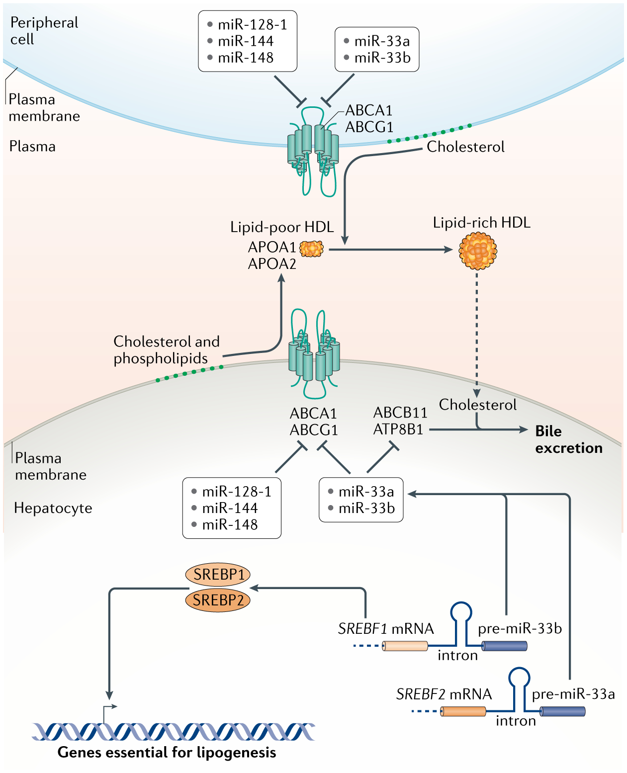
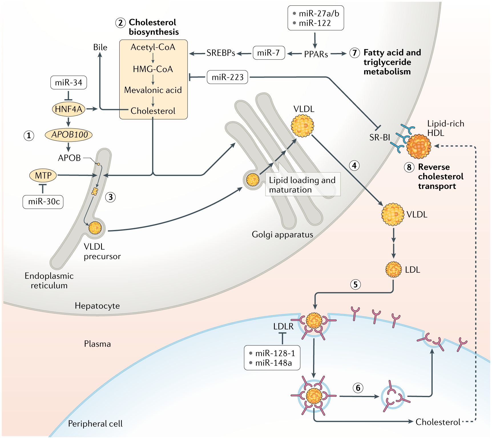
In animals, systemic control of metabolism is conducted by metabolic tissues and relies on the regulated circulation of a plethora of molecules, such as hormones and lipoprotein complexes. MicroRNAs (miRNAs) are a family of post-transcriptional gene repressors that are present throughout the animal kingdom and have been widely associated with the regulation of gene expression in various contexts, including virtually all aspects of systemic control of metabolism. Here we focus on glucose and lipid metabolism and review current knowledge of the role of miRNAs in their systemic regulation. We survey miRNA-mediated regulation of healthy metabolism as well as the contribution of miRNAs to metabolic dysfunction in disease, particularly diabetes, obesity and liver disease. Although most miRNAs act on the tissue they are produced in, it is now well established that miRNAs can also circulate in bodily fluids, including their intercellular transport by extracellular vesicles, and we discuss the role of such extracellular miRNAs in systemic metabolic control and as potential biomarkers of metabolic status and metabolic disease.


Conflict of interest statement

Competing interests

The authors declare no competing interests.

Figures

Fig. 1 |
Fig. 1 |. Insulin synthesis and secretion in the islet β-cell and its control by mirNas.
The influx of glucose and its effects on synthesis and secretion of insulin in pancreatic β-cells. Numbers 1–9 refer to the different steps of insulin release. Glucose enters the cell via the transporter GLUT2 (1). It is then metabolized via glycolysis in the cytosol and the tricarboxylic acid (TCA) cycle in mitochondria to generate ATP (2). This increases the ratio of ATP to ADP in the cell (3). A K+ ion channel senses the elevated ATP/ADP ratio and closes (4), resulting in plasma membrane depolarization (5). This is sensed by a voltage-gated calcium channel, which opens, leading to Ca2+ influx (6). Elevation of calcium level triggers rearrangement of the actin cytoskeleton and docking of insulin secretory granules to the plasma membrane, leading to their fusion and release. In parallel, ERK senses the high ATP/ADP ratio and activates the transcription factor NEUROD1 to potentiate insulin gene transcription (8). Following the first phase of insulin release is a second, more sustained phase, which involves substantial cytoskeletal rearrangements and long-range transport of insulin vesicles from intracellular stores to the plasma membrane. Binding of glucagon-like peptide 1 (GLP1) to GLP1R augments these processes via the cAMP–protein kinase A signal transduction pathway (9). MicroRNAs (miRNAs) expressed in β-cells that are implicated in regulating insulin are mostly involved in antagonizing the various steps in the synthesis and release of insulin. They attenuate GLP1 signal transduction, inhibit insulin gene transcription, reduce calcium influx to the cytosol and lower secretory granule docking and release. However, a small subset of miRNAs have also been shown to promote insulin synthesis. AC, adenylyl cyclase.
Fig. 2 |
Fig. 2 |. Insulin signal transduction and metabolic responses in peripheral tissues and their control by mirNas.
Cells bind circulating insulin via the insulin receptor, which activates insulin receptor substrate (IRS). This in turn recruits association of the regulatory p85 and catalytic p110α subunits of phosphoinositide 3-kinase (PI3K), which becomes activated. Activated PI3K catalyses the addition of phosphate groups to the 3′-OH position in the inositol ring of phosphoinositides to generate phosphatidylinositol 3,4,5-trisphosphate (PIP3). Phosphatase and tensin homologue (PTEN) converts PIP3 to hypophosphorylated isoforms. Binding of PIP3 to AKT stimulates its kinase activity, and two of its targets are glycogen synthase kinase 3 (GSK3) and the transcription factor FOXO1, both of which are repressed by phosphorylation. Phosphorylated GSK3 is blocked from inactivating glycogen synthase, resulting in synthesis of glycogen. Phosphorylated FOXO1 is blocked from transcribing genes encoding enzymes in the gluconeogenic pathway, resulting in dampening of gluconeogenesis. AKT additionally promotes glucose uptake by increasing the levels of glucose transporters at the cell membrane. Shown are the various microRNAs (miRNAs) that regulate insulin signal transduction in cells and miRNAs that regulate glycogenesis and gluconeogenesis. HNF1B, hepatocyte nuclear factor 1β; ORP8, oxysterol-binding protein-related protein 8.
Fig. 3 |
Fig. 3 |. HDL biogenesis and control by mirNas in hepatocytes.
High-density lipoproteins (HDLs) are generated by the association of apolipoproteins APOA1 and APOA2 with lipids. Hepatocytes assemble extracellular lipid-poor HDL particles, which are then enriched with lipids, mostly cholesterol, derived from peripheral cells to make lipid-rich HDL. Loading of lipids into HDLs occurs via the ATP-binding cassetter transporters ABCA1 and ABCG1. The levels of these transporters are inhibited by a number of microRNAs (miRNAs) as shown. Two of these miRNAs, miR-33a and miR-33b, perform this function in concordance with two transcription factors, SREBP1 and SREBP2, which stimulate the synthesis of enzymes in lipogenic pathways. Since the precursors of miR-33a and miR-33b are located in the introns of the genes encoding SREBP2 and SREBP1 respectively, both the proteins and the miRNAs are co-expressed in tandem. miR-33a and miR-33b also inhibit expression of ABCB11 and ATP8B1, which in hepatocytes transport sterols into bile for excretion, allowing clearance of cholesterol derived from lipid-rich HDL returned to the liver after loading in the periphery. Altogether, the coordinated actions of these miRNAs and transcription factors result in accumulation of lipids within hepatocytes.
Fig. 4 |
Fig. 4 |. Control of lipid metabolism and LDL biogenesis/turnover by mirNas.
The biogenesis and turnover of low-density lipoproteins (LDL) is shown in steps 1–6. Apolipoprotein B-100 (APOB100) is synthesized and inserted into the endoplasmic reticulum of hepatocytes (1). In parallel, sterol biosynthesis converts the metabolite acetyl-CoA into cholesterol (2). Microsomal triglyceride transfer protein (MTP) transfers lipids to APOB100 to initiate assembly of very-low-density lipoprotein (VLDL) complexes in the endoplasmic reticulum (3). VLDL is shuttled to the Golgi apparatus, where it matures via lipidation, and then is secreted from hepatocytes (4). In the circulatory system, VLDL is oxidized into LDL, and this species reaches cells of the periphery. There, LDL binds to LDL receptor (LDLR) and is endocytosed (5). Sterols and other lipids are transferred out of the endosomes of peripheral cells, where they are utilized (6). The endosomes are recycled to the plasma membrane to redeposit LDLR on the surface. MicroRNAs (miRNAs) regulate virtually all steps of the LDL pathway, as indicated. Moreover, some miRNAs also regulate enzymes that control fatty acid and triglyceride metabolism in the liver, mostly by promoting these processes (7). However, miR-223 represses genes involved in cholesterol biosynthesis. In addition, miR-223 inhibits reverse transport of cholesterol from the peripheral cells back to the liver by repressing hepatocyte expression of the scavenger receptor SR-BI, which binds to cholesterol-loaded HDL and transports cholesterol into the liver (8). The net effect of the action of miR-223 is to reduce hepatic cholesterol levels. HNF4A, hepatocyte nuclear factor 4α; HMG-CoA, 3-hydroxy-3-methylglutaryl-CoA; PPARs, peroxisome proliferator-activated receptors; SREBPs, sterol regulatory element-binding proteins.

Similar articles

  • MicroRNAs in metabolic disease.
    Fernández-Hernando C, Ramírez CM, Goedeke L, Suárez Y. Fernández-Hernando C, et al. Arterioscler Thromb Vasc Biol. 2013 Feb;33(2):178-85. doi: 10.1161/ATVBAHA.112.300144. Arterioscler Thromb Vasc Biol. 2013. PMID: 23325474 Free PMC article. Review.
  • The miRNA Interactome in Metabolic Homeostasis.
    Hartig SM, Hamilton MP, Bader DA, McGuire SE. Hartig SM, et al. Trends Endocrinol Metab. 2015 Dec;26(12):733-745. doi: 10.1016/j.tem.2015.09.006. Epub 2015 Oct 20. Trends Endocrinol Metab. 2015. PMID: 26492831 Free PMC article. Review.
  • Insight into miRNAs related with glucometabolic disorder.
    Zhang BH, Shen CA, Zhu BW, An HY, Zheng B, Xu SB, Sun JC, Sun PC, Zhang W, Wang J, Liu JY, Fan YQ. Zhang BH, et al. Biomed Pharmacother. 2019 Mar;111:657-665. doi: 10.1016/j.biopha.2018.12.123. Epub 2019 Jan 3. Biomed Pharmacother. 2019. PMID: 30611990 Review.
  • The Role of miRNAs in Metabolic Diseases.
    Macvanin M, Obradovic M, Zafirovic S, Stanimirovic J, Isenovic ER. Macvanin M, et al. Curr Med Chem. 2023;30(17):1922-1944. doi: 10.2174/0929867329666220801161536. Curr Med Chem. 2023. PMID: 35927902 Review.
  • Integrative roles of microRNAs in lipid metabolism and dyslipidemia.
    Sedgeman LR, Michell DL, Vickers KC. Sedgeman LR, et al. Curr Opin Lipidol. 2019 Jun;30(3):165-171. doi: 10.1097/MOL.0000000000000603. Curr Opin Lipidol. 2019. PMID: 30985366 Free PMC article. Review.

Cited by

References

    1. Carthew RW & Sontheimer EJ Origins and mechanisms of miRNAs and siRNAs. Cell 136, 642–655 (2009). - PMC - PubMed
    1. Bartel DP MicroRNAs: target recognition and regulatory functions. Cell 136, 215–233 (2009). - PMC - PubMed
    1. Filipowicz W, Bhattacharyya SN & Sonenberg N Mechanisms of post-transcriptional regulation by microRNAs: are the answers in sight? Nat. Rev. Genet 9, 102–114 (2008). - PubMed
    1. Baek D et al. The impact of microRNAs on protein output. Nature 455, 64–71 (2008). - PMC - PubMed
    1. Selbach M et al. Widespread changes in protein synthesis induced by microRNAs. Nature 455, 58–63 (2008). - PubMed

Publication types

LinkOut - more resources